

⚕️ Written by Dr. Sarah Mitchell, MD, MPH  •  📋 Evidence-Based Articles  •  🔍 Medically Reviewed

⚠️ Not a substitute for professional medical advice

Author: HealthAuthorityLife Editorial Team

  • Interstitial Cystitis: Symptoms, Triggers, and Natural Relief

    Interstitial Cystitis: Symptoms, Triggers, and Natural Relief

    Key Takeaways

    • Interstitial cystitis (IC) is a chronic condition causing bladder pressure and pain that does not stem from a typical bacterial infection.
    • Identifying specific IC diet triggers, such as acidic or caffeinated foods, is a foundational step in symptom management.
    • Chronic inflammation within the bladder lining significantly impacts nerve sensitivity and overall pelvic floor health.
    • Gut health and the microbiome are increasingly recognized as essential factors in modulating the systemic inflammation that drives bladder flare-ups.
    • Natural relief for IC often involves a multi-pronged approach, focusing on lifestyle modifications, stress reduction, and dietary adjustments.

    Living with a body that feels constantly under siege is an exhausting reality for millions. For those struggling with the persistent, often debilitating discomfort of interstitial cystitis, the journey to wellness can feel like an endless search for answers in a medical landscape that often overlooks the nuance of chronic pelvic pain. Often referred to as painful bladder syndrome, this condition creates a cycle of urgency, frequency, and pelvic tenderness that disrupts sleep, professional life, and personal well-being. Unlike a standard urinary tract infection that resolves with a course of antibiotics, this condition requires a deep, long-term strategy involving internal balance, dietary vigilance, and inflammation management. The healthauthoritylife Editorial Team has curated this comprehensive guide to help you understand the mechanisms behind your symptoms and explore sustainable pathways toward finding relief.

    What Is Interstitial Cystitis and Why Does It Happen

    Interstitial cystitis is a chronic condition characterized by recurrent discomfort or pain in the bladder and surrounding pelvic region. Unlike acute infections, which are caused by invasive bacteria that can be easily cultured, interstitial cystitis typically involves a complex, non-infectious inflammatory response. The bladder wall is protected by a specialized layer called the glycosaminoglycan (GAG) layer—a substance that acts as a shield, preventing irritants found in urine from penetrating the underlying tissue. In many individuals managing this condition, this protective lining appears to be compromised or “leaky.” When the barrier is thin or damaged, the highly acidic and electrolyte-rich content of urine makes direct contact with the sensitive bladder wall nerves, resulting in the telltale, searing pain associated with the syndrome.

    Experts often describe the condition as a systemic issue rather than a localized bladder problem alone. While the bladder is the site of the most intense symptoms, the root cause may involve multiple overlapping systems. Some research suggests that the bladder lining damage is secondary to a more generalized dysfunction in the way the body processes pain signals. The nerves in the pelvic floor can become hyper-sensitized, essentially “learning” to perceive normal bladder fullness as extreme pain. This is why individuals often find that even when their bladder is empty, the sensation of urgency remains.

    Why does this happen to some people and not others? There is no singular cause, but rather a constellation of factors that likely trigger the onset. Genetic predisposition may play a role, as some families report multiple members suffering from bladder pain disorders or other autoimmune conditions. Additionally, some theories posit that a previous, severe urinary tract infection might have altered the local immune environment, triggering a chronic inflammatory cascade that never fully shut off after the original bacteria were cleared. Others believe that pelvic floor dysfunction—muscles that are too tight or in constant spasm—creates an environment of restricted blood flow, further damaging the bladder tissue over time. Ultimately, understanding interstitial cystitis requires shifting the focus from “finding a cure” to “managing the terrain.” By addressing the factors that weaken the bladder’s mucosal lining and calming the nervous system’s response to pelvic sensory input, many people find that they can reclaim a significant portion of their quality of life.

    Early Warning Signs and Common Symptoms of IC

    The progression of IC symptoms is rarely linear. It often begins as subtle, easily dismissed discomfort—perhaps a slight sense of pressure after drinking coffee or a mild sensation of urgency that feels like a standard “full bladder.” However, as the condition progresses, these signs become more persistent and disruptive. One of the primary indicators is urinary frequency. While a healthy bladder can comfortably hold urine for several hours, someone experiencing an IC flare may find themselves needing to visit the restroom six, eight, or even twenty times in a single day, often with very little output per trip.

    The pain associated with the syndrome is distinct and usually manifests as a deep pressure or burning sensation in the suprapubic area, just above the pubic bone. This pain is not constant in every patient; for many, it is cyclical, worsening as the bladder fills and often providing temporary, albeit brief, relief immediately after emptying. The intensity of this pain can range from a dull, persistent ache to sharp, stabbing sensations that can make daily activities, such as sitting at a desk or driving a car, difficult to tolerate. Furthermore, many individuals report pain during sexual intercourse, which can add a layer of emotional and relational strain to the physical challenge.

    It is common for patients to experience “flares,” which are periods where symptoms intensify significantly for days or weeks at a time. These flares are often triggered by specific environmental, emotional, or dietary factors. For instance, a period of high emotional stress might cause the pelvic floor muscles to tighten involuntarily, which in turn compresses the bladder and spikes symptoms. Understanding your specific pattern of IC symptoms is the first step toward effective management. Keeping a bladder diary—a simple log tracking your fluid intake, symptom intensity, and potential triggers—is widely considered by specialists to be one of the most effective tools for identifying the specific “tell-tale” signs that precede your unique flare-ups. By noticing that your urgency increases specifically after consumption of certain foods or during specific times of your hormonal cycle, you can begin to anticipate and mitigate the severity of these episodes before they reach a peak of discomfort.

    Approach Mechanism Best For
    Elimination Dieting Removing known acidic or reactive chemical irritants. Long-term maintenance and identifying specific triggers.
    Pelvic Floor Therapy Manual release of tightened, hypertonic musculature. Those with comorbid muscle tension or referred pain.
    Stress Reduction Lowering cortisol and systemic nervous system arousal. Reducing the frequency and duration of sudden flares.

    The Role of Chronic Inflammation in Bladder Health

    Inflammation is a natural biological process meant to protect the body from injury or infection, but when it becomes chronic, it shifts from a repair mechanism to a destructive force. In the context of the bladder, chronic inflammation causes the tissue to remain in a constant state of irritation, preventing the healing of the epithelial lining. When the bladder remains inflamed, the immune cells in the area continue to release signaling molecules that keep pain nerves on high alert. This creates a feedback loop: the nerves send pain signals to the brain, the brain perceives danger, and the body may respond by further restricting blood flow or tensing pelvic muscles, which only adds to the inflammatory load.

    Managing the inflammatory response is critical for those seeking natural relief for IC. This involves looking beyond the bladder and examining systemic health markers. Oxidative stress, which occurs when there is an imbalance between free radicals and antioxidants in the body, is a primary driver of inflammation. High levels of systemic oxidative stress can weaken the integrity of cell membranes throughout the body, including the bladder’s protective GAG layer. By incorporating an anti-inflammatory lifestyle, you are not just treating the bladder; you are providing the foundational support the body needs to repair damaged tissues.

    There are several ways to lower this inflammatory burden. Nutrition is a massive component, specifically focusing on foods that provide antioxidants and phytonutrients. However, systemic inflammation is also affected by factors such as sleep quality, chronic stress, and environmental toxin exposure. When the body is sleep-deprived, inflammatory markers like C-reactive protein (CRP) tend to rise, which can directly exacerbate bladder tenderness the following day. Similarly, emotional stress triggers the release of cortisol, which, in excess, interferes with the body’s ability to manage inflammation. Consequently, a comprehensive approach to bladder health must include stress management techniques, such as mindfulness, deep breathing, or restorative movement, which help down-regulate the nervous system and give the bladder tissue a chance to rest and recover. Viewing IC as an inflammatory state rather than just a local irritation empowers patients to take a holistic view of their health, focusing on lowering the “background noise” of systemic inflammation that contributes to daily pain.

    Common Dietary Triggers That Flare Interstitial Cystitis

    Dietary modification is often the cornerstone of initial treatment strategies for interstitial cystitis. Because everything consumed eventually makes its way through the renal system, the urine’s chemical composition is directly influenced by diet. For a sensitive bladder, this means that even minor shifts in acidity or chemical makeup can turn a peaceful day into a painful flare-up. The most common IC diet triggers are those that are inherently acidic or contain substances known to irritate the bladder lining directly. Coffee, for example, is one of the most frequently cited culprits. Both the caffeine and the high acidity of coffee are notorious for triggering urgency and burning sensations in the bladder.

    Beyond caffeine, many individuals find that alcohol—particularly red wine and beer—serves as a potent irritant. Alcohol acts as a diuretic, which increases the frequency of bladder emptying and can cause the bladder to become dehydrated or concentrated with acidic byproducts. Citrus fruits and juices, such as lemons, oranges, and grapefruit, are also high on the “avoid” list for most people. While these are healthy for many, the citric acid can be incredibly harsh on a damaged bladder wall. Furthermore, artificial sweeteners, specifically aspartame and saccharin, have been linked to increased symptoms for a significant number of people. These substances may alter the chemical environment of the urine, making it more abrasive as it sits in the bladder.

    Managing dietary triggers is not about permanent deprivation, but rather about identifying your unique threshold. Everyone’s sensitivity level is different. Some individuals may be able to tolerate a small cup of decaffeinated coffee, while others may react to a single slice of aged cheese or a small amount of soy sauce. The key to effective management is the elimination-reintroduction process. By removing all common triggers for a set period—typically two to four weeks—you allow the bladder lining time to settle and the inflammation to subside. Once the baseline is established, you can carefully reintroduce one potential trigger at a time, noting any symptoms over the following 48 hours. This methodical approach allows you to curate a personalized “safe” list, ensuring you are not restricting your diet unnecessarily, but protecting your bladder from the specific foods that cause you the most distress.

    The Connection Between Gut Health and Bladder Pain

    In recent years, the medical community has increasingly acknowledged the “gut-bladder axis”—the intricate link between the health of the digestive tract and the function of the bladder. The human gut contains trillions of microbes that regulate everything from immune responses to systemic inflammation. When the gut microbiome is out of balance, a condition known as dysbiosis, it can lead to increased intestinal permeability, often called “leaky gut.” When this happens, bacterial byproducts and undigested food particles may enter the bloodstream, triggering an immune response and generalized systemic inflammation that can settle in the bladder.

    The proximity of the gut and the bladder in the pelvic cavity also means that digestive issues can directly impact the bladder’s physical comfort. Chronic constipation, for instance, is a major issue for those with bladder pain. A rectum full of stool puts mechanical pressure on the bladder, which restricts its capacity and can cause the bladder to signal the brain to “empty” more frequently. Furthermore, chronic constipation changes the pelvic floor muscle dynamics, often leading to tension that worsens the discomfort of IC symptoms. When the gut is sluggish, the nerves surrounding the bladder can become irritated, creating a sense of urgency that is actually a result of bowel pressure rather than true bladder filling.

    Supporting gut health is, therefore, a powerful strategy for how to manage interstitial cystitis naturally. This begins with fiber intake, which helps ensure regular bowel movements and reduces the mechanical pressure on the bladder. Additionally, consuming fermented foods or high-quality probiotics can help rebalance the microbiome, potentially lowering the systemic inflammatory signals that keep the bladder in a state of distress. Many individuals also find relief by focusing on easily digestible foods during a flare, reducing the workload on their digestive system and minimizing the potential for inflammatory flare-ups. By treating the digestive tract as an ally in bladder health, you create a much more stable environment, reducing the “hidden” triggers that keep the bladder in a state of chronic, painful sensitivity. Paying attention to your digestion is not just for gut health—it is a proactive, foundational step in silencing the bladder’s distress signals.

    Natural Anti-Inflammatory Supplements for IC Support

    Managing the systemic inflammation associated with interstitial cystitis often requires a multifaceted approach. While pharmacological interventions exist, many individuals living with painful bladder syndrome turn to nutritional supplementation to support bladder wall integrity and reduce reactive inflammation. When selecting supplements, the focus should remain on ingredients that soothe the mucosal lining or provide antioxidant support to neutralize oxidative stress within the pelvic region.

    Quercetin is perhaps the most widely recognized supplement in the IC community. As a bioflavonoid found in many fruits and vegetables, quercetin acts as a natural mast cell stabilizer. Since mast cell activation is thought to play a role in the hypersensitivity of the bladder lining, stabilizing these cells may help reduce the release of histamines that trigger burning and frequency. Many people find success by combining quercetin with bromelain, an enzyme that may improve the absorption of quercetin while offering additional systemic anti-inflammatory benefits.

    Another staple in the natural management of IC is D-Mannose. While primarily known for its use in supporting urinary tract health, some with IC find it helpful in creating a protective environment. Furthermore, Aloe Vera, specifically in freeze-dried, supplement form, is frequently utilized to provide a soothing, mucilaginous coating to the bladder wall. By mimicking the GAG (glycosaminoglycan) layer—which is often compromised in IC patients—aloe supplements can act as a physical buffer against the irritating effects of acidic urine.

    It is important to note that the efficacy of these supplements can be highly individual. What provides relief for one person may cause a flare in another due to the specific acidity or chemical profile of the supplement. When introducing new supplements, experts suggest a “start low and go slow” approach to monitor for potential bladder reactions.

    Supplement Primary Mechanism Best For
    Quercetin Mast Cell Stabilization Reducing histamine-related bladder burning
    Aloe Vera (Freeze-dried) Mucosal Lining Support Soothing internal bladder wall irritation
    Omega-3 Fatty Acids Systemic Anti-inflammatory Long-term reduction of chronic inflammation
    Magnesium Glycinate Muscle Relaxation Reducing pelvic floor tension and spasms

    Lifestyle Habits to Reduce Bladder Pressure and Frequency

    The daily habits you cultivate play a significant role in managing bladder inflammation causes and reducing the severity of IC symptoms. Because the bladder is highly sensitive to external pressure, physical environment and posture become key components of your management strategy. Simple adjustments can significantly lower the tactile triggers that lead to urgency.

    Clothing choices are often overlooked but are essential for comfort. Tight-fitting clothing—especially high-waisted jeans, restrictive shapewear, or tight leggings—can exert unnecessary pressure on the suprapubic area. For those with a hypersensitive bladder, this constant external compression can exacerbate feelings of urgency and frequency. Switching to looser, breathable fabrics that do not constrict the pelvic girdle is a practical, immediate change that many find provides instant relief.

    Hydration management is another critical habit. While it is tempting to limit water intake to reduce the need for frequent urination, this can actually backfire. Concentrated, highly acidic urine is much more irritating to a damaged bladder lining than dilute urine. The goal is to maintain a consistent, steady intake of water throughout the day to keep urine pH neutral and diluted. Avoiding “gulping” large amounts of fluid all at once—which can cause a rapid, painful bladder expansion—and instead sipping small amounts steadily can help maintain a comfortable bladder volume without triggering spasms.

    Furthermore, evaluating your seating posture can alleviate pelvic floor strain. Prolonged sitting on hard surfaces can compress the soft tissues of the pelvic floor. Utilizing a “donut” cushion or a coccyx relief pillow can shift the weight away from the perineum and pelvic floor muscles. If your work requires long hours at a desk, setting a timer to stand up, stretch, and readjust every thirty minutes can prevent the pelvic floor from “locking” in a state of hypertonicity.

    Stress Management Techniques for Pelvic Floor Relaxation

    The connection between the nervous system and the pelvic floor is profound. In many individuals with IC, stress does not just manifest mentally; it physically migrates to the pelvic region. When the body enters a “fight or flight” state, the pelvic floor muscles often tighten involuntarily. Since these muscles surround the bladder and urethra, this chronic tension can mimic or worsen IC symptoms, leading to increased frequency and a feeling of incomplete emptying.

    Diaphragmatic breathing is a cornerstone technique for unlocking this tension. Unlike shallow chest breathing, deep diaphragmatic breathing encourages the pelvic floor to naturally lengthen and relax with each inhale. To practice this, place one hand on your chest and the other on your abdomen. As you inhale slowly through your nose, aim to move only your hand on your abdomen, allowing the pelvic floor to descend gently. As you exhale, allow the muscles to return to their natural state. Repeating this for five to ten minutes in a quiet space can help signal to the nervous system that it is safe to down-regulate, effectively reducing the internal pressure on the bladder.

    Mindfulness-based stress reduction (MBSR) and guided progressive muscle relaxation are also highly effective. Progressive muscle relaxation involves systematically tensing and then releasing different muscle groups, with specific focus on the glutes, thighs, and pelvic floor. By teaching your brain to identify the difference between tension and relaxation, you gain better conscious control over your pelvic muscles, which helps prevent the guarding patterns that often trigger IC flares.

    Lastly, movement therapies such as restorative yoga or Yin yoga are often recommended. These practices focus on long-held, gentle stretches that target the hips and lower back, areas that hold tension deeply connected to the pelvic floor. Avoid high-impact exercises like running or heavy lifting during a flare, as these can jar the bladder. Instead, opt for gentle movement that keeps the pelvis mobile and fluid, helping to prevent the stagnation of tension in the lower body.

    When to See a Specialist for Bladder Pain

    While natural management strategies are powerful, there is a clear threshold where professional medical intervention becomes necessary. Interstitial cystitis is a diagnosis of exclusion, meaning your doctor must first rule out other conditions—such as chronic urinary tract infections, bladder stones, pelvic organ prolapse, or even bladder cancer—that can present with identical symptoms.

    You should seek a specialist, typically a urologist or a urogynecologist with specific expertise in interstitial cystitis, if you notice “red flag” symptoms. These include the presence of blood in your urine (hematuria), unexplained weight loss, persistent fever, or pain that radiates severely to the kidneys or lower back. Furthermore, if your pain levels are preventing you from maintaining a functional daily life, disrupting your sleep on a nightly basis, or causing significant psychological distress, professional medical support is non-negotiable.

    Specialists can offer diagnostic procedures such as cystoscopy with hydrodistention, which allows them to inspect the bladder lining for “Hunner’s lesions” or signs of glomerulations (pinpoint hemorrhages). They may also provide access to specialized treatments, such as bladder instillations (medication delivered directly into the bladder), physical therapy via a pelvic floor specialist, or prescription medications that help stabilize the bladder muscle or decrease nerve sensitivity. Early intervention by a qualified specialist is often the key to preventing the condition from progressing to a more rigid, chronic state.

    Frequently Asked Questions

    Can interstitial cystitis go away on its own?

    For many people, IC is a chronic condition that fluctuates over time rather than disappearing entirely. However, many individuals experience long periods of remission where symptoms are minimal or nonexistent, especially when they successfully identify and manage their specific triggers through diet and lifestyle changes.

    Is IC diet the same for everyone?

    No, the IC diet is highly individual. While there are common “known offenders”—such as caffeine, alcohol, citrus, and spicy foods—each person has a unique threshold. Keeping a detailed food diary to track your specific reactions is the most reliable way to create a personalized diet that minimizes flare-ups.

    How can I tell the difference between a UTI and an IC flare?

    UTIs are caused by bacteria and typically involve a positive urine culture, often accompanied by a fever or cloudy, foul-smelling urine. IC flares are non-bacterial and, while the pain is similar, there is typically no infection present. If you suspect an infection, it is vital to have a doctor perform a urinalysis to rule out a true UTI.

    Does pelvic floor physical therapy really help with bladder pain?

    Yes, many experts consider pelvic floor physical therapy (PFPT) to be one of the most effective treatments for IC. Because the bladder and pelvic muscles share nerve pathways, relaxing the muscles through specialized manual therapy can significantly reduce the urgency, frequency, and pain associated with the condition.

    Are there specific types of water that are better for IC?

    Some people with IC find that highly acidic or highly alkaline water can trigger symptoms. Many patients prefer spring water with a neutral pH (around 7.0 to 7.4). It is generally recommended to avoid carbonated water or mineral waters with high levels of additives, as these can irritate the already sensitive bladder lining.

    How long does an IC flare typically last?

    There is no standard duration for an IC flare; it varies from person to person. A flare may last a few hours, several days, or, in more severe cases, weeks. The duration often depends on how quickly the initial trigger is identified and removed, as well as the implementation of calming strategies like pelvic floor relaxation and proper hydration.

    Conclusion

    Living with interstitial cystitis requires a balanced approach that combines self-awareness, dietary vigilance, and professional guidance. By understanding your unique symptom profile, you can shift from a reactive mindset to a proactive one, effectively managing your health on your own terms. While IC is undoubtedly challenging, it is also manageable; many people lead full, active, and vibrant lives by mastering the art of symptom control through the strategies outlined in this guide.

    Remember that you are your own best advocate. Start by keeping a detailed log of your daily habits, triggers, and the effectiveness of your natural relief strategies. If you are struggling to find relief, do not hesitate to reach out to a urology specialist who understands the complexity of painful bladder syndrome. Your journey to comfort is a marathon, not a sprint—take it one day at a time, celebrate your small wins, and remain consistent with your care. If you are ready to take control of your bladder health, begin by tracking your diet this week and scheduling a consultation with a pelvic health professional to discuss your personalized management plan.

    By healthauthoritylife Editorial Team

    This article is for informational purposes only and is not a substitute for professional medical advice. Always consult your doctor before making changes to your diet, exercise routine, or treatment plan.


  • Why Hashimoto’s Causes Cold Intolerance & How to Stay Warm

    Why Hashimoto’s Causes Cold Intolerance & How to Stay Warm

    Key Takeaways

    • Hashimoto’s cold intolerance is primarily driven by a slowed metabolic rate caused by insufficient thyroid hormone production.
    • Thyroid hormone regulation acts as the body’s internal thermostat, governing how efficiently your cells produce heat.
    • Chronic cold hands and feet are classic clinical indicators of suboptimal thyroid function often associated with Hashimoto’s.
    • Metabolic health in Hashimoto’s patients is deeply linked to adrenal function, as the stress response directly impacts hormonal conversion.
    • Recognizing early thermal sensitivity allows for proactive interventions that can support better temperature regulation and overall vitality.

    For those living with Hashimoto’s thyroiditis, the world can often feel like an unrelenting winter, even in the middle of July. It is a common, yet frequently misunderstood experience: sitting in a climate-controlled room while wearing a thick sweater, or feeling the blood drain from your fingers until they turn ghost-white at the slightest chill. This persistent sensation of being perpetually cold is not merely a personality quirk or a lack of blood circulation; it is a profound biological signal from your endocrine system. When your immune system attacks your thyroid, it disrupts the body’s ability to manage its most vital functions, chief among them being the ability to maintain a stable internal temperature. Understanding the physiological mechanisms behind Hashimoto’s cold intolerance is the first step toward regaining control over your comfort, your metabolism, and your quality of life.

    The Connection Between Thyroid Function and Body Temperature

    The thyroid gland is often compared to the conductor of an orchestra, or more accurately in this context, the furnace of the human body. Its primary job involves the secretion of triiodothyronine (T3) and thyroxine (T4), hormones that travel through the bloodstream to reach nearly every cell in the body. These hormones serve as the master switches for your metabolic rate. When your thyroid is functioning optimally, these hormones signal your cells—specifically the mitochondria—to produce energy, and a natural byproduct of this energy production is heat. This process, known as thermogenesis, is how your body maintains its standard core temperature of approximately 98.6 degrees Fahrenheit, regardless of the environment around you.

    Thyroid hormone regulation is a delicate feedback loop. Your hypothalamus, the brain’s control center, monitors the level of circulating thyroid hormones. If levels dip, the hypothalamus releases thyrotropin-releasing hormone (TRH), which prompts the pituitary gland to secrete thyroid-stimulating hormone (TSH). Under normal conditions, this system is exceptionally efficient. However, in Hashimoto’s thyroiditis, the autoimmune attack gradually degrades the functional tissue of the thyroid gland. As fewer cells remain to produce T4, the body’s thermostat begins to malfunction. Because the hormone signal is weak, the cellular “furnace” does not receive the command to burn fuel at the appropriate intensity. This leads to a systemic decline in metabolic heat production.

    The result is a phenomenon where the body becomes hypersensitive to environmental changes. A healthy individual might adapt to a drop in room temperature by shivering or increasing metabolic heat through subtle adjustments. For those with compromised thyroid function, these adaptive mechanisms are often sluggish or altogether absent. The body prioritizes vital organ function over the warmth of extremities, which is why cold hands and feet are the first to signal a decline in thyroid health. This is not just a sensation of being “chilly”; it is a measurable physiological state where the body is actively conserving energy because it is struggling to meet the energy demands of basic thermogenesis. Recognizing this connection is vital, as it shifts the focus from simply “feeling cold” to understanding that the body is signaling a deeper hormonal imbalance that requires attention to metabolic health.

    Why Hashimoto’s Patients Experience Chronic Cold Intolerance

    Chronic cold intolerance in the context of Hashimoto’s is more than just a temporary response to a drafty window. It represents a persistent systemic state where the body’s internal set point for temperature has shifted downward. When the immune system mistakenly identifies thyroid tissue as a foreign invader, it creates inflammation and destruction that prevents the gland from meeting the body’s metabolic demand. As the thyroid capacity diminishes, the peripheral conversion of T4 (the inactive hormone) into T3 (the active, heat-producing hormone) often becomes inefficient. This is particularly problematic because T3 is the form of thyroid hormone that directly influences mitochondrial activity and the activation of brown adipose tissue, which is responsible for generating heat.

    Furthermore, many Hashimoto’s patients experience fluctuations in hormone levels due to periodic “flare-ups” of autoimmune activity. These flares can release surges of stored hormone from damaged thyroid cells, followed by a deeper trough of deficiency. This roller-coaster effect makes consistent body temperature regulation nearly impossible. The body never quite knows how much “fuel” it will have available to produce heat from one day to the next. This unpredictability forces the autonomic nervous system to stay in a state of high alert, managing blood flow to conserve core heat at the expense of comfort in the skin and extremities.

    Beyond the lack of T3, structural changes in the blood vessels can exacerbate this intolerance. Low thyroid hormone levels are often linked to altered vascular reactivity. Research and clinical observation suggest that the smooth muscles in the blood vessels of Hashimoto’s patients may be more sensitive to cold, leading to excessive vasoconstriction. This effectively shuts off blood flow to the fingers and toes more aggressively than in someone with a healthy metabolic rate. When you combine this vascular constriction with a reduced base metabolic rate, you create the “perfect storm” for chronic coldness. It is a feedback loop: the body feels cold, constricts blood vessels to protect the core, and as a result, the extremities become even colder, reinforcing the feeling of systemic chilliness. Breaking this cycle requires a multi-faceted approach that addresses hormone replacement, inflammation reduction, and peripheral circulation support.

    Hypothyroidism and Its Impact on Metabolic Rate

    Metabolic health and Hashimoto’s are inseparable. The metabolic rate is essentially the speed at which your body converts the food you eat and the oxygen you breathe into energy. In a hypothyroid state, the engine of your metabolism is essentially idling, even when you are exerting yourself. This is why many people with thyroid-induced cold sensitivity also struggle with fatigue, sluggish digestion, and unexplained weight gain. The body, sensing a shortage of energy, becomes remarkably efficient at energy conservation, but this “thriftiness” manifests as a profound inability to generate heat.

    This metabolic slowdown affects the entire body. The digestive tract, which requires significant energy to process nutrients, often slows down, leading to bloating and constipation. The heart rate may decrease, leading to reduced circulation to the periphery. The skin, as the body’s largest organ, may become dry and thin, losing its ability to act as an effective thermal insulator. These systemic changes are direct consequences of the body trying to survive in a low-energy environment caused by hypothyroidism. When the metabolic rate drops, the body’s baseline “cost of living” decreases, which might seem like a survival advantage in times of famine but is debilitating in modern life.

    To combat this, it is essential to support the body’s metabolic processes holistically. It is not enough to simply “warm up” with external heat; one must provide the body with the precursors it needs to generate its own warmth. This includes addressing nutrient deficiencies—such as selenium, zinc, and iron—which are crucial for the proper conversion of T4 to T3. Without these cofactors, even if the thyroid gland is functioning, the conversion process to active, heat-generating T3 will falter. The following table highlights common approaches to managing these metabolic challenges and provides guidance on who might benefit most from each.

    Strategy Focus Area Best For
    Targeted Nutritional Support Selenium, Zinc, and Vitamin D levels Supporting enzyme pathways for T4 to T3 conversion
    Controlled Thermogenesis Gradual, consistent physical activity Improving blood flow to chronically cold extremities
    Adrenal Rhythm Balancing Stress reduction and sleep hygiene Those with Hashimoto’s who also report chronic burnout
    Thyroid Medication Optimization T4/T3 combination therapies Patients who remain symptomatic despite TSH normalization

    By shifting the focus from treating the cold sensation as a symptom to treating the metabolic slowdown as the root cause, you move toward a more sustainable management plan. It is about encouraging the “furnace” to turn back on by ensuring that the cellular machinery has the correct inputs, the right hormonal signals, and a supportive environment to perform its thermogenic duty.

    Recognizing the Early Signs of Thyroid-Induced Cold Sensitivity

    The transition from a healthy metabolic state to one dominated by Hashimoto’s cold intolerance is rarely an overnight event. It is usually a gradual accumulation of small, subtle shifts in how your body responds to temperature. Many individuals ignore these early warning signs, attributing them to age, stress, or a changing environment. However, catching these signs early can provide a window of opportunity to intervene with lifestyle and clinical adjustments before the autoimmune process causes more extensive damage. One of the most common early indicators is a change in the ability to tolerate air-conditioned environments. If you find yourself consistently needing a sweater or a desk heater when colleagues are comfortable, this is a significant diagnostic clue.

    Another early sign is the “after-shower chill.” People with healthy thyroid function typically have enough metabolic reserve to warm up quickly after getting out of a shower. If you find that it takes an hour or more for your body to return to a baseline sense of warmth, or if you feel a deep, penetrating cold in your bones that a towel or robe cannot fix, it suggests your body lacks the metabolic “bounce” required for temperature recovery. Furthermore, many note that their hands and feet feel unnaturally cold to the touch even when the rest of their body feels relatively warm. This localized temperature dysregulation is a classic sign that the body is diverting heat to the core and sacrificing peripheral circulation.

    In addition to these physical cues, consider the patterns of your daily energy. Does your cold intolerance correlate with your energy crashes? Often, the “afternoon slump” in Hashimoto’s patients coincides with a drop in body temperature. You might notice that by 3:00 PM, your hands are like ice, and your focus begins to wane. This is a clear indicator of the connection between thyroid hormone levels and energy production. Tracking these observations—perhaps using a simple daily journal—can be invaluable when discussing your health with your healthcare team. You are looking for patterns that suggest your internal “thermostat” is failing to maintain the consistency required for vibrant health. When you can pinpoint exactly when and how the cold sets in, you are better equipped to implement targeted solutions, whether that means adjusting your nutrition, managing stress levels, or re-evaluating your current thyroid treatment protocol.

    How Adrenal Health Influences Your Internal Thermostat

    You cannot talk about Hashimoto’s cold intolerance without discussing the adrenal glands. Often called the body’s “stress glands,” the adrenals produce cortisol, the hormone responsible for managing our response to stress. In the delicate dance of endocrine health, the thyroid and the adrenals are tightly coupled. When the body is under chronic stress—which is common for those dealing with an autoimmune condition—the adrenal glands may become either overactive or fatigued. This state of adrenal imbalance has a direct, detrimental impact on your thyroid health, further complicating your ability to regulate body temperature.

    When the body is under stress, the adrenal glands prioritize the production of cortisol to keep you alert and capable of “fighting” the stressor. However, this production can inhibit the conversion of T4 to T3. Since T3 is the primary hormone that powers thermogenesis, this conversion block effectively turns the heat down in your body. Furthermore, cortisol itself plays a role in regulating body temperature; high levels of cortisol can lead to nighttime sweats, while low levels, often seen in later stages of adrenal dysfunction, contribute to an inability to maintain core warmth. This is why many Hashimoto’s patients describe feeling “tired but wired” while simultaneously being unable to get warm.

    The adrenal-thyroid connection is a major factor in why some patients find it so difficult to stay warm even when their thyroid blood tests look “normal.” If your adrenals are exhausted, your cells may become less sensitive to thyroid hormones, a state sometimes referred to as cellular thyroid resistance. Your blood levels of T3 might be sufficient, but the “thermostat” in your cells is not “hearing” the signal. Healing this relationship requires a focus on stabilizing the adrenal rhythm. This involves gentle strategies such as prioritizing deep, restorative sleep, managing the intensity of exercise, and ensuring consistent blood sugar levels throughout the day to prevent unnecessary stress responses. By calming the adrenal response, you allow the body to stop diverting its limited resources toward stress management and instead redirect them toward the vital function of thermogenesis and metabolic health. As you begin to stabilize your stress response, you may find that your internal thermostat naturally begins to function with more reliability, making those chilly mornings significantly easier to navigate.

    Nutrient Deficiencies That Exacerbate Cold Intolerance

    When dealing with Hashimoto’s, the body often struggles with nutrient absorption, creating a “perfect storm” for metabolic slowdown. Even if you are eating enough calories, specific micronutrient deficiencies can impair your thyroid’s ability to convert hormones and generate heat. Thyroid hormone regulation is heavily dependent on specific cofactors; without them, the cellular machinery responsible for thermogenesis—the process of heat production—stutters.

    Iron deficiency is perhaps the most common culprit behind persistent cold hands and feet in those with hypothyroidism. Iron is an essential component of the enzyme thyroid peroxidase, which is necessary for the production of thyroid hormones. Furthermore, iron is required for the conversion of T4 (the inactive hormone) into T3 (the active, heat-generating hormone). When ferritin stores are low, your body may struggle to maintain a stable internal temperature, leading to a state of chronic chilly sensations.

    Selenium and zinc also play pivotal roles in metabolic health. Selenium is critical for the deiodinase enzymes that catalyze the conversion of T4 to T3. Without adequate selenium levels, even if you are taking thyroid medication, your body may fail to utilize it effectively to warm up the tissues. Zinc, meanwhile, is essential for the production of thyroid-releasing hormone (TRH) and is necessary for the integrity of thyroid hormone receptors. A deficiency in either mineral often leaves patients feeling like they are living in a permanent winter, regardless of the room temperature.

    Vitamin B12 and Vitamin D represent two other major areas of concern. Vitamin B12 is vital for maintaining healthy red blood cells; when levels are low, anemia can occur, reducing the body’s ability to transport oxygen—which is needed for heat production—to the extremities. Vitamin D is technically a steroid hormone rather than a vitamin, and it acts as a gatekeeper for thyroid health. Chronic low Vitamin D levels have been clinically observed alongside Hashimoto’s, and because it influences immune system modulation, normalizing these levels is often a primary step in improving the body’s internal thermostat.

    Lifestyle Habits to Naturally Support Your Thyroid Metabolism

    While medication serves as a baseline, lifestyle habits act as the scaffold that supports your metabolic health. The goal is to reduce the “allostatic load”—the physiological wear and tear resulting from chronic stress—which is known to suppress the hypothalamic-pituitary-thyroid (HPT) axis. When you are under chronic stress, your body prioritizes cortisol over thyroid hormone production, effectively turning down your metabolic “furnace” to conserve energy.

    Circadian rhythm regulation is a surprisingly effective tool for temperature management. Your thyroid function is tied to your natural sleep-wake cycle. By exposure to natural morning sunlight and maintaining a consistent bedtime, you help regulate the circadian rhythm, which in turn helps stabilize the hormones that control your basal metabolic rate. Poor sleep quality is directly linked to lower body temperatures; focusing on “sleep hygiene” by keeping the bedroom cool (paradoxically, this helps sleep quality), dark, and quiet can ensure your body has the restorative time it needs to produce hormones efficiently.

    Gentle movement is another pillar of metabolic support. While it is tempting to hibernate when you feel cold, inactivity can worsen the issue by slowing circulation. The key for Hashimoto’s patients is finding the “Goldilocks zone”—exercise that is intense enough to stimulate blood flow and warm the muscles but not so strenuous that it triggers an inflammatory flare-up. Low-impact activities like restorative yoga, walking, or light resistance training help move blood to the extremities and improve insulin sensitivity, which is a key player in how the body generates heat.

    Finally, stress management through practices like breathwork or meditation is not just “self-care”—it is metabolic support. Chronic activation of the sympathetic nervous system (fight-or-flight) can lead to peripheral vasoconstriction, where blood vessels in your hands and feet narrow, making you feel colder. Techniques that engage the parasympathetic nervous system (rest-and-digest) help dilate these blood vessels, allowing for better blood flow and warmth throughout the body.

    Dietary Changes to Improve Thermogenesis in Hashimoto’s

    Dietary strategies for improving cold intolerance focus on two things: providing the fuel required for the thyroid to function and reducing the inflammatory burden that makes thyroid hormone signaling difficult. Because the digestive system often slows down in hypothyroidism, focus on foods that are easy to digest while remaining nutrient-dense.

    The thermic effect of food is a real phenomenon—the energy required to digest certain nutrients actually creates heat. Protein has the highest thermic effect of all macronutrients. Incorporating high-quality, anti-inflammatory proteins like wild-caught fish, grass-fed meats, or properly cooked legumes can help “stoke the fire” of your metabolism. Conversely, eating large amounts of raw, cold foods (like iced smoothies or heavy salads) can sometimes feel difficult for a sluggish digestive system to process. Many people with Hashimoto’s find that switching to warm, cooked meals—like soups, stews, and roasted root vegetables—provides immediate physical comfort and reduces the digestive strain.

    Avoiding blood sugar crashes is equally important for temperature regulation. When your blood sugar drops, your body’s stress response kicks in to stabilize it, which can cause cold flashes and fatigue. Eating balanced meals that include healthy fats (like avocados, coconut oil, or olive oil) helps provide a steady, slow-burning source of energy that supports sustained warmth throughout the day.

    The following table compares different dietary approaches often explored by those with Hashimoto’s to manage energy and warmth:

    Dietary Strategy Mechanism for Warmth Best For
    Cooking Meals Reduces digestive energy expenditure; warms the gut. Those with bloating/slow digestion.
    Increasing Protein Higher thermic effect of food (TEF) increases heat. Maintaining lean muscle & energy.
    Healthy Fats Provides sustained fuel for metabolism. Avoiding blood sugar-induced cold.
    Ginger/Spices Promotes circulation/vasodilation. Improving cold hands and feet.
    Anti-Inflammatory Lowers systemic stress on the thyroid. Reducing overall Hashimoto’s flare-ups.

    When to Consult Your Doctor About Persistent Coldness

    It is important to acknowledge that while feeling cold is a common symptom of Hashimoto’s, it can also be a sign that your current treatment plan requires adjustment. If you find that your cold intolerance is becoming increasingly severe, or if it is accompanied by other warning signs, it is time to schedule an appointment with your healthcare provider.

    First, evaluate if your thyroid levels are truly optimized. Many patients find that their “normal” laboratory range for TSH is not their “optimal” range for symptom resolution. If your TSH is climbing toward the upper end of the reference range, you may be experiencing subclinical hypothyroidism, meaning your body is struggling to maintain metabolic function even if your results fall technically within the normal bracket. Discussing a medication dosage adjustment or a change in medication type (such as the addition of T3, if appropriate) with your doctor can often resolve persistent coldness.

    Secondly, watch for “red flag” symptoms that suggest something other than just thyroid function might be at play. If you experience persistent chest pain, shortness of breath, sudden numbness in a limb, or extreme fatigue that interferes with your ability to carry out daily activities, do not ignore these signs. Additionally, if you have been managing your Hashimoto’s well but suddenly feel colder, you may have developed an secondary issue, such as anemia, adrenal insufficiency, or a vitamin deficiency that requires blood work to diagnose.

    Keep a “symptom log” for a week before your appointment. Note the times of day you feel coldest, what you ate, the ambient temperature, and any stress you experienced. Providing your doctor with specific data makes it much easier for them to determine if your coldness is a manageable side effect of Hashimoto’s or if it signals that your body is in need of more comprehensive metabolic support.

    Managing Daily Environments for Better Temperature Regulation

    Sometimes, the best approach to cold intolerance is a mix of physiological support and tactical environmental management. Because your internal thermostat is not as sensitive as it should be, you need to rely on external aids to maintain a comfortable baseline temperature.

    Layering is the most obvious, but often misused, strategy. Instead of one thick, heavy sweater, use several thin layers. The space trapped between the layers acts as an insulator, keeping your body heat closer to your skin. Natural fibers like wool and silk are far superior to synthetic fabrics; wool remains a good insulator even if it happens to get damp, and it has excellent thermoregulatory properties. Silk provides a lightweight base layer that traps body heat without adding unnecessary bulk.

    Investing in heated gear can be a game-changer for those who spend time in colder climates or work in drafty offices. Heated vests, rechargeable hand warmers, and electric blankets for your feet while you work can provide the extra thermal boost needed to prevent the “numb finger” sensation that often accompanies Hashimoto’s. These tools take the burden off your metabolism to produce all the heat, allowing your body to allocate its limited energy toward other processes like immune system regulation and tissue repair.

    Finally, consider the role of your home environment. Using a humidifier in the winter is essential, as dry air makes the body feel colder due to increased evaporation of moisture from the skin. Furthermore, check for small air leaks around windows or doors. Even a minor draft can trigger a localized cooling effect that keeps your peripheral blood vessels in a state of constriction. By creating an environment that is supportive rather than demanding, you minimize the amount of energy your body wastes on fighting the cold.

    Frequently Asked Questions

    Why do my hands and feet stay cold even in the summer?

    When you have Hashimoto’s, your thyroid hormone levels may be low, which directly impacts blood circulation. Your body prioritizes keeping your core organs warm, and if it lacks sufficient thyroid hormones to run the metabolic process efficiently, it restricts blood flow to the extremities, leading to perpetually cold hands and feet even in warmer weather.

    Can stress really make my cold intolerance worse?

    Yes, significantly. Chronic stress triggers the release of cortisol, which interferes with the conversion of T4 to the active T3 thyroid hormone. Furthermore, stress triggers the sympathetic nervous system, which causes your blood vessels to narrow (vasoconstriction) as a survival mechanism, further reducing the flow of warm blood to your skin and extremities.

    Is there a specific diet that cures cold intolerance?

    There is no single “cure-all” diet, but many people find relief by focusing on an anti-inflammatory, nutrient-dense eating pattern. Prioritizing cooked, warm meals, ensuring adequate protein intake for thermogenesis, and correcting mineral deficiencies like iron, selenium, and zinc can significantly improve how your body generates and retains heat.

    Should I start taking supplements to warm up?

    Before starting any supplements, you should have your blood levels tested by a healthcare provider. Taking high doses of supplements like iron or selenium without a verified deficiency can be harmful. Always consult your doctor to ensure that any supplements you choose are appropriate for your specific health needs and do not interfere with your thyroid medication.

    Will exercise help or hurt my cold sensitivity?

    Moderate, consistent exercise usually helps by increasing circulation and improving metabolic rate. However, extreme or high-intensity exercise can sometimes spike cortisol levels and trigger inflammation, which may temporarily make cold intolerance worse. Aim for low-impact movement that leaves you feeling energized rather than drained.

    If my thyroid labs are “normal,” why am I still freezing?

    Many laboratory “normal” ranges are quite broad, and your personal “optimal” range might be different. Additionally, the test only measures the amount of hormone in your blood, not necessarily how well those hormones are being utilized by your cells. You may want to discuss symptoms with your doctor to explore if you need a medication adjustment or if other factors, like low iron or blood sugar imbalances, are contributing to the coldness.

    Conclusion

    Living with Hashimoto’s requires a proactive approach to your health, especially when it comes to the often-debilitating symptom of cold intolerance. By understanding that your coldness is a reflection of your underlying metabolic health, you can shift from simply “enduring” the chill to actively supporting your body’s internal furnace. Through targeted nutrient support, mindful lifestyle adjustments, and strategic environmental management, it is entirely possible to regain control over your temperature regulation and find comfort again.

    Remember that you do not have to settle for feeling cold all the time. Your symptoms are valid, and they are your body’s way of communicating that it needs more support. Start by tracking your symptoms, consulting with your medical team to optimize your thyroid hormone levels, and implementing small, sustainable changes to your diet and habits. Taking these steps can lead to profound improvements in your quality of life, energy levels, and overall sense of well-being. Start today—your journey to feeling warm and energized begins with the next small, informed decision you make.

    By healthauthoritylife Editorial Team

    This article is for informational purposes only and is not a substitute for professional medical advice. Always consult your doctor before making changes to your diet, exercise routine, or treatment plan.

  • Chronic Epigenetic Aging: How to Slow Down Your Biological Clock

    Chronic Epigenetic Aging: How to Slow Down Your Biological Clock

    Key Takeaways

    • Epigenetic aging reflects your biological age rather than the number of birthdays you have celebrated, offering a more accurate picture of your internal health.
    • DNA methylation acts as a molecular switch, determining how genes are expressed and driving the progression of cellular aging.
    • Environmental stressors, inflammation, and metabolic dysfunction are primary culprits that accelerate your biological clock.
    • While chronological age is fixed, many aspects of your biological age are malleable through specific dietary and lifestyle interventions.
    • Understanding the difference between your genetic code and your epigenome is the first step toward reclaiming control over your health span.

    You have likely noticed that two individuals of the same chronological age can look, feel, and perform vastly differently. While one person may exhibit signs of vitality and robust energy, another might struggle with chronic fatigue or age-related physical decline. This discrepancy is not merely a matter of luck or genetics; it is the visible manifestation of epigenetic aging. Your internal biological clock does not tick in lockstep with the calendar on your wall. Instead, it responds to the cumulative impact of your environment, habits, and physiological stressors. By understanding the intricate mechanisms of your epigenome, you can begin to make informed decisions that help you slow down aging and optimize your cellular health for a longer, more vibrant life.

    What Is Epigenetic Aging and Why Does It Matter?

    At its core, epigenetic aging refers to the chemical modifications occurring on your DNA that alter how genes are expressed without changing the underlying genetic code. Think of your DNA as the hardware of a computer—the static, unchanging blueprint that dictates your eye color, height, and predispositions. Epigenetics, by contrast, acts as the software. It determines which programs are running, which are muted, and which are functioning at optimal capacity. Over time, these molecular markers accumulate or diminish, leading to a drift in how your cells communicate and repair themselves.

    This process matters profoundly because it serves as a biomarker for your health span—the number of years you spend in good health—rather than your life span. When your epigenetic markers start to veer away from an optimal state, cellular efficiency drops. Systems that once regenerated with ease may begin to lag, leading to systemic inflammation and a loss of tissue integrity. Experts generally agree that this drift is the silent engine behind many chronic conditions associated with later life.

    Monitoring these markers is no longer purely theoretical. Modern science allows us to measure biological age through specific patterns of chemical tags on your DNA. If your biological age is higher than your chronological age, it indicates that your cells are experiencing stress at an accelerated rate. By identifying this early, you gain the agency to implement targeted longevity tips before the effects manifest as debilitating illness. This is not about vanity or simply chasing youth; it is about extending your period of high functionality and vitality. When you view aging as a manageable biological process rather than an inevitable decay, the entire framework of your health strategy changes from reactive treatment to proactive optimization.

    Many individuals find that once they understand the “why” behind their cellular aging, they become more motivated to change their daily habits. If you consider that every meal, every hour of sleep, and every interaction with environmental toxins sends a signal to your genes, you realize that you are constantly “programming” your body’s future state. Recognizing that epigenetic aging is a dynamic, fluid process is the most empowering shift you can make for your long-term wellness.

    Epigenetic Aging vs. Chronological Aging Explained

    Distinguishing between chronological and biological clocks is essential for anyone interested in functional health. Chronological aging is the objective measure of time—the duration since your birth. It is a linear, unchangeable progression. No matter what you eat or how much you exercise, you will remain one year older for every rotation of the Earth around the sun. This metric is useful for legal and social purposes, but it often fails to provide a meaningful indicator of how well your body is actually functioning at a cellular level.

    Epigenetic aging, or biological aging, is non-linear and highly responsive to your environment. Your biological clock can be influenced by diet, stress levels, physical activity, sleep hygiene, and exposure to environmental toxins. In some cases, a person may be sixty years old chronologically but possess the metabolic efficiency and cellular health of a forty-year-old. Conversely, individuals exposed to chronic stress or poor nutrition may see their biological age outpace their chronological age by several years.

    Comparison Point Chronological Aging Epigenetic Aging Best For
    Definition Time since birth Molecular status of cells General logistics
    Malleability Fixed/Static Dynamic/Malleable Health optimization
    Key Driver Solar calendar Lifestyle & Environment Predictive health

    The gap between these two ages represents your “longevity potential.” When you learn to slow down aging, you are effectively closing the gap where your biological age has crept ahead of your chronological age. This approach shifts the conversation from merely “living longer” to “staying healthy longer.” While chronological aging is a countdown, epigenetic aging is more like a thermostat. It can be turned up or down depending on the internal climate you create for your cells.

    Experts often point out that we have been conditioned to accept the decline that comes with aging as a biological inevitability. However, when we look at the research regarding epigenetics and health, we see that cellular senescence—the state where cells stop dividing and begin to secrete harmful inflammatory compounds—is not always an immediate occurrence. It is the result of thousands of micro-decisions. By tracking how your biological age responds to changes in your routine, you can observe which interventions are actually working for your unique physiology, rather than relying on generalized advice that may not address your specific cellular needs.

    Common Drivers of Accelerated Biological Aging

    Understanding what forces your body to age faster is just as important as knowing how to slow the process. Accelerated biological aging does not typically happen due to a single event, but rather through the accumulation of chronic “micro-insults” to your system. Primary among these is chronic systemic inflammation. Often described as “inflammaging,” this state of constant, low-grade immune activation forces your cells to focus on defense rather than repair and rejuvenation. When your immune system is perpetually on high alert, your resources are diverted away from long-term maintenance, leading to faster telomere shortening and epigenetic instability.

    Metabolic dysfunction is another major accelerant. When your cells struggle to process nutrients efficiently—particularly glucose—it leads to a state of oxidative stress. High levels of circulating sugar and insulin can damage proteins and lipids, which in turn signals your cells to alter their epigenetic markers in ways that are generally unfavorable for longevity. This is why managing blood glucose stability is frequently cited as a foundational pillar for those attempting to reverse biological age or at least stabilize it.

    Environmental toxicity also plays a significant role in how your biological clock ticks. We are exposed to a myriad of pollutants, endocrine disruptors, and synthetic chemicals through our water, air, and household products. While the body has robust detoxification pathways, these systems can become overwhelmed, especially when they are already burdened by poor nutrition or high cortisol levels. The epigenome is particularly sensitive to these chemicals, which can leave permanent marks on your DNA, effectively muting genes that are essential for repair and cellular health.

    Furthermore, psychological stress acts as a potent biological signal. Chronic stress triggers the persistent release of hormones like cortisol, which have profound effects on the epigenome. This is not just a “feeling”; it is a physiological cascade that directly impacts the methylation patterns of your genes. When you are in a state of high stress, your body prioritizes immediate survival over long-term longevity. While this served our ancestors well when facing acute physical threats, it is devastating for the modern human living under constant, low-level anxiety. By identifying these drivers—inflammation, metabolic imbalance, toxins, and chronic stress—you can begin to systematically remove the obstacles preventing your body from maintaining its optimal biological state.

    The Role of DNA Methylation in Your Health Span

    To truly grasp how to manipulate your biological clock, one must understand DNA methylation. It is one of the best-studied epigenetic mechanisms and serves as a primary marker for the aging process. DNA methylation involves the addition of a methyl group—a carbon atom surrounded by three hydrogen atoms—to the DNA molecule. When these methyl groups attach to specific parts of a gene, they typically act as a “lock,” preventing that gene from being read or expressed. Conversely, when they are removed, the gene can be “unlocked” and expressed.

    Throughout your life, the patterns of these methyl tags change in a predictable, age-related fashion. Scientists have identified “epigenetic clocks” that look at these specific methylation sites to accurately estimate your biological age. As we age, some regions of our DNA become overly methylated (hyper-methylated), while others become under-methylated (hypo-methylated). This shift in balance is what drives cellular aging. When important genes responsible for DNA repair or cell cycle regulation are turned off by excessive methylation, your cells lose their ability to fix damage, and their functionality declines.

    The beauty of this mechanism lies in its reversibility. Because DNA methylation is a chemical process rather than a genetic mutation, it can be influenced by the substances you put into your body. Certain nutrients act as methyl donors, providing the raw materials your body needs to maintain healthy methylation patterns. For example, folate, vitamin B12, and choline are essential for the biochemical cycles that keep your epigenome running correctly. If you are deficient in these nutrients, your body cannot effectively regulate its methylation cycles, leading to accelerated cellular aging.

    Understanding your health span through the lens of methylation is a proactive approach to wellness. It moves the focus from “treating symptoms” to “supporting the system.” By ensuring that your cells have the right cofactors for healthy methylation, you are effectively providing the instructions your DNA needs to keep your tissues young and responsive. While you cannot change your genes, you can change the methylation patterns that decide how they are used. This understanding is the cornerstone of modern longevity science, offering a tangible target for those who wish to influence their biological future.

    Lifestyle Habits That Accelerate Your Cellular Clock

    If you want to understand how to slow down aging, you must first become a detective of your own life. Many habits that are considered “normal” in modern society are actually subtle triggers for accelerated biological aging. The most pervasive of these is a sedentary lifestyle. Movement is not just about burning calories or building muscle; it is a critical signaling mechanism for your genes. When you remain seated for long periods, your body enters a low-energy state that downregulates the expression of genes involved in metabolic health and muscle maintenance. Regular movement, even in short bursts, sends a signal to your body that it needs to remain active, flexible, and capable of repair.

    Sleep deprivation is another major culprit. During deep sleep, the body performs a massive amount of “housekeeping” work, including clearing out metabolic waste products from the brain and repairing DNA damage that occurred during the day. When you consistently cut your sleep short, you interrupt these vital cycles. This not only causes immediate fatigue but also creates a cumulative epigenetic burden. Over months and years, this lack of restorative time manifests as a measurable increase in your biological age. Prioritizing consistent, high-quality sleep is one of the most effective longevity tips available, as it provides the foundation upon which all other health interventions are built.

    Dietary choices also carry immense weight. Diets high in ultra-processed foods, refined sugars, and inflammatory vegetable oils force the body to deal with constant metabolic volatility. This creates a cascade of oxidative stress that puts immense pressure on your methylation cycles. Furthermore, chronic caloric surplus—eating more energy than your body requires—constantly activates growth-related pathways that, while useful during development, can prevent the body from entering the “repair and maintenance” mode necessary for long-term health. Intermittent fasting or simply aligning your eating patterns with your circadian rhythm can help shift your cells into this repair state.

    Finally, social isolation and lack of purpose are increasingly recognized as factors that accelerate aging. Human beings are social creatures, and chronic loneliness triggers a stress response that is just as physically damaging as poor diet. The neurochemical environment created by positive social connection and a sense of purpose actually modulates gene expression, favoring pathways that support longevity and immune function. By curating a lifestyle that minimizes these negative drivers, you are not just “surviving”; you are creating a biological environment where your cells can thrive for as long as possible. The goal is to build a life where your daily habits actively work against the forces of decay, preserving your health span for the years ahead.

    Nutrition Strategies to Support Healthy Epigenetic Expression

    The foundational approach to managing your biological clock begins with what you put on your plate. Epigenetic expression—the way your genes are switched on or off—is highly sensitive to dietary inputs. When we talk about cellular aging, we are often referring to the accumulation of “epigenetic noise,” where cells lose their ability to interpret their own genetic instructions correctly. Nutrients act as signaling molecules that can direct your cells to prioritize repair and longevity pathways rather than decay.

    One of the most potent ways to influence your epigenome is through the consumption of methyl donors. Methylation is a biochemical process that acts as an “off” switch for genes that may contribute to chronic inflammation or accelerated aging. Foods rich in folate, vitamin B12, and choline—such as leafy greens, lentils, cruciferous vegetables, and organ meats—provide the necessary raw materials for the methylation cycle to function optimally. By ensuring a steady supply of these micronutrients, you help stabilize your DNA methylation patterns, effectively keeping your “biological clock” synchronized.

    Furthermore, polyphenols found in colorful plant foods play a significant role in activating sirtuins. Sirtuins are a family of proteins that act as master regulators of cellular health, responsible for DNA repair and metabolic efficiency. Resveratrol, found in berries and grapes, and quercetin, found in onions and apples, are prime examples of dietary compounds that appear to mimic the longevity benefits of caloric restriction. By incorporating a diverse array of phytonutrients, you are essentially providing your cells with the “epigenetic instructions” to maintain a more youthful state.

    It is also critical to address the role of glucose control in epigenetic health. High blood sugar levels trigger chronic insulin spikes, which can accelerate the process of glycation. Glycation occurs when excess sugar molecules attach to proteins, forming advanced glycation end-products (AGEs). These compounds are particularly detrimental to the epigenome, as they can cause cellular stress and promote pro-inflammatory gene expression. Prioritizing low-glycemic, whole-food carbohydrates and healthy fats helps keep blood sugar levels stable, thereby preventing the “epigenetic drag” associated with metabolic dysfunction.

    Targeted Supplements for Enhancing Longevity

    While a nutrient-dense diet is the cornerstone of longevity, targeted supplementation can address specific metabolic bottlenecks that hinder the body’s natural repair mechanisms. As we age, the systemic levels of certain cofactors naturally decline, making supplementation a pragmatic tool for those looking to influence their biological age.

    NADH and its precursor, NMN (nicotinamide mononucleotide), have gained significant attention for their role in NAD+ production. NAD+ is an essential coenzyme found in all living cells, and it is a vital substrate for sirtuins. Because NAD+ levels naturally fall over time, many researchers suggest that boosting these levels may help maintain the cellular energy required for DNA repair and epigenetic maintenance.

    Another area of interest involves compounds that support “mitophagy”—the process by which the body clears out damaged mitochondria. Urolithin A, a metabolite produced by the gut microbiome after consuming foods like pomegranates, has been studied for its ability to improve mitochondrial function. When mitochondria are efficient, the cell experiences less oxidative stress, which in turn preserves the integrity of the epigenome.

    Supplement Mechanism of Action Best for
    NMN/NR NAD+ Precursor Cellular energy and metabolic efficiency
    Resveratrol Sirtuin Activator DNA repair and anti-inflammatory support
    Methyl-Folate Methyl Donor Maintaining healthy DNA methylation cycles
    Curcumin Antioxidant/Anti-inflammatory Reducing chronic systemic “inflammaging”
    Omega-3 Fatty Acids Cell Membrane Fluidity Neurological health and cardiovascular resilience

    It is essential to approach supplementation with a “less is more” mindset. The goal is to provide the body with the support it needs to reach homeostasis, rather than overwhelming biological systems with megadoses. Always prioritize high-quality, bioavailable forms of these supplements, and monitor how your body responds to changes over time.

    How Exercise Modulates Your Biological Age

    Exercise is perhaps the most powerful “medicine” available for altering the trajectory of your biological age. Unlike dietary changes, which influence the epigenome primarily through chemical signaling, physical activity forces the body to adapt to mechanical and metabolic stress. This process, known as hormesis, is the biological phenomenon where low-dose stress improves the body’s capacity to handle larger challenges.

    Cardiovascular exercise, particularly aerobic conditioning, has been shown to increase the production of nitric oxide, which supports endothelial health and blood flow. Improved circulation ensures that oxygen and essential nutrients reach cells efficiently, which is vital for the repair of epigenetic markers. Furthermore, consistent aerobic activity stimulates the production of BDNF (Brain-Derived Neurotrophic Factor), a protein that supports neuroplasticity and cognitive longevity.

    Strength training, on the other hand, provides a unique set of benefits related to muscle preservation. As we age, the loss of muscle mass, or sarcopenia, is a major driver of biological decline. Muscle tissue is metabolically active; it acts as a “sink” for glucose, helping to keep blood sugar levels in check and preventing the glycation processes that damage the epigenome. Resistance training also stimulates the release of myokines—signaling proteins released by muscle fibers—that have systemic anti-inflammatory effects throughout the body.

    HIIT (High-Intensity Interval Training) offers a time-efficient method to trigger profound epigenetic shifts. Research suggests that high-intensity exercise can improve mitochondrial biogenesis—the creation of new, healthy mitochondria—more effectively than moderate, steady-state exercise. By pushing the body to its metabolic limit periodically, you signal to your genes that efficient energy production is a survival priority. The combination of varied training modalities ensures that you are challenging your biological clock from multiple angles, maximizing the likelihood of a more youthful, resilient epigenetic profile.

    Sleep and Stress Management: Keys to Epigenetic Repair

    The cellular processes of repair and maintenance are largely nocturnal activities. During sleep, your body enters a restorative state where damaged DNA is identified and corrected, and the brain clears out metabolic waste products that accumulated throughout the day. Chronic sleep deprivation is fundamentally an “epigenetic disruptor,” as it prevents these critical cleanup pathways from executing correctly, leading to an accelerated accumulation of biological age markers.

    Consistent sleep quality influences the circadian rhythm, which is the master clock that dictates when specific genes are turned on or off. When this rhythm is disrupted—due to blue light exposure, erratic schedules, or ambient stress—the entire cascade of gene expression becomes desynchronized. Maintaining a consistent wake-sleep cycle helps anchor these genetic processes, ensuring that cellular repair mechanisms are active at the correct time of day.

    Stress management is equally paramount. The “fight-or-flight” response, mediated by the hormone cortisol, was never intended to be a permanent state of existence. When cortisol levels remain high for extended periods, it creates a state of systemic inflammation that actively promotes the “aging” of cells. Chronic psychological stress has been associated with the shortening of telomeres—the protective caps on the ends of chromosomes. While telomere shortening is distinct from, but related to, epigenetic aging, both are indicative of a cell that is under significant duress.

    Practices such as mindfulness meditation, breathwork, and deep relaxation techniques are not merely self-care rituals; they are interventions that can modulate the autonomic nervous system. By shifting the body from a sympathetic (stress) state to a parasympathetic (rest and digest) state, you lower systemic inflammation and provide the biological environment necessary for epigenetic stability. Learning to manage the stressors of modern life is as vital to your longevity as the food you eat or the exercise you perform; it is the final pillar in the architecture of healthy aging.

    Frequently Asked Questions

    Can you truly reverse your biological age?

    While we cannot stop the passage of chronological time, research suggests that biological age is plastic. By optimizing lifestyle factors—including diet, exercise, and sleep—many individuals can slow down the rate of aging and, in some cases, see improvements in biomarkers that correlate with a younger biological profile.

    What is the most accurate way to measure biological age?

    Currently, the most accurate methods involve testing for “epigenetic clocks” using blood or saliva samples to analyze DNA methylation patterns. While consumer-facing tests are becoming more available, it is important to choose reputable providers and view the results as a general guide rather than a clinical diagnosis.

    Do genetics play a bigger role than lifestyle in aging?

    While genetics do influence longevity, many experts believe that lifestyle choices account for a significant portion of how your genes are expressed. You may have the genetic potential for longevity, but your daily habits dictate whether those genes are “switched on” or suppressed.

    How quickly can I see changes in my biological age markers?

    Biological age is not something that changes overnight. Significant shifts in your epigenetic markers often require sustained, consistent lifestyle modifications over several months to years. Think of it as a long-term investment in your cellular health.

    Is intermittent fasting good for epigenetic health?

    Intermittent fasting can be a powerful tool for longevity because it induces autophagy, a process where cells “clean house” by removing damaged components. However, it should be approached with care and individualized based on your personal health status and nutritional needs.

    Do I need expensive supplements to slow down aging?

    No. While certain supplements can provide a boost, the foundational elements—whole foods, consistent movement, restorative sleep, and stress management—provide the vast majority of the benefits. Supplements are designed to support a healthy lifestyle, not replace the need for one.

    Conclusion

    The journey toward managing your biological clock is one of intentionality and consistency. Epigenetic aging is not a fixed destiny; it is a dynamic process that responds to the environment you create for your cells every single day. By prioritizing nutrient-dense nutrition, targeted supplementation, regular movement, and high-quality recovery, you are providing your body with the instructions it needs to thrive. The goal is not just to live longer, but to extend your healthspan—the number of years you spend in vibrant, functional, and disease-free health. Start by implementing small, sustainable changes today, and your future self will thank you for the cellular resilience you are building now. Stay informed, stay active, and embrace the science of longevity as a long-term commitment to your well-being.

    By healthauthoritylife Editorial Team

    This article is for informational purposes only and is not a substitute for professional medical advice. Always consult your doctor before making changes to your diet, exercise routine, or treatment plan.
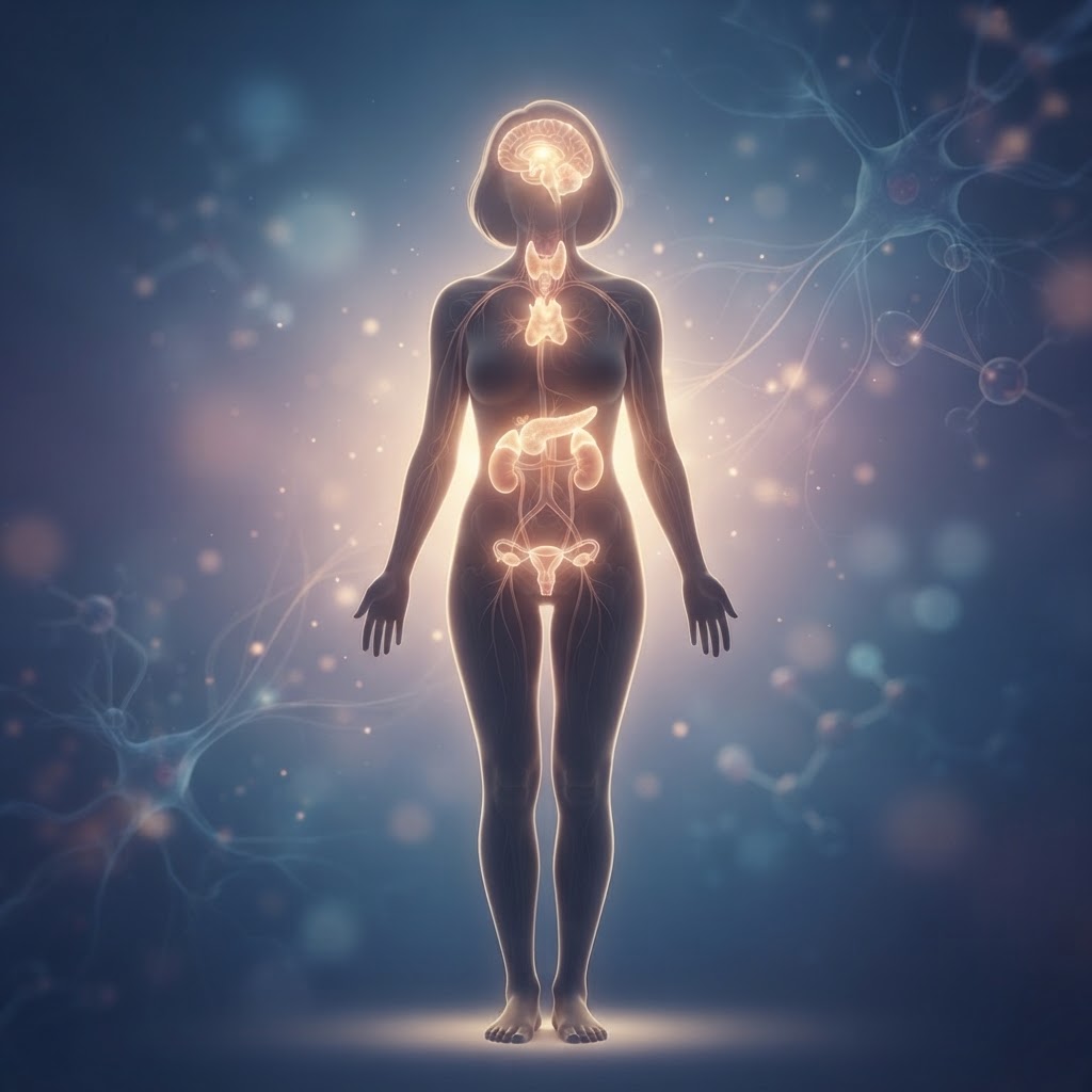
  • Raynaud’s Disease: Early Symptoms, Causes, and Natural Relief

    Raynaud’s Disease: Early Symptoms, Causes, and Natural Relief

    Key Takeaways

    • Raynaud’s phenomenon involves exaggerated vasospasm in response to cold or stress, primarily affecting the extremities.
    • Distinguishing between primary and secondary forms is critical for understanding underlying health risks.
    • Early Raynaud’s disease symptoms often manifest as sudden color changes in fingers, ranging from white to blue or red.
    • Vasospasm is the fundamental physiological mechanism that temporarily restricts blood flow to peripheral tissues.
    • Effective Raynaud’s syndrome management centers on proactive trigger avoidance and lifestyle adjustments to improve blood flow naturally.

    Have you ever stepped into a chilly grocery store aisle or experienced a moment of intense stress, only to find your fingers turning stark white, feeling numb, or pulsing with an uncomfortable, throbbing heat? While many people simply dismiss these sensations as an extreme reaction to cold, they may actually be early indicators of a vascular condition known as Raynaud’s. For those living with this sensitivity, the world feels like a constant battle against temperature drops and emotional triggers. Understanding the nuances of this condition is the first step toward reclaiming your comfort and taking control of your circulatory health. In this comprehensive guide, the healthauthoritylife Editorial Team explores the intricacies of Raynaud’s, from the initial warning signs to practical, science-backed strategies for natural relief.

    What Is Raynaud’s Phenomenon?

    Raynaud’s phenomenon is a vascular condition characterized by the narrowing of small blood vessels in response to cold temperatures or emotional stress. When these vessels undergo what is known as a vasospasm, blood flow to the fingers, toes, and occasionally the ears, nose, or lips is severely restricted. This isn’t just “feeling cold”—it is a distinct physiological reaction that causes the affected areas to undergo dramatic color changes. These episodes can last anywhere from a few minutes to several hours, and the physical discomfort—ranging from a cold, numb sensation to an aching, tingling pain—can significantly disrupt daily activities.

    At its core, the condition is a hyper-reactive state of the sympathetic nervous system. In a typical person, when the body detects cold, the blood vessels in the extremities constrict slightly to preserve core body temperature and protect vital organs. For individuals with Raynaud’s, this response is vastly exaggerated. The body essentially overreacts, “shutting off” the supply to the fingers and toes far more aggressively than necessary. This constriction leads to the characteristic color changes often described as the “triphasic response”: first, the area turns white (pallor) as blood flow is cut off; second, it may turn blue (cyanosis) as the tissues lose oxygen; and finally, as the vessel relaxes and blood rushes back in, the area turns bright red (hyperemia) and often feels warm, throbbing, or painful.

    Understanding the physiological mechanism behind this phenomenon is essential for effective Raynaud’s syndrome management. It is not necessarily a sign of a failing heart or systemic circulatory disease in every case; rather, it represents a localized sensitivity of the peripheral vasculature. Many people find that their symptoms remain mild and manageable throughout their lives, often seeing improvement through lifestyle changes designed to improve blood flow naturally. However, because these symptoms can mimic or overlap with other connective tissue disorders, awareness is key. By recognizing that these episodes are rooted in the way your nervous system perceives external environmental stimuli, you can begin to anticipate attacks and adopt habits—such as heat retention techniques and stress reduction—that keep the vessels relaxed and blood circulating more freely.

    Primary vs. Secondary Raynaud’s: What You Need to Know

    Clinicians and researchers generally categorize this condition into two distinct types: Primary and Secondary. Understanding the difference between the two is perhaps the most important step in assessing your health. Primary Raynaud’s, also called Raynaud’s disease, is the most common form. It occurs on its own and is not associated with any other underlying medical condition. It typically develops in late adolescence or early adulthood, and it is often considered a “nuisance” condition because, while uncomfortable, it rarely causes permanent damage to the tissues of the fingers or toes. Those with primary symptoms often find that the episodes are symmetrical, meaning both hands are affected similarly, and they respond quite well to lifestyle modifications.

    Secondary Raynaud’s, or Raynaud’s phenomenon, is a more serious iteration. It is caused by an underlying health issue, such as an autoimmune disease, structural vascular problems, or occupational trauma. For example, conditions like scleroderma, lupus, rheumatoid arthritis, or Sjogren’s syndrome are frequently linked to secondary Raynaud’s. Unlike the primary form, secondary Raynaud’s often presents later in life and can be more severe. The restricted blood flow may be intense enough to cause skin sores, ulcers, or, in rare and extreme cases, tissue death (gangrene) if not managed properly. This is why professionals emphasize that anyone experiencing new or worsening symptoms should seek a medical evaluation to rule out secondary causes.

    To help distinguish between the two approaches, the following table outlines the general characteristics of how these conditions are managed and observed.

    Approach/Feature Primary Raynaud’s Secondary Raynaud’s Best For
    Lifestyle Management Highly effective, focus on warmth Supplemental to medical therapy Daily symptom prevention
    Clinical Monitoring Rarely needed beyond exams Frequent, required for underlying disease Long-term health safety
    Targeted Relief Warmth, gloves, movement Medication + specialized protection Immediate comfort during flare-ups
    Risk Profile Low risk of tissue damage Potential for digital ulcers/damage Determining urgency of care

    If you suspect you have Raynaud’s, observe the symmetry of your symptoms. If your episodes involve only one hand, or if you notice skin breakdown and non-healing sores, it is imperative to move beyond simple lifestyle hacks and consult a specialist. Distinguishing the primary from the secondary form allows you to determine whether your goal is purely symptomatic relief or if you need a comprehensive plan to manage an overarching, systemic health concern.

    Common Early Symptoms and Warning Signs

    Recognizing the early warning signs of Raynaud’s disease symptoms can feel like learning to read a subtle language your body is speaking. Many individuals ignore the initial bouts, attributing the numbness or coldness to a drafty room or being naturally “cold-blooded.” However, early detection can significantly enhance your quality of life. The earliest symptom is usually an intense, localized sensitivity to cold. While everyone’s hands get cold in the winter, the person with Raynaud’s will notice their fingers becoming painful or turning colors while others in the same environment remain perfectly comfortable. This disproportionate response is a hallmark sign.

    The sequence of color changes is arguably the most recognizable indicator. Initially, you might notice that a finger—or even just the tip of a finger—turns completely white. This happens because the artery in that specific area has constricted so tightly that the blood flow is cut off entirely. The sensation accompanying this is often a dull ache or complete numbness; the finger may feel like a wooden appendage that doesn’t belong to you. As the spasm continues, the finger might transition to a bluish, purplish hue. This is the stage where the tissue is being deprived of oxygenated blood. It can be visually alarming, particularly if you have not experienced it before.

    As the episode ends and the blood vessels finally begin to relax, the “reperfusion” phase occurs. This is often the most painful part of the experience. As the blood rushes back into the narrow vessels, the finger turns bright, beet red. This is frequently accompanied by a burning, tingling, or throbbing sensation, which many describe as a “pins and needles” feeling similar to when a limb “falls asleep.” While these episodes usually occur in the hands, they can also affect the toes, and some people report experiencing these sensations in their ears, nose, or lips. If you find yourself frequently checking your hands for color changes after moving from a heated car into the cold air, or even after grabbing a cold soda from the refrigerator, you are likely witnessing the onset of Raynaud’s phenomenon.

    It is important to track how often these episodes occur and what seems to trigger them. Some people experience these symptoms only once or twice a year, while others deal with them daily during the winter months. Keeping a simple log of your symptoms—noting the time, the environment, and your emotional state—can provide valuable data that helps you recognize patterns. Early intervention often involves basic behavioral changes, such as layering clothing, using hand warmers, or practicing stress-management techniques before a potential trigger even occurs. By paying attention to these early physical cues, you can stay ahead of the curve, minimizing the severity and frequency of the spasms.

    The Role of Vasospasm in Raynaud’s Episodes

    To truly understand how to manage Raynaud’s, we must demystify the physiological process known as vasospasm. Vasospasm is the sudden, involuntary constriction of blood vessels. Under normal physiological conditions, your body uses vasospasm as a protective tool. If you cut your finger, your vessels constrict to slow down bleeding. If you walk into a snowstorm, your vessels constrict in your extremities to force blood toward your vital organs—your heart, lungs, and brain—to maintain your core temperature. This is a survival mechanism that serves us well in extreme circumstances. However, in the context of Raynaud’s, the body’s “thermostat” is essentially broken or overly sensitive, triggering this survival reaction in completely safe or moderate environments.

    The “spasm” refers to the contraction of the smooth muscles that line the walls of the tiny arteries—the arterioles—in your fingers and toes. When these muscles tighten, the diameter of the vessel shrinks, leaving little to no room for blood to pass through. This is why the skin goes pale; there is literally no blood being delivered to the capillaries at the surface of the skin. Once the trigger—be it cold or stress—is removed, the smooth muscle relaxes, the vessel opens, and blood flows back in. This rapid transition from empty to full is exactly what creates that intense, throbbing sensation of heat and pain. It is essentially a localized flood of blood returning to tissue that had been momentarily “starved.”

    Research suggests that in those with Raynaud’s, the nerves that control these blood vessels are hypersensitive to stimuli. While the cold is the most common trigger, the hormonal response to stress (specifically the release of adrenaline and norepinephrine) can also cause the vessels to narrow. This is why you might find that you experience a flare-up during a high-pressure meeting or a stressful argument, even if you are in a comfortably warm room. The chemical signals being sent to your blood vessels to “prepare for danger” are being misinterpreted, resulting in the same constriction that occurs in the cold. Understanding this link between the nervous system and vascular tone is vital because it moves the focus of treatment away from just “getting warm” and toward “calming the nervous system.”

    For those seeking to improve blood flow naturally, the goal is to stabilize the vascular system. Because vasospasms are triggered by a range of internal and external signals, your management strategy should be multifaceted. This includes everything from dietary adjustments that promote vasodilation (the widening of blood vessels) to techniques that lower your baseline stress level. By consistently working to keep the nervous system in a “rest and digest” state, you can effectively raise the threshold required to trigger a vasospasm, potentially leading to fewer, shorter, and less intense episodes. The physiology of the vasospasm reminds us that our circulation is deeply connected to our environment and our internal well-being; treating the phenomenon requires a holistic look at how we navigate both.

    Identifying Common Triggers for Cold Sensitivity

    Identifying your specific triggers is the cornerstone of successful Raynaud’s syndrome management. Because the condition is a reaction to stimuli, the more you can control your exposure to those stimuli, the more freedom you will have. Most people are quick to blame the cold, but that is rarely the whole story. Cold sensitivity can manifest in unexpected ways. For instance, the transition between temperature zones—like stepping out of a house maintained at 72 degrees into 40-degree weather—is often a more potent trigger than simply spending time in the cold itself. The shock of the rapid temperature shift causes the nervous system to react much more aggressively than a gradual exposure would.

    Beyond the obvious weather-related triggers, there are several “stealth” triggers that many people overlook. High levels of emotional stress or anxiety are top-tier offenders. When you are stressed, your body diverts blood flow away from the extremities to the large muscles, a vestige of our ancestral “fight or flight” response. If you have Raynaud’s, your vessels are already prone to constriction, and the added pressure of adrenaline can tip you over the edge into a full-blown episode. Practicing mindfulness, deep breathing exercises, or gentle movement can help modulate this reaction. Recognizing that a stressful phone call might lead to white, painful fingers allows you to preemptively warm your hands or move your body to encourage circulation.

    Certain dietary habits can also act as triggers. For example, nicotine is a potent vasoconstrictor, meaning it causes blood vessels to narrow. For anyone with circulation issues, nicotine use is universally discouraged by experts as it significantly exacerbates the frequency and severity of episodes. Caffeine is another substance that can have a variable effect; while it may increase alertness, it can also cause blood vessels to tighten for some individuals. Monitoring your intake of coffee, tea, and caffeinated sodas can help you determine if they are contributing to your sensitivity. Instead, many find that incorporating herbal infusions or staying strictly hydrated with room-temperature water supports better overall flow.

    Finally, consider the environmental factors in your home or workplace. Are you sitting with your wrists resting on a cold desk? Is there an air conditioning vent blowing directly onto your hands? These small, constant environmental stressors can keep your body in a state of high alert. Simple fixes, like a desk mat to insulate your wrists, wearing fingerless gloves while typing, or keeping a small space heater nearby, can make a profound difference. By viewing your daily routine through the lens of a “trigger audit,” you can identify the subtle ways your environment is working against your circulation. Managing Raynaud’s isn’t about avoiding the world; it’s about curating your interaction with it to keep your blood flowing smoothly and your body at ease.

    Natural Strategies to Improve Peripheral Circulation

    Improving peripheral circulation is the cornerstone of managing Raynaud’s phenomenon. Because the condition is characterized by an exaggerated response of the blood vessels to cold or emotional stress, the primary goal is to keep the vascular system primed and responsive. By focusing on systemic blood flow, you can often reduce the frequency and intensity of vasospastic episodes.

    One of the most effective natural strategies is the implementation of targeted movement. When you are sedentary, the blood has to work harder to return from the extremities to the heart. Incorporating “vasodilatory” exercise, such as brisk walking, swimming, or stationary cycling, helps maintain cardiovascular flexibility. Specifically, rhythmic arm swinging or “windmilling” can use centrifugal force to encourage blood flow into the fingers during a cold snap.

    Temperature contrast therapy, often referred to as hydrotherapy, can also train your blood vessels to remain resilient. By alternating between warm and cool water immersion—or simply using warm and cool compresses—you challenge the muscular walls of your arteries to constrict and dilate. This “exercise” for your blood vessels can lead to improved baseline circulation over time. Always ensure the water is not extreme in temperature to avoid triggering a flare-up or damaging the skin.

    Breathwork is another often overlooked tool. Deep, diaphragmatic breathing activates the parasympathetic nervous system, which helps counteract the “fight-or-flight” response that often triggers Raynaud’s attacks. When you breathe deeply, you lower the overall stress levels in your body, which prevents the sympathetic nervous system from causing unnecessary vasoconstriction in the hands and feet.

    Nutritional Supplements and Herbs for Vascular Health

    Supporting vascular health through nutrition requires a focus on vasodilation—the widening of blood vessels. Many individuals look toward natural supplements that encourage the production of nitric oxide, a molecule that helps the inner lining of blood vessels (the endothelium) relax. While it is essential to consult with a healthcare professional before starting any regimen, several supplements are frequently discussed in the context of vascular support.

    Magnesium is widely considered a key player in circulatory health. It acts as a natural muscle relaxant and can prevent the involuntary spasms in blood vessels that lead to the color changes associated with Raynaud’s. Magnesium glycinate or citrate are common forms, often taken to support both cardiovascular function and nervous system stability.

    Ginkgo biloba is a traditional herb recognized for its potential to improve blood flow to the extremities. It is believed to help thin the blood slightly and improve the elasticity of the vessel walls. Similarly, Omega-3 fatty acids, found in high-quality fish oils, are renowned for their anti-inflammatory properties. By reducing inflammation within the blood vessel walls, Omega-3s may help maintain smooth, uninterrupted blood flow.

    Another popular botanical is ginger. Known for its thermogenic properties, ginger can create a warming effect within the body, which helps to naturally stimulate peripheral circulation. Whether consumed as a tea or taken in concentrated supplement form, ginger is often used as a preventative measure during the colder months.

    Supplement/Herb Primary Mechanism Best For
    Magnesium Muscle relaxation/Vascular tone Preventing vessel spasms
    Ginkgo Biloba Improved blood rheology Enhancing peripheral microcirculation
    Omega-3 Fatty Acids Anti-inflammatory support Vascular wall health
    Ginger Extract Thermogenic stimulation Warming the body internally

    Lifestyle Habits to Prevent Raynaud’s Flare-Ups

    Managing Raynaud’s syndrome requires a proactive lifestyle approach. Preventing a flare-up is far easier than trying to warm up fingers that have already turned white or blue. The most critical habit is the “layering” principle. It is not just about wearing a thick coat; it is about protecting the core. If the body’s core temperature drops, the brain automatically restricts blood flow to the extremities to preserve internal heat. By keeping your torso, neck, and head warm, you signal to your body that it is safe to maintain circulation to your hands and feet.

    Hand protection should be treated as a year-round priority for those with severe symptoms. This means keeping gloves or mittens easily accessible, even in grocery store freezer aisles or air-conditioned offices. Opt for high-quality, moisture-wicking fabrics rather than simple cotton, which can trap sweat and lead to a cooling effect when moisture evaporates.

    Another vital lifestyle habit is the management of stimulants. Nicotine is a potent vasoconstrictor, meaning it causes blood vessels to narrow significantly. For individuals with Raynaud’s, smoking—or even exposure to secondhand smoke—is a major trigger for episodes. Additionally, heavy caffeine consumption can affect the vascular system; while it might provide an initial energy boost, its impact on heart rate and blood pressure can sometimes be counterproductive for those prone to vasospasm.

    Stress management routines must become non-negotiable. Because emotional stress triggers the same vascular response as cold, finding a way to decompress is essential. Practices like yoga, meditation, or even simple daily journaling can help manage the body’s sympathetic nervous system response, creating a more balanced internal environment that is less likely to overreact to environmental stressors.

    When to See a Doctor for Persistent Circulation Issues

    While primary Raynaud’s is often manageable through lifestyle changes, secondary Raynaud’s can be an indicator of underlying health concerns. It is crucial to monitor your symptoms closely. You should seek medical evaluation if you notice that your Raynaud’s symptoms appear suddenly after the age of 30, or if the episodes become increasingly asymmetric—for example, if only one hand is affected while the other remains warm.

    Furthermore, if you experience painful sores, ulcers, or areas of thickened, scarred skin on your fingertips, you must schedule an appointment with a professional. These signs can indicate prolonged, severe tissue ischemia (lack of oxygenated blood), which requires clinical intervention to prevent permanent damage. Similarly, if your circulation issues are accompanied by joint pain, unexplained rashes, fever, or persistent fatigue, these could be symptoms of autoimmune conditions such as lupus, scleroderma, or rheumatoid arthritis.

    A doctor can perform specific diagnostic tests, such as a nailfold capillaroscopy—a simple, non-invasive exam where a microscope is used to look at the tiny blood vessels at the base of your fingernail. This can help differentiate between benign primary Raynaud’s and secondary forms that require more specialized treatment plans, such as prescription calcium channel blockers or other vasodilatory medications.

    Frequently Asked Questions

    Is Raynaud’s disease considered a lifelong condition?

    For many, primary Raynaud’s is a chronic condition, but it is often manageable. Symptoms may fluctuate throughout a person’s life, sometimes improving during warmer months or as one makes consistent lifestyle adjustments. Secondary Raynaud’s, however, depends on the management of the underlying condition causing it.

    Can stress really trigger a Raynaud’s attack even if it is warm?

    Yes. Because Raynaud’s is driven by the autonomic nervous system, emotional distress can trigger the same “fight-or-flight” response as cold temperatures. This causes the blood vessels to constrict, leading to an attack regardless of the ambient temperature.

    Are there certain foods I should avoid to help my circulation?

    While no single food causes an attack, highly processed foods, excess sodium, and excessive sugar can contribute to inflammation and poor vascular health. Many people find that reducing stimulants like caffeine and avoiding nicotine has a positive impact on their symptom frequency.

    Do I need to live in a warm climate to manage Raynaud’s?

    Moving to a warm climate can certainly reduce the number of environmental triggers, but it is not a requirement for management. Many individuals successfully manage the condition in cold climates by using smart layering, portable hand warmers, and focusing on internal circulatory health.

    Are women more likely to develop Raynaud’s?

    Statistical data consistently shows that Raynaud’s occurs more frequently in women than in men. While the exact reasons are not fully understood, hormonal factors and differences in the baseline vascular response to cold are often cited as potential contributing elements.

    Can natural remedies replace medical treatment for Raynaud’s?

    Natural remedies are generally considered complementary. While they can significantly reduce the frequency and severity of episodes for many, they should not replace a doctor’s guidance, especially if you have secondary Raynaud’s or are experiencing tissue damage.

    Conclusion

    Raynaud’s phenomenon is a complex condition that sits at the intersection of environmental sensitivity and vascular resilience. While the sudden changes in finger color and the resulting discomfort can be alarming, understanding the underlying mechanisms is the first step toward reclaiming control. By integrating consistent physical movement, nutrient-dense vascular support, and mindful lifestyle adjustments, you can significantly diminish the impact that cold and stress have on your daily life. Remember that your body is a dynamic system—by nourishing your blood vessels and managing your triggers, you can foster better circulation and improve your overall well-being. If you find your symptoms worsening or notice physical changes in your skin, do not hesitate to reach out to a healthcare professional for personalized guidance.

    Are you ready to take charge of your vascular health? Start by identifying your specific triggers today and incorporating one of our suggested circulation-boosting habits into your morning routine.

    By healthauthoritylife Editorial Team

    This article is for informational purposes only and is not a substitute for professional medical advice. Always consult your doctor before making changes to your diet, exercise routine, or treatment plan.

  • Adrenal Insufficiency: Symptoms, Causes, and Natural Support

    Adrenal Insufficiency: Symptoms, Causes, and Natural Support

    Key Takeaways

    • Adrenal insufficiency is a distinct, medically recognized condition where the glands fail to produce sufficient hormones, unlike the non-clinical concept of adrenal fatigue.
    • Recognizing early symptoms like persistent fatigue, low blood pressure, and salt cravings is critical for timely medical intervention.
    • Understanding the distinction between primary (Addison’s disease) and secondary insufficiency is essential for accurate diagnosis and long-term endocrine system support.
    • Chronic, unmanaged stress creates a physiological burden that may exacerbate symptoms and hinder endocrine recovery.
    • Standardized blood and stimulation testing, interpreted by an endocrinologist, remains the gold standard for diagnosing adrenal dysfunction.

    The endocrine system serves as the body’s master communication network, and at the heart of this complex web are the adrenal glands—small but mighty structures perched atop your kidneys. Responsible for secreting life-sustaining hormones like cortisol and aldosterone, these glands dictate how we respond to stress, manage blood pressure, and maintain electrolyte balance. When these glands fail to produce enough of these essential chemicals, the results can be systemic and debilitating. Navigating the nuances of adrenal health requires a clear understanding of clinical realities versus popular wellness trends. In this comprehensive guide, the healthauthoritylife Editorial Team explores the clinical landscape of adrenal insufficiency, how to differentiate it from other fatigue-related conditions, and how to approach care with precision and scientific backing.

    What Is Adrenal Insufficiency and How It Differs from Adrenal Fatigue

    Adrenal insufficiency (AI) is a clinically diagnosed medical condition. It occurs when the adrenal glands do not produce an adequate amount of essential hormones, primarily cortisol and, in some cases, aldosterone. Cortisol is often referred to as the “stress hormone,” but its function is far broader; it is vital for regulating blood sugar levels, reducing inflammation, and maintaining cardiovascular function. When the body cannot manufacture these hormones in sufficient quantities, the consequences are profound, affecting every organ system. AI can be life-threatening if left unmanaged, as it compromises the body’s ability to respond to physiological stress, trauma, or severe illness.

    There is often confusion in the wellness community regarding “adrenal fatigue,” a term frequently used to describe a broad spectrum of exhaustion, brain fog, and body aches. It is important to note that the medical community generally does not recognize “adrenal fatigue” as an official diagnosis. Unlike adrenal insufficiency—which can be measured through specific blood tests, stimulation tests, and hormonal assays—adrenal fatigue is often used to characterize a vague sense of burnout. While the symptoms may feel similar, the underlying biology is entirely different. Adrenal insufficiency involves measurable damage or functional failure of the hypothalamic-pituitary-adrenal (HPA) axis or the adrenal glands themselves.

    Managing adrenal health effectively requires separating these two concepts. If you are experiencing genuine adrenal insufficiency, you are dealing with a deficiency that requires medical supervision, often involving hormone replacement therapy. If you are experiencing what is commonly labeled as “adrenal fatigue,” you are likely dealing with a state of physiological exhaustion or burnout that stems from chronic lifestyle stressors, poor sleep, or metabolic imbalances. The healthauthoritylife Editorial Team emphasizes that confusing these two can delay necessary medical care for those with true insufficiency. Always prioritize professional testing to determine if your symptoms originate from a clinical hormonal deficit or from lifestyle-related systemic stress.

    To better understand the differences in approach, the following table breaks down how different wellness and medical strategies correlate to these conditions:

    Approach Best For Clinical Goal
    Hormone Replacement Therapy Confirmed Adrenal Insufficiency Replacing missing hormones
    Lifestyle & Stress Management Adrenal Fatigue/Burnout HPA axis regulation
    Endocrine System Support General Wellness Balanced hormonal signaling
    Salt/Electrolyte Supplementation Addison’s disease natural care Maintaining fluid balance

    Common Early Symptoms You Should Not Ignore

    Early symptoms of adrenal insufficiency are often subtle and can mimic other common conditions, such as chronic fatigue syndrome, depression, or even simple dehydration. Because of this, it is common for individuals to live with these issues for months or even years before receiving a formal diagnosis. Paying close attention to how your body responds to daily stressors is the first step in effective management. The most hallmark early sign is a profound, unexplained fatigue that is not alleviated by rest. While everyone experiences tiredness, individuals with adrenal issues often feel a specific type of exhaustion—one that begins in the morning and fluctuates throughout the day, often peaking when the body’s cortisol levels should naturally be highest.

    Another classic warning sign involves your blood pressure and cardiovascular response. Because aldosterone regulates the body’s fluid and salt balance, a deficiency can lead to low blood pressure, particularly upon standing. This phenomenon, known as orthostatic hypotension, may cause you to feel dizzy, lightheaded, or faint when moving quickly from a seated to a standing position. If you find yourself consistently needing to move slowly to avoid feeling woozy, this is a physiological indicator that your endocrine system is struggling to maintain proper blood pressure regulation.

    Salt cravings are another frequent, yet overlooked, symptom. Because the body may be losing excessive amounts of sodium through urine, it will instinctively signal for high-salt foods. If you find that you are constantly reaching for saltier snacks, it might be more than just a preference—it could be a biological compensation mechanism. Furthermore, gastrointestinal distress is common. Many people with early-stage adrenal insufficiency report nausea, decreased appetite, and unintended weight loss. Unlike typical digestive issues, these symptoms often persist regardless of dietary changes.

    Skin changes can also offer clues. In primary adrenal insufficiency, specifically, a person might notice hyperpigmentation, or darkening of the skin, particularly in areas like the skin folds, scars, and inside the mouth (buccal mucosa). This occurs due to elevated levels of melanocyte-stimulating hormone, a byproduct of the same precursor that the body pumps out in an attempt to get the struggling adrenal glands to work harder. While not everyone will experience skin darkening, it is a significant clinical marker that necessitates a prompt conversation with a healthcare provider. Ignoring these signs can lead to an “adrenal crisis,” a medical emergency where the body lacks the hormonal response to handle significant stress. Recognizing these symptoms early allows for proactive management and protects your long-term health.

    Primary vs Secondary Adrenal Insufficiency: Understanding the Causes

    Distinguishing between primary and secondary adrenal insufficiency is crucial because the underlying pathology determines the entire course of treatment. Primary adrenal insufficiency, most commonly known as Addison’s disease, occurs when the adrenal glands themselves are damaged. This damage prevents the glands from producing the necessary hormones despite having the signal from the brain to do so. In many parts of the world, autoimmune destruction is the most frequent cause. In this scenario, the body’s immune system mistakenly attacks the adrenal cortex, the outer layer of the gland that produces cortisol and aldosterone. Once this tissue is damaged, the body can no longer produce these hormones naturally.

    Other causes of primary insufficiency include infections that affect the adrenal glands, such as tuberculosis or fungal infections, as well as surgical removal of the glands or hemorrhage due to certain blood-thinning medications. When addressing Addison’s disease natural care, it is vital to remember that because the damage is structural, the body will always require some form of exogenous hormone support to maintain life-sustaining balance. While lifestyle habits can support overall health, they cannot replace the hormonal output of a gland that has been structurally compromised.

    Secondary adrenal insufficiency is fundamentally different. In this case, the adrenal glands are physically healthy, but they aren’t receiving the signal to produce hormones. This signaling failure originates in the brain, specifically the pituitary gland. The pituitary gland produces a hormone called adrenocorticotropic hormone (ACTH), which acts as a messenger to tell the adrenals to release cortisol. If the pituitary is not producing enough ACTH—due to a tumor, radiation therapy, or trauma—the adrenal glands essentially “go to sleep.”

    A very common, often overlooked cause of secondary insufficiency is the long-term use of corticosteroid medications. Medications like prednisone, dexamethasone, or even high-dose inhaled steroids used for asthma or chronic inflammation can suppress the pituitary gland. When these medications are taken for a prolonged period, the brain stops sending the signal to the adrenals because it assumes there is already enough cortisol present. If these medications are stopped abruptly, the pituitary may not “wake up” quickly enough to resume production, leading to a state of secondary insufficiency. This is why medical professionals always stress the importance of tapering off steroid treatments slowly. Understanding whether your issue is primary or secondary allows you and your care team to determine whether the focus should be on replacing the hormone directly or, in some secondary cases, working to restore the signaling pathways between the brain and the adrenal glands.

    How Chronic Stress Impacts Your Adrenal Glands Over Time

    While acute stress is a natural, healthy response—designed to help us escape immediate danger through the “fight or flight” mechanism—chronic, unremitting stress is a different beast entirely. When you are exposed to ongoing stressors, whether they are work-related, emotional, or environmental, your HPA axis remains in a state of high alert. Under normal conditions, the body releases a burst of cortisol to handle a stressor and then returns to a baseline state of equilibrium. However, in the modern world, the stressor often does not go away. This leads to a persistent, non-stop elevation of cortisol that can eventually overwhelm the body’s internal regulation systems.

    The impact of this chronic demand is cumulative. Over time, the constant signaling from the brain to the adrenal glands can lead to a state of dysregulation. While the glands may continue to produce cortisol, the body’s tissues may become desensitized to it, or the feedback loops that tell the brain to “stop the stress signal” become stuck. This is a primary driver in what many refer to as metabolic exhaustion. Managing adrenal health in the context of chronic stress is about breaking this feedback loop. It is not just about the glands themselves; it is about retraining your nervous system to exit the fight-or-flight state.

    Chronic stress also depletes the micronutrients required for healthy endocrine function. Cortisol production is an energy-intensive process that relies heavily on nutrients like Vitamin C, magnesium, B-complex vitamins, and specific minerals. When you are constantly stressed, your body burns through these resources rapidly, leaving your endocrine system without the raw materials it needs to synthesize hormones effectively. This deficiency creates a vicious cycle: the body is stressed, it demands more hormones, it depletes its nutrient stores, and then it is unable to produce the hormones needed to calm that stress back down.

    Furthermore, chronic stress disrupts the circadian rhythm, which is tied directly to your cortisol patterns. Ideally, cortisol should be highest in the morning to provide energy for the day and drop to its lowest point by nighttime to facilitate sleep. Chronic stress often flips this pattern, causing cortisol levels to remain elevated at night, which prevents deep, restorative sleep. This lack of sleep, in turn, puts more stress on the body the next day. To support your adrenals, you must address the stress upstream. This means implementing consistent sleep hygiene, engaging in regular nervous system regulation—such as breathwork or meditation—and ensuring your nutritional intake is optimized to support the high demand the adrenal glands face during challenging times.

    Diagnostic Tests and When to See an Endocrinologist

    If you suspect that your symptoms are related to adrenal insufficiency, the most important step is to seek a professional evaluation. Self-diagnosis is dangerous in the context of endocrine health because the symptoms are non-specific and the consequences of mismanaging a hormonal deficiency can be severe. An endocrinologist is the specialist best equipped to navigate the complex diagnostic pathway of adrenal function. They understand the intricacies of how the pituitary and adrenal glands interact and can distinguish between temporary imbalances and permanent insufficiency.

    The diagnostic process typically begins with a morning cortisol blood test. Because cortisol levels naturally fluctuate throughout the day, doctors prefer to test early in the morning, between 6:00 AM and 8:00 AM, when levels should be at their natural peak. If this blood test shows levels that are abnormally low, further investigation is required. However, a single blood test is rarely enough to provide a definitive diagnosis. It is merely a snapshot in time and can be influenced by recent sleep, stress, or medications.

    To gain a clearer picture, an endocrinologist will often utilize a stimulation test. The most common is the ACTH stimulation test. In this procedure, a healthcare provider injects a synthetic form of ACTH, the hormone that typically signals the adrenals to produce cortisol. They then measure the cortisol levels in your blood at specific intervals—typically 30 and 60 minutes after the injection. If your adrenal glands are healthy, they will respond to the synthetic ACTH by producing a robust surge of cortisol. If the glands fail to produce a significant increase, this provides strong evidence of adrenal insufficiency. This test is the gold standard for confirming a diagnosis.

    You should prioritize seeing an endocrinologist if you have persistent, unexplained symptoms like extreme fatigue, unexplained weight loss, persistent low blood pressure, or hyperpigmentation. Do not wait for a crisis to occur. If you are taking long-term steroid medication for conditions such as asthma, rheumatoid arthritis, or autoimmune disorders, you should be under the regular care of an endocrinologist to monitor your adrenal function, as the risk of secondary insufficiency is significantly higher. Early detection allows for the implementation of necessary hormone replacement strategies, which are highly effective at restoring quality of life. By focusing on evidence-based diagnostic procedures, you ensure that any endocrine system support you pursue is grounded in clinical reality rather than speculation.

    The Role of Nutrition in Supporting Healthy Adrenal Function

    When managing adrenal health, nutrition serves as the primary foundation for stabilizing the endocrine system. Because the adrenal glands are highly sensitive to blood sugar fluctuations, a diet that minimizes glucose spikes is essential. When blood sugar drops rapidly, the body triggers a cortisol release to mobilize stored energy, which further stresses already taxed adrenal glands. Consequently, the cornerstone of dietary support is the consumption of balanced meals containing high-quality proteins, healthy fats, and complex, fiber-rich carbohydrates at regular intervals throughout the day.

    Focusing on nutrient density ensures that your body has the raw materials required for hormone synthesis. The adrenal glands have a high requirement for Vitamin C, which is utilized heavily during the stress response. Incorporating whole foods such as bell peppers, citrus fruits, and berries can provide a consistent intake of this vital nutrient. Furthermore, electrolytes play a pivotal role. Those dealing with adrenal issues often struggle with mineral balance, particularly sodium and potassium. While conventional medical advice often pushes for low salt intake, individuals with suboptimal adrenal function may actually require a slight increase in high-quality sea salt intake to maintain blood pressure and fluid balance, provided they are under appropriate clinical guidance.

    Anti-inflammatory eating patterns are equally critical. Chronic inflammation acts as a persistent stressor on the hypothalamic-pituitary-adrenal (HPA) axis. By emphasizing colorful vegetables, omega-3 fatty acid sources like wild-caught salmon or flaxseeds, and avoiding processed vegetable oils or refined sugars, you reduce the inflammatory load. It is also beneficial to limit stimulants. Caffeine, while providing a temporary energy boost, acts by forcing the adrenals to release more cortisol and adrenaline. Over time, this consumption creates a cycle of dependency and exhaustion, making it significantly harder to regulate your natural energy patterns.

    Lifestyle Strategies to Regulate Your Cortisol Rhythm

    Your cortisol rhythm is governed by the circadian clock, which ideally dictates high cortisol levels in the morning to initiate wakefulness and lower levels at night to facilitate restorative sleep. In cases of adrenal insufficiency, this rhythm often becomes inverted or flattened. Lifestyle modifications serve as the primary tool for recalibrating this internal clock. One of the most effective strategies is the practice of “light hygiene.” Exposing your eyes to natural sunlight within 30 minutes of waking helps signal the HPA axis to produce its morning cortisol pulse, while dimming lights and reducing blue light exposure in the evening signals the body to prepare for melatonin production.

    Physical activity, while generally healthy, must be approached with caution. High-intensity interval training or heavy endurance training can act as a massive stressor that, in an already compromised endocrine system, leads to further depletion. For those focused on managing adrenal health, restorative movement is the superior choice. Activities like yoga, Pilates, Qigong, or gentle walking promote lymphatic drainage and parasympathetic nervous system activation without triggering a massive sympathetic stress response. The goal is to move the body in a way that feels energizing rather than draining.

    Stress management techniques are not mere luxuries; they are essential clinical strategies. Techniques such as box breathing, progressive muscle relaxation, or mindfulness-based stress reduction (MBSR) act as a “brake” on the stress response. By regularly engaging these modalities, you teach your nervous system to exit the fight-or-flight state more efficiently. Additionally, establishing firm boundaries around your time and commitments is a necessary lifestyle change. Protecting your restorative time is just as important as the nutrition you consume, as the body cannot heal if it is constantly in a state of hyper-vigilance.

    Essential Micronutrients and Adaptogens for Adrenal Support

    Supporting the endocrine system often involves the strategic use of targeted nutrients and herbal allies. Adaptogens are a unique class of herbs that assist the body in adapting to stress by modulating the HPA axis. They help “adapt” the body’s response—lowering cortisol when it is too high and supporting energy production when it is too low. However, because different adaptogens have different energetic properties, choosing the right one requires an understanding of your specific symptomatic profile.

    Below is a comparison of common botanical and nutritional supports used for endocrine health.

    Support Category Common Examples Primary Benefit Best For
    Stimulating Adaptogens Rhodiola Rosea Boosts mental focus and energy Morning use for low-energy states
    Calming Adaptogens Ashwagandha Reduces cortisol and anxiety Evening use for stress and sleep
    B-Vitamin Complex B5 (Pantothenic Acid) Supports hormone synthesis Daily maintenance of adrenal function
    Magnesium Supplements Magnesium Glycinate Promotes muscle and nerve relaxation Reducing nervous tension
    Mineral Support Adrenal Glandulars Provides building blocks for repair Advanced stages of exhaustion

    In addition to herbs, B-vitamins—specifically B5, B6, and B12—are known as the “anti-stress” vitamins. Pantothenic acid (B5) is a necessary precursor for the production of adrenal steroid hormones. Without adequate B-vitamin status, the body’s ability to convert nutrients into cellular energy is severely compromised. Magnesium also deserves special mention. Chronic stress leads to rapid depletion of magnesium stores, which in turn causes further nervous system irritability, creating a vicious cycle. Supplementing with highly bioavailable forms of magnesium can help stabilize the nervous system and improve sleep quality, which is paramount for endocrine recovery.

    Common Mistakes That Can Worsen Adrenal Imbalance

    The pursuit of healing can sometimes backfire if certain pitfalls are not avoided. One of the most common mistakes is the “all-or-nothing” approach to lifestyle changes. When someone discovers they have adrenal imbalances, they often attempt to overhaul their diet, exercise routine, and sleep habits overnight. This sudden influx of changes is, in itself, a significant stressor on the system. Healing from endocrine exhaustion is a marathon, not a sprint; it requires consistent, incremental adjustments rather than drastic, unsustainable changes.

    Another error is the reliance on stimulants to push through fatigue. Using energy drinks, excessive caffeine, or even pre-workout supplements to mask symptoms of exhaustion prevents you from listening to the signals your body is sending. By overriding the fatigue, you push the adrenal glands to perform tasks they are not currently capable of, leading to deeper levels of depletion. This also applies to suppressing symptoms with over-the-counter medications that do not address the root cause of the hormonal imbalance.

    Furthermore, failing to address the gut-brain axis is a frequent oversight. The health of your microbiome significantly impacts your endocrine system. If there is undiagnosed gut dysbiosis, food sensitivities, or chronic infections, the immune system remains in a state of high alert. This chronic immune activation signals the adrenals to continue producing cortisol to combat the inflammation. Treating adrenal health in a vacuum, without considering the gut and immune system, often results in slow or nonexistent progress. True recovery necessitates a comprehensive look at the body as an interconnected system.

    Creating a Long-Term Holistic Plan for Hormone Balance

    A successful, long-term plan for managing endocrine health is built on the principle of self-monitoring and gradual progression. Start by establishing a baseline. Keep a journal of your energy levels, sleep quality, and mood throughout the day. This provides invaluable data on how specific foods, activities, or stressors impact your personal cortisol rhythm. Over time, you will begin to notice patterns that allow you to make proactive adjustments rather than reactive ones.

    The second pillar of a long-term plan is routine stabilization. The endocrine system thrives on predictability. Aim to eat, sleep, and move at roughly the same times every day. This consistency reduces the “cognitive load” and physical stress of uncertainty on your body. If you find your energy crashing at 3:00 PM every day, don’t reach for caffeine; instead, schedule a 10-minute period of deep breathing or a high-protein, low-sugar snack exactly 30 minutes before that slump occurs. Proactive management stops the downward slide of energy before it happens.

    Lastly, ensure you have a supportive care team. Managing complex hormonal issues is rarely a solo endeavor. Working with professionals who understand the nuance of endocrine support—whether they are functional medicine practitioners, nutritionists, or endocrinologists—will help you refine your protocol. They can provide the objective testing, such as salivary or blood cortisol panels, that helps differentiate between temporary stress-induced fatigue and formal medical conditions like Addison’s Disease. Holistic care means respecting your body’s limits while providing it with every possible resource to regain its balance.

    Frequently Asked Questions

    How do I know if I have adrenal fatigue or true Addison’s Disease?

    Addison’s disease is a diagnosed medical condition involving permanent adrenal insufficiency that requires hormone replacement therapy, while “adrenal fatigue” is a non-clinical term used to describe a collection of symptoms resulting from chronic stress. To differentiate, a doctor will typically perform blood tests to check levels of cortisol, ACTH, and aldosterone. If you suspect an endocrine disorder, seek medical testing to rule out autoimmune or structural issues.

    Can stress really cause physical symptoms for years?

    Yes, chronic stress keeps the body in a constant state of sympathetic nervous system arousal. Over time, this causes systemic inflammation, blood sugar dysregulation, and persistent alterations in cortisol production. These physiological changes can manifest as fatigue, brain fog, digestive issues, and immune system suppression that persist as long as the underlying stress-response mechanism remains unmanaged.

    Are adaptogens safe for everyone to take?

    While adaptogens are generally considered gentle, they can interact with medications, particularly those for thyroid issues, blood pressure, or blood sugar management. Some adaptogens can be stimulating, while others are calming, and their effects vary significantly based on an individual’s unique hormone profile. Always consult a healthcare provider before adding these substances to your regimen to ensure they are appropriate for your health history.

    Why does my energy crash every afternoon if I slept well?

    An afternoon energy crash is often a sign of blood sugar instability rather than just a lack of sleep. If your breakfast or lunch was too high in refined carbohydrates or sugar, your blood glucose levels rise and then drop sharply, prompting the adrenals to release cortisol to stabilize them. This “cortisol spike” often leaves you feeling drained once the effect wears off later in the day.

    Does salt intake matter for adrenal health?

    In cases of significant adrenal insufficiency, the body may struggle to retain sodium, which can lead to low blood pressure and dizziness. Many health experts suggest that individuals with sub-optimal adrenal function benefit from high-quality sea salt in their diet to support fluid balance. However, if you have high blood pressure or heart conditions, you should strictly monitor your sodium intake under the supervision of a physician.

    How long does it typically take to recover from adrenal exhaustion?

    Recovery is highly individual and depends on the duration and intensity of the stress that led to the imbalance. Because the endocrine system involves complex feedback loops, healing is a gradual process. Many people start to notice incremental improvements in energy and mood within a few weeks of consistent dietary and lifestyle changes, but full endocrine restoration can take several months or even longer.

    Conclusion

    Managing your endocrine health is not about finding a single “magic bullet,” but rather about creating an environment in which your body feels safe enough to regulate its own rhythms. By honoring the need for balanced blood sugar, adopting restorative movement, and utilizing the right nutritional tools, you can break the cycle of chronic stress that keeps the adrenal glands in a state of depletion. True wellness comes from listening to your body’s subtle cues and consistently choosing actions that support, rather than exhaust, your internal systems. Start small, be patient with your progress, and prioritize the stability of your nervous system as your greatest asset. If you are feeling overwhelmed, remember that you do not have to do this alone; seek out practitioners who can help you develop a personalized, science-backed approach to endocrine support.

    By healthauthoritylife Editorial Team

    This article is for informational purposes only and is not a substitute for professional medical advice. Always consult your doctor before making changes to your diet, exercise routine, or treatment plan.
  • Hashimoto’s Hair Loss: Why It Happens and How to Regrow Naturally

    Hashimoto’s Hair Loss: Why It Happens and How to Regrow Naturally

    Key Takeaways

    • Hashimoto’s hair loss is primarily driven by a combination of hormonal imbalances, chronic inflammation, and systemic nutrient depletion.
    • Autoimmune hair loss in thyroid patients often presents as diffuse thinning rather than localized patches, though patterns can vary.
    • Optimizing thyroid hormone levels is the foundational step for halting thyroid hair thinning, but it is rarely the only solution required.
    • Addressing underlying nutritional gaps like ferritin, B12, and zinc is essential for supporting the follicular regeneration cycle.
    • Successful Hashimoto’s thyroiditis hair regrowth strategies involve a multi-pronged approach targeting inflammation, gut health, and stress regulation.

    For many individuals living with Hashimoto’s thyroiditis, the visible manifestation of their condition often feels just as burdensome as the systemic fatigue or metabolic symptoms. Hair loss is a common, deeply distressing symptom that frequently leaves patients feeling like they have lost a piece of their identity. When the body is under a constant state of autoimmune attack, the scalp follicles are rarely the top priority for the brain. Instead, systemic resources are diverted toward fighting inflammation, leaving hair brittle, thin, and prone to excessive shedding. Understanding that this is not merely a cosmetic concern, but a clinical signal from your endocrine and immune systems, is the first step toward regaining control. By looking beyond superficial topical treatments and addressing the systemic drivers of Hashimoto’s hair loss, it is possible to stop thyroid hair thinning and support the body’s natural capacity for restoration.

    The Connection Between Hashimoto’s and Hair Loss

    The relationship between Hashimoto’s thyroiditis and hair loss is deeply rooted in the systemic disruption caused by autoimmunity. When the immune system mistakenly attacks the thyroid gland, it creates a cascade of hormonal instability that affects virtually every cell in the body. Because hair follicles have one of the highest turnover rates of any tissue in the human system, they are hypersensitive to these shifts in the internal environment. Many people struggling with thyroid-related shedding report that their hair becomes dry, coarse, and incredibly brittle long before they notice significant thinning on their brush or in the shower drain.

    At the heart of the connection is the role of thyroid hormones in regulating the metabolism of the hair follicle itself. Thyroid hormones are responsible for stimulating the regeneration of hair matrix cells. When Hashimoto’s causes the thyroid to function sluggishly—or causes extreme volatility in hormone levels—the metabolic signaling to these follicles is significantly blunted. This often manifests as diffuse thinning, where the hair density decreases evenly across the scalp, rather than the defined bald spots typically associated with other forms of autoimmune hair loss like alopecia areata. However, because Hashimoto’s is an autoimmune condition, some patients do experience co-occurring alopecia areata, which further complicates the clinical picture.

    Beyond the direct hormonal impact, the immune system’s constant state of activation in Hashimoto’s creates a high-cortisol, high-inflammatory environment. The body views this autoimmune flare-up as a form of physiological stress. In response, the body enters a survival mode, preserving energy for vital organs and downregulating “non-essential” processes, such as hair growth. Furthermore, the persistent inflammation can lead to scalp sensitivity and micro-inflammation around the follicle, which effectively pushes the hair out of its active growth phase prematurely. Understanding this connection is vital; if you treat only the scalp with topical serums while leaving the underlying autoimmune process unchecked, you are likely only addressing the symptoms rather than the root cause. A comprehensive approach acknowledges that hair health is a lagging indicator of systemic health—when your thyroid and immune system find their balance, your hair often follows suit.

    How Hypothyroidism Affects the Hair Growth Cycle

    To grasp why Hashimoto’s patients face such unique hair challenges, one must look at the hair growth cycle: anagen (growth), catagen (transition), and telogen (shedding). Under normal, healthy conditions, the majority of your hair is in the anagen phase, which can last for years. In the setting of hypothyroidism, this cycle is dramatically disrupted. Many experts suggest that thyroid hormone deficiency shortens the anagen phase, meaning hair spends less time actively growing and moves into the catagen and telogen phases much faster than intended.

    When the hair enters the telogen phase prematurely, it leads to a condition known as telogen effluvium, which is common in autoimmune cases. In this state, the follicle stops production and the hair shaft sits dormant in the scalp for a period before shedding. Because the hair is shifting into this resting phase faster than the body can restart the growth cycle, the total volume of hair on the head begins to dwindle noticeably. Patients often notice this several months after a period of high stress or an unmanaged thyroid flare, as there is a time lag between the metabolic shift and the actual release of the hair shaft.

    The severity of this disruption is often tied to the duration and intensity of the hypothyroidism. When thyroid hormone levels remain low for extended periods, the hair matrix cells simply do not receive the metabolic “green light” they need to proliferate. This results in hair that is not only thinner in volume but also potentially thinner in diameter, appearing wispy or frizzy. Furthermore, low thyroid function can decrease the blood flow to the scalp. Since the hair follicle relies on a steady supply of nutrients and oxygen delivered via the bloodstream to maintain growth, reduced peripheral circulation—a common feature of a sluggish metabolism—further suffocates the follicle. Reversing this requires more than just replacing thyroid hormone; it requires creating a metabolic environment where the hair cycle can once again spend the majority of its time in the growth phase. Regrowing hair after thyroid stabilization is a slow process that requires patience, as the hair must first exit its dormant phase and build up enough biological resources to produce a healthy, thick shaft again.

    Approach Mechanism of Action Best For
    Hormone Optimization Restores thyroid signaling to hair matrix cells Addressing root systemic deficiency
    Nutrient Repletion Corrects ferritin, B12, and mineral gaps Improving hair shaft thickness and strength
    Anti-inflammatory Diet Reduces cytokine activity and scalp inflammation Long-term autoimmune management
    Topical Scalp Care Increases localized blood flow Supporting existing follicle health

    The Role of Nutritional Deficiencies in Thyroid Hair Thinning

    Nutritional deficiencies are among the most common, yet frequently overlooked, contributors to thyroid-related hair loss. In Hashimoto’s thyroiditis, the digestive tract is often compromised. Many patients suffer from hypochlorhydria, or low stomach acid, which is a direct consequence of long-term hypothyroidism. When stomach acid is insufficient, the body cannot adequately break down proteins or absorb critical micronutrients from food, even if the person is eating a nutrient-dense diet. This creates a hidden form of malnutrition that keeps the body from supporting non-essential tasks like hair growth.

    Ferritin, the stored form of iron, is perhaps the most significant nutrient for hair regrowth in thyroid patients. Even if a patient’s serum iron is within the “normal” range, low ferritin levels can lead to significant shedding. The hair follicles require a high level of iron to synthesize the proteins necessary for hair production. When ferritin stores are depleted, the hair follicle is often the first place the body stops sending iron, leading to rapid thinning. Many experts recommend that those dealing with hair loss work with their healthcare team to ensure ferritin levels are optimized within an ideal, rather than just “normal,” range.

    Beyond iron, other critical nutrients often fall by the wayside. Vitamin B12 is essential for red blood cell production, and a deficiency here can starve the hair follicles of oxygen. Zinc and selenium are also vital; selenium is needed to convert the inactive T4 thyroid hormone to the active T3 version, while zinc is required for the structural integrity of the hair shaft. Because selenium and zinc levels are often naturally lower in people with Hashimoto’s, supplementation—carefully managed and monitored—is often a key component of hair regrowth protocols. Finally, Vitamin D deficiency is nearly ubiquitous among autoimmune patients. Vitamin D plays a significant role in the hair follicle cycle, and low levels have been linked to various forms of hair loss. Assessing these levels through comprehensive blood work is essential, as guessing with supplements without knowing your baseline can lead to imbalances. When you pair hormone optimization with a targeted approach to correcting these specific nutritional deficiencies, you create the substrate required for the body to start repairing damaged follicles and encouraging new, healthy growth.

    Autoimmune Inflammation and Follicular Damage

    The damage to hair follicles in Hashimoto’s is not just metabolic—it is immunological. Autoimmune inflammation essentially creates a hostile environment for the scalp. Chronic systemic inflammation leads to elevated levels of pro-inflammatory cytokines, which are signaling molecules that govern the immune response. When these cytokines are circulating at high levels, they can target the hair follicle itself. This is often referred to as “perifollicular inflammation,” a condition where the area immediately surrounding the hair follicle becomes irritated and inflamed, essentially interfering with the hair’s ability to anchor itself to the scalp.

    For many patients, this inflammation manifests as itching, burning, or increased scalp sensitivity. If ignored, this chronic micro-inflammation can eventually lead to scarring or miniaturization of the follicle. Once a follicle undergoes significant miniaturization, it becomes increasingly difficult to stimulate it back to its original thickness. This is why aggressive management of inflammation is a non-negotiable part of any Hashimoto’s hair regrowth plan. Reducing the “inflammatory load” involves more than just diet; it requires identifying and removing triggers that keep the immune system on high alert. These triggers vary by individual but often include hidden food sensitivities—such as gluten or dairy—as well as environmental toxins and chronic, unmanaged stress.

    Furthermore, stress itself acts as a massive inflammatory trigger. The HPA (hypothalamic-pituitary-adrenal) axis, which governs our stress response, is intimately linked to the thyroid. When you are under chronic stress, your adrenal glands produce excess cortisol. High cortisol levels can suppress T3 production and interfere with the immune system’s regulation, potentially exacerbating the autoimmune attack on the thyroid. This creates a vicious cycle: the thyroid disease causes stress, the stress causes inflammation, and the inflammation causes more hair loss. Interrupting this loop requires a deliberate focus on nervous system regulation. Practices such as mindfulness, rhythmic breathing, and prioritizing restorative sleep are not just “wellness trends” for Hashimoto’s patients; they are vital interventions to lower systemic inflammation and provide the hair follicles with the stability they need to exit the resting phase and start the growth process again. By focusing on calming the immune fire, you allow the body to shift resources back to peripheral tissues like the hair, skin, and nails.

    Testing to Identify Underlying Drivers of Hair Loss

    Navigating the complexity of Hashimoto’s hair loss requires a systematic approach to diagnostic testing. Because hair loss is rarely caused by a single factor, relying solely on standard TSH testing is often insufficient for those looking to uncover the root causes of their shedding. To get a complete picture of why your hair is thinning, you need to look beyond basic thyroid markers and investigate the systemic factors that are influencing your follicles. A truly comprehensive assessment involves a deep dive into hormonal, nutrient, and immune markers to pinpoint the exact weak links in your recovery.

    First and foremost, a complete thyroid panel is essential. This includes more than just TSH; it must encompass Free T3, Free T4, Reverse T3, and thyroid antibodies (TPO and TgAb). High levels of Reverse T3, for example, can indicate that your body is shunting thyroid hormone away from its active use, which can contribute to hair thinning even if your TSH appears within the normal range. Understanding the “Free” levels of your thyroid hormones is the only way to know how much hormone is actually available for your tissues to use, rather than what is bound up and inactive in your bloodstream.

    Next, a thorough metabolic and nutritional screen is necessary. As mentioned previously, ferritin is a critical marker, and most experts agree that looking for an optimal level—rather than the bottom of the lab range—is key for hair health. You should also test for Vitamin D (25-hydroxyvitamin D), serum B12, homocysteine, and zinc levels. Some clinicians also recommend checking for markers of inflammation, such as high-sensitivity C-reactive protein (hs-CRP) or ESR, to get an idea of the total systemic inflammatory burden. If the inflammation markers are elevated, it confirms that your immune system is likely still in an overactive state, and efforts should be prioritized toward reducing that inflammation rather than just “treating” the hair.

    Finally, consider functional testing if standard blood work does not reveal a clear culprit. Gut health testing can be incredibly revealing for Hashimoto’s patients, as the health of the intestinal lining and the microbiome directly influences both immune regulation and nutrient absorption. If you have underlying gut dysbiosis or “leaky gut,” you may be malabsorbing the very nutrients you need to grow healthy hair, regardless of how many supplements you take. By using this testing data, you can move away from the “trial and error” approach and create a precise, targeted strategy to stop thyroid hair thinning and begin the long-term process of follicular recovery and regrowth.

    Optimizing Thyroid Hormone Levels for Scalp Health

    The relationship between Hashimoto’s thyroiditis and hair loss is deeply rooted in the metabolic demands of the hair follicle. Hair follicles are among the most metabolically active tissues in the body, requiring a steady supply of thyroid hormones—specifically triiodothyronine (T3)—to regulate the hair growth cycle. When thyroid function is suboptimal, the follicles often enter the telogen (resting) phase prematurely, leading to thinning, brittleness, and slowed regrowth. To address the root cause of thyroid hair thinning, achieving an optimal hormonal environment is non-negotiable.

    Managing Hashimoto’s effectively requires looking beyond standard TSH (Thyroid Stimulating Hormone) testing. While TSH provides a snapshot of the pituitary gland’s communication with the thyroid, it often fails to account for peripheral conversion issues where T4 (the storage hormone) is not adequately converted into the active T3 hormone that tissues—including your scalp—require. Many individuals report that hair regrowth only begins when their Free T3 levels are shifted into the upper quadrant of the laboratory reference range. Working with a knowledgeable practitioner to investigate the entire thyroid panel, including Free T4, Free T3, and Reverse T3, can reveal why your hair follicles are essentially “starving” for metabolic energy despite what a standard screening might suggest.

    Furthermore, Hashimoto’s is an autoimmune condition, meaning the body is constantly fighting inflammation. This chronic immune activation consumes valuable resources. If the body is in a state of high inflammation, it prioritizes essential organ function over non-essential tissue growth like hair and nails. Optimizing your hormone levels, therefore, is not just about taking a pill; it is about reducing the workload on the thyroid by managing autoimmune triggers, thereby allowing the body to redirect its energy toward structural integrity and hair production.

    Anti-Inflammatory Dietary Approaches for Hair Regrowth

    Dietary intervention is perhaps the most powerful tool for modulating the autoimmune response associated with Hashimoto’s. When inflammation is high, the body produces pro-inflammatory cytokines that can contribute to follicular miniaturization. A nutritional strategy aimed at hair regrowth must focus on stabilizing blood sugar, reducing systemic inflammation, and supporting gut health, as the health of the thyroid is inextricably linked to the integrity of the digestive lining.

    The foundation of an anti-inflammatory diet for Hashimoto’s often involves the removal of known triggers. Many people find relief by adopting a gluten-free lifestyle. Research has frequently explored the molecular mimicry between gluten proteins and thyroid tissue; when the immune system attacks gluten, it may inadvertently damage thyroid cells. By removing gluten, you reduce the inflammatory signaling that keeps the immune system in a state of hyper-arousal. Similarly, identifying and removing dairy, soy, and refined sugars can lead to a significant reduction in antibody levels, which is a precursor to stabilizing hair health.

    Beyond elimination, focus on “nutrient density.” Your hair follicles require a specific profile of amino acids, minerals, and vitamins to synthesize keratin. Incorporating bone broth, wild-caught fatty fish (for Omega-3 fatty acids), and a rainbow of antioxidant-rich vegetables provides the raw materials necessary for follicle recovery. Here is a comparison of dietary strategies often used by those seeking to support thyroid-related hair regrowth:

    Dietary Approach Mechanism of Action Best For
    Autoimmune Protocol (AIP) Eliminates common dietary triggers to reduce immune load. Reducing acute inflammation and thyroid antibodies.
    Mediterranean Diet Focuses on healthy fats, lean proteins, and fiber. Long-term hormonal balance and antioxidant support.
    Blood Sugar Balancing Stabilizes insulin to prevent hormonal fluctuations. Reducing cortisol-driven hair loss and insulin resistance.
    Anti-Inflammatory Whole Foods Prioritizes phytonutrients and anti-inflammatory spices. General follicle support and sustained health.

    Essential Supplements to Support Healthy Hair Follicles

    While food should be your primary source of nutrition, the specific demands of a damaged autoimmune system often require targeted supplementation to fill in the gaps. Because Hashimoto’s patients are prone to malabsorption—often due to low stomach acid—certain nutrients are frequently found to be deficient in this demographic.

    Iron (Ferritin): Low ferritin (stored iron) is one of the most common causes of hair thinning in women. Even if you are not technically “anemic,” a low-normal ferritin level can prevent oxygen from reaching the follicles. It is crucial to have your levels tested before supplementing, as excess iron can be harmful, but maintaining an optimal range is vital for hair retention.

    Selenium: This trace mineral is essential for the conversion of T4 to T3 and acts as a powerful antioxidant that protects the thyroid gland from oxidative stress. Many experts suggest that adequate selenium intake can help lower thyroid peroxidase (TPO) antibodies, which helps quiet the immune attack on the thyroid gland, potentially stabilizing hair loss.

    Zinc: Zinc is involved in the synthesis of hair proteins and plays a role in follicle protection. It is a cofactor for thyroid hormone conversion, making it a “double-duty” nutrient for Hashimoto’s patients. However, because it can deplete copper, it should ideally be balanced by a professional.

    Vitamin D3 and K2: Vitamin D is technically a pro-hormone and acts as an immunomodulator. Most people with Hashimoto’s are deficient in Vitamin D, and there is a direct correlation between low levels and hair shedding. Pairing D3 with K2 ensures that calcium is directed to the bones rather than the arteries, supporting overall systemic health.

    B-Complex (specifically B12 and Biotin): B-vitamins are essential for the production of red blood cells, which carry the oxygen your follicles need to grow. While biotin is the most famous for hair health, it is only effective if the underlying thyroid and B12 levels are also addressed.

    Lifestyle Habits to Reduce Stress and Promote Hair Density

    Cortisol, the primary stress hormone, is an “enemy” of hair regrowth. Chronic stress keeps the body in a “fight or flight” state, which depletes the body of the nutrients needed for hair synthesis and redirects energy away from non-essential functions like growing hair. In Hashimoto’s patients, the adrenal glands are already under pressure due to the thyroid-adrenal axis imbalance. When you are stressed, your body may convert more T4 into Reverse T3 (an inactive form of thyroid hormone) rather than the active T3 your hair needs.

    Mitigating this starts with daily nervous system regulation. Practices such as deep-breathing exercises, restorative yoga, and meditation are not just “nice-to-haves”; they are physiological interventions that lower cortisol levels. Even ten minutes of focused breathing can shift the body from a sympathetic (stress) state to a parasympathetic (rest and digest) state, which is where true healing and follicular regeneration occur.

    Sleep hygiene is equally critical. During deep sleep, the body performs maintenance tasks, including the repair of cellular damage and the regulation of hormone levels. Aiming for 7-9 hours of quality, uninterrupted sleep is a non-negotiable step in halting Hashimoto’s hair loss. If you struggle with sleep, assess your bedroom for light and noise pollution, and consider keeping your room cool to encourage the natural drop in body temperature necessary for restorative rest.

    Finally, consider the scalp environment. Chronic tension, tight hairstyles, and the use of harsh, chemical-laden hair products can exacerbate thinning. Massaging the scalp gently daily can stimulate blood flow to the follicles, encouraging a healthier supply of oxygen and nutrients. Treat your scalp as an extension of your skin—it requires the same gentle, nourishing care you would give to your face.

    Frequently Asked Questions

    Can hair loss caused by Hashimoto’s be reversed permanently?

    Yes, for many individuals, hair regrowth is possible once the underlying autoimmune activity is managed and thyroid hormone levels are brought into an optimal physiological range. While the process can take several months, as the hair growth cycle itself is slow, addressing the root causes like nutritional deficiencies, inflammation, and hormonal imbalance typically leads to noticeable improvements in hair density and thickness.

    How long does it take to see results after changing my diet?

    Because the human hair growth cycle is quite lengthy, you should expect to wait at least 3 to 6 months before seeing significant changes in your hair thickness. Hair grows roughly half an inch per month; therefore, the hair that begins to regrow today will take time to reach a visible length and strength. Consistency with an anti-inflammatory diet is the key to providing your follicles with the sustained nutrition they need to thrive.

    Are there specific shampoos that stop thyroid hair loss?

    While topical products like caffeine shampoos or those containing rosemary oil can help stimulate blood flow to the scalp and improve the appearance of thickness, they will not “stop” thyroid hair loss on their own. Hair loss in Hashimoto’s is a systemic issue, not just a topical one. Products should be used as supportive tools alongside medical and nutritional interventions rather than as a cure-all.

    Why do my thyroid levels look normal, but my hair is still falling out?

    This is a common frustration. “Normal” thyroid levels on a standard lab report often reflect a range that is statistically average, not necessarily optimal for hair health. If your Free T3 is on the low end of the spectrum, or if you have high levels of thyroid antibodies (TPO/Tg), your body may still be struggling with sub-clinical hypothyroidism or autoimmune-driven inflammation that is significant enough to disrupt your hair follicles.

    Should I take biotin for Hashimoto’s hair thinning?

    Biotin can be a helpful addition to a comprehensive treatment plan, but it is rarely enough to address the problem on its own. Furthermore, high doses of biotin can sometimes interfere with thyroid blood test results, causing them to appear inaccurate. Always inform your healthcare provider if you are taking biotin before you have your thyroid hormones tested, and ensure that you are also supporting your thyroid with necessary nutrients like selenium and zinc.

    Does stress really make Hashimoto’s hair loss worse?

    Yes, stress is a major exacerbating factor. Stress triggers the release of cortisol, which interferes with the conversion of thyroid hormones and keeps the body in a pro-inflammatory state. Because Hashimoto’s is an autoimmune condition, this chronic state of physiological stress can trigger further autoimmune “flares,” which in turn leads to increased shedding and reduced hair density.

    Conclusion

    Navigating Hashimoto’s hair loss can feel overwhelming, but it is important to remember that you are not powerless. Your hair is an external marker of your internal health; when you address the inflammation, hormonal imbalances, and nutritional deficits associated with Hashimoto’s, you are essentially signaling to your body that it is safe to prioritize growth once again. By focusing on root-cause strategies—such as optimizing thyroid hormone levels, adopting an anti-inflammatory diet, filling nutritional gaps, and managing systemic stress—you can support your body’s natural ability to repair and regrow.

    The journey toward restoration is rarely linear, but with time, patience, and a commitment to these evidence-based lifestyle changes, many individuals find that they can reclaim both their health and the health of their hair. Start small, track your progress, and work closely with healthcare professionals who understand the complexities of thyroid health. You have the tools to influence your healing process—take the first step today by prioritizing the nourishment your body so clearly needs.

    By healthauthoritylife Editorial Team

    This article is for informational purposes only and is not a substitute for professional medical advice. Always consult your doctor before making changes to your diet, exercise routine, or treatment plan.

  • Epiretinal Membrane: Symptoms, Causes, and Natural Support

    Epiretinal Membrane: Symptoms, Causes, and Natural Support

    Key Takeaways

    • An epiretinal membrane is a thin layer of scar-like tissue that develops on the surface of the macula, potentially distorting central vision.
    • While common symptoms include wavy lines and blurred central vision, many people experience very mild changes that do not require surgical intervention.
    • The aging process is the primary cause, as the vitreous gel inside the eye naturally shrinks and separates from the retina.
    • Managing oxidative stress through targeted eye health nutrition is a foundational strategy for supporting long-term retinal health.
    • While surgical intervention is standard for severe cases, lifestyle adjustments and nutritional support are vital components of any comprehensive eye care plan.

    When you look at the world around you, your central vision is likely the most precious tool you possess. It allows you to read a book, recognize the faces of loved ones, and navigate the intricate details of daily life. However, when the delicate surface of your retina begins to change, those once-clear images can become distorted or blurred. An epiretinal membrane, often referred to as a macular pucker, is a condition that affects this critical area of the eye. By understanding the causes of epiretinal membrane and recognizing the early warning signs, you can take proactive steps to maintain your ocular health. Whether you are currently experiencing symptoms or simply looking to protect your eyesight for the future, this guide offers a deep dive into the mechanisms of this condition and how to nurture your vision through lifestyle and nutritional support.

    What Is an Epiretinal Membrane?

    At its core, an epiretinal membrane (ERM) is a thin, delicate sheet of fibrous tissue that develops on the surface of the macula, which is the center part of the retina responsible for sharp, detailed vision. To visualize this, consider the anatomy of the eye: the interior is filled with a clear, jelly-like substance called the vitreous. In a healthy eye, this vitreous gel remains attached to the surface of the retina. As we age, the vitreous naturally begins to shrink and pull away from the retina. In most cases, this separation happens smoothly and without incident. However, in some individuals, the process leaves behind tiny remnants of cellular debris or triggers the formation of a thin, scar-like membrane on the retinal surface.

    This membrane functions much like a thin film of plastic wrap that has been wrinkled and placed over a high-definition camera lens. Because the membrane is composed of biological tissue, it has the potential to contract over time. As it tightens, it physically pulls on the retina, causing the macula to wrinkle or “pucker.” This is why clinicians often refer to the condition as a macular pucker. Unlike conditions involving deep retinal disease, an epiretinal membrane typically sits on the surface of the retina, meaning it does not generally penetrate the underlying layers. The severity of the condition depends largely on how much this membrane thickens and how significantly it distorts the underlying macula.

    It is important to emphasize that having an epiretinal membrane does not automatically mean that a person will suffer from profound vision loss. In many cases, these membranes remain thin and stable for years, causing little to no perceptible change in vision. For others, however, the contraction can lead to progressive challenges. The condition is most common in individuals over the age of 50, as the natural physical changes to the vitreous are a standard part of the human aging process. It is neither a result of lifestyle choices nor a disease in the traditional sense, but rather an anatomical shift that requires observation and, when necessary, management. Understanding that this is a localized change—rather than a systemic infection or a degenerative disease—is the first step in managing your expectations and focusing on how to improve vision naturally by supporting the health of the surrounding ocular tissue.

    Common Symptoms of Macular Pucker

    Recognizing the specific epiretinal membrane symptoms is essential for early detection, although these signs are often subtle enough to go unnoticed during the initial stages. The most hallmark experience reported by patients is a feeling that their vision is “wavy” or “crooked.” If you were to look at a grid of straight lines—such as a crossword puzzle or tiles on a bathroom wall—you might notice that the lines appear bent, distorted, or slightly tilted. This is because the pucker in the macula disrupts the perfect, flat plane that your retina requires to interpret images accurately.

    Beyond distortion, many people report a general, mild blurring in the center of their field of vision. It is crucial to note that an epiretinal membrane typically does not affect your peripheral vision. If you find that your side vision is significantly impaired, it is usually an indicator of a different ocular concern. In the early stages, this blurriness might only be noticeable when reading fine print or trying to thread a needle. Over time, as the membrane contracts further, the blurriness may become more constant, making tasks that require high-contrast visual acuity more challenging.

    Another symptom that individuals often describe is a “patchy” feeling in their vision, as if there is a translucent film or a smudge in the middle of their sight that cannot be wiped away. In more advanced instances, some patients may notice that colors appear slightly less vibrant or that images seem smaller than they actually are, a phenomenon known as micropsia. These symptoms can be highly individual; some people with a visible membrane on an eye exam report zero symptoms, while others with a similar appearance may notice significant visual interference. This discrepancy highlights the complexity of the eye and the importance of professional monitoring. If you notice a sudden change, such as a rapid increase in distortion, it is vital to have your eyes examined to rule out other, more acute retinal complications.

    Primary Causes and Risk Factors

    When exploring the causes of epiretinal membrane, it becomes clear that biology—specifically the aging process—is the primary driver. As mentioned, the shrinking of the vitreous humor is the root cause for the vast majority of cases. As the vitreous ages, it becomes more liquid and loses its structural integrity, eventually pulling away from the retina in an event known as posterior vitreous detachment (PVD). Most of the time, the vitreous detaches cleanly. However, if some cellular material is left behind on the retina, or if the retina experiences a tiny, microscopic tear during the separation process, the eye’s natural healing response is triggered. This healing process involves the proliferation of glial cells—the structural cells of the retina—which migrate to the surface to “patch” the area, resulting in the formation of an epiretinal membrane.

    While age is the most significant risk factor, there are other underlying causes of epiretinal membrane that warrant consideration. Individuals who have a history of ocular conditions, such as severe inflammation (uveitis), trauma to the eye, or previous retinal surgery, are at a higher risk of developing these membranes. Even if the original injury occurred years ago, the chronic inflammatory response can stimulate the growth of fibrous tissue. Additionally, conditions like diabetic retinopathy or retinal vein occlusion can alter the environment of the retina, making the formation of a macular pucker more likely.

    It is a common misconception that reading too much or excessive screen time causes these membranes to form. While digital eye strain can cause fatigue and dry eyes, it does not physically cause the vitreous to detach or scar tissue to form on the macula. However, systemic health factors, such as blood sugar regulation and cardiovascular health, can influence overall retinal integrity. Because the retina is one of the most metabolically active tissues in the body, its health is tightly linked to systemic circulation. Those concerned about the progression of an epiretinal membrane should focus on comprehensive health, ensuring that they are managing any chronic conditions that could contribute to ocular inflammation. By reducing the overall burden on the eye’s tissues, one may effectively mitigate the inflammatory signals that encourage membrane growth.

    Approach Mechanism Best For
    Regular Ocular Monitoring Serial imaging to track membrane stability. Asymptomatic cases and early-stage identification.
    Antioxidant Supplementation Reducing oxidative markers in the vitreous. Proactive support and maintenance of retinal health.
    Pars Plana Vitrectomy Surgical removal of the membrane and vitreous. Significant visual impairment affecting daily life.
    Lifestyle Modification Blood sugar control and systemic inflammation management. Preventing secondary complications and supporting tissue health.

    How Epiretinal Membrane Affects Vision Quality

    The impact of an epiretinal membrane on your daily vision quality is primarily characterized by a loss of “functional acuity.” Functional acuity refers to your ability to perform tasks that require fine detail, such as reading text on a smartphone, identifying street signs while driving, or distinguishing subtle nuances in colors. Because the macula is responsible for your sharpest vision, any degree of wrinkling or distortion at this site acts as a filter that degrades the clarity of every image you perceive.

    For many, the first sign of a decline in quality is the need for more light. When the macula is puckered, it does not process light as efficiently as a smooth retinal surface. Patients often report that they find themselves turning up the brightness on their screens or seeking out rooms with more natural sunlight to read comfortably. This is because the eye is attempting to overcome the diminished contrast caused by the membrane’s presence. Furthermore, because the retina is slightly shifted or “folded” by the membrane, the brain struggles to map the image correctly. This can lead to a sense of visual fatigue; your brain is constantly working harder to “de-warp” the images sent by your eyes, which can result in headaches or feelings of exhaustion after reading or working at a computer for extended periods.

    While the progression of this condition is typically slow, the day-to-day fluctuations can be frustrating. On some days, you might find that your vision feels relatively clear, while on other days, the distortion seems more pronounced. These fluctuations are often tied to secondary factors like eye dryness, fatigue, or the natural variations in how the vitreous gel sits on the retina. It is important to emphasize that this condition is generally considered “non-progressive” in terms of reaching total blindness; it is a localized mechanical issue rather than a degenerative disease that destroys retinal cells. Therefore, while it impacts the quality of your vision, it does not usually result in the complete loss of sight. By understanding these subtle impacts, you can better communicate with your eye care professional about how the condition is affecting your quality of life, which is the primary metric for determining if surgical intervention like a vitrectomy might eventually be considered.

    The Role of Oxidative Stress in Retinal Health

    To understand the long-term maintenance of your eyes, one must look at the impact of oxidative stress. The retina is an incredibly energy-demanding tissue; it processes high levels of light, which inherently generates free radicals—unstable molecules that can damage cellular structures. In a healthy eye, an internal antioxidant defense system neutralizes these molecules before they can cause damage. However, when the eye is compromised by an epiretinal membrane, the delicate environment of the macula may become more susceptible to this oxidative burden. Excessive oxidative stress can encourage the ongoing production of inflammatory cytokines, which may potentially stimulate the fibrous tissue of an epiretinal membrane to thicken or grow.

    This is where the concept of eye health nutrition becomes a vital strategy. The goal of supporting your retinal health is not necessarily to “remove” an existing membrane—which requires physical removal by a surgeon—but rather to stabilize the local environment, reduce inflammation, and prevent further degradation of the retinal surface. Nutrients such as lutein, zeaxanthin, and omega-3 fatty acids are well-recognized in clinical literature for their role in supporting retinal integrity. Lutein and zeaxanthin act like internal sunglasses, filtering harmful blue light and reducing the oxidative load on the macula. Omega-3 fatty acids, commonly found in fish oil and certain plant-based seeds, are potent anti-inflammatory agents that help maintain the structural fluidity of retinal cells.

    Beyond specific nutrients, the overall approach to how to improve vision naturally involves mitigating systemic oxidative stress. This includes maintaining steady blood sugar levels, as high glucose concentrations are known to increase oxidative stress throughout the body, including the eyes. Furthermore, protecting the eyes from direct, intense UV exposure is a practical step that experts generally agree can lower the inflammatory load. By adopting a diet rich in colorful, antioxidant-packed fruits and vegetables and ensuring adequate intake of essential fatty acids, you are providing your eyes with the raw materials needed for repair and resilience. While these lifestyle choices cannot undo the mechanical pull of a pre-existing membrane, they serve as a protective barrier, ensuring that the surrounding retinal tissue remains as healthy and functional as possible. When paired with regular, professional eye exams, these nutritional and lifestyle tips form the backbone of a robust, comprehensive plan for long-term eye health.

    Dietary Changes to Protect Your Macula

    Nutrition serves as the foundational architecture for long-term retinal health. Because the macula—the tiny, highly sensitive part of the retina responsible for central vision—is under constant oxidative stress from light exposure and metabolic processes, it requires a robust supply of specific micronutrients. For individuals managing a chronic epiretinal membrane (ERM), dietary intervention is not necessarily about dissolving the membrane, but about fortifying the surrounding retinal tissue to withstand the mechanical stress of the pucker.

    The primary goal of a macula-supportive diet is the reduction of systemic inflammation and the increase of antioxidant density. A diet rich in leafy greens, vibrant orange and yellow vegetables, and cold-water fish provides the raw materials the eye uses to maintain the integrity of its photoreceptor cells.

    • Lutein and Zeaxanthin: These carotenoids are often referred to as “nature’s sunglasses.” They accumulate in the macula and filter harmful blue light, potentially reducing the oxidative burden on the retinal tissue. Kale, spinach, collard greens, and orange peppers are the most potent sources.
    • Omega-3 Fatty Acids: Long-chain omega-3s, particularly DHA, are structural components of the retina. These healthy fats help maintain fluid cell membranes, which is essential for optimal signaling between the retina and the brain. Integrating wild-caught salmon, mackerel, sardines, and flaxseeds can help manage the inflammatory markers that may exacerbate retinal conditions.
    • Vitamin C and E: As potent antioxidants, these vitamins neutralize free radicals that damage retinal proteins. Citrus fruits, strawberries, almonds, and sunflower seeds are excellent additions to a retina-supportive meal plan.
    • Zinc: Zinc acts as a vital cofactor for many enzymes that repair tissue. While it is essential, it must be balanced; excessive zinc intake can interfere with copper absorption, so focusing on food sources like legumes, seeds, and lean meats is typically preferred over high-dose supplementation.

    Furthermore, managing blood glucose levels is critical. Chronic spikes in blood sugar can cause microvascular damage that weakens the retinal infrastructure, making it more susceptible to the mechanical tugging caused by a macular pucker. Minimizing refined carbohydrates and focusing on whole, complex grains helps keep ocular blood flow stable and healthy.

    Essential Supplements for Eye Tissue Support

    While food should always be the primary source of nutrients, supplements can fill gaps, especially for those with digestive or absorption challenges. When selecting supplements for eye health, the focus should be on high-quality, bioavailable formulations that mirror the findings of long-term ocular health studies.

    Below is a comparison of common supplemental components and their primary roles in supporting retinal and macular longevity.

    Supplement Component Primary Function Best For
    Lutein & Zeaxanthin Blue light filtration and macular pigment density. Long-term protection against oxidative light stress.
    DHA/EPA Omega-3s Structural integrity of photoreceptor cells. Maintaining retinal fluid balance and reducing inflammation.
    Astaxanthin Advanced antioxidant support for the retina. Neutralizing singlet oxygen radicals in the back of the eye.
    Bilberry Extract Capillary wall strengthening and blood flow support. Improving microcirculation around the macula.

    When incorporating these into your daily regimen, consistency is key. Ocular tissues do not rebuild overnight; it takes several months for carotenoids to accumulate in the retina to levels that provide meaningful protection. Additionally, always prioritize “whole food” supplements—extracts that retain the synergistic cofactors found in nature rather than highly processed synthetic isolates.

    Lifestyle Habits to Reduce Eye Strain

    The eyes are dynamic organs, and for someone with an epiretinal membrane, the quality of visual input matters. When the retina is stretched or distorted by a pucker, the visual system has to work harder to interpret images, which can lead to rapid visual fatigue, headaches, and general malaise. Developing habits that mitigate eye strain can preserve your visual stamina throughout the day.

    The 20-20-20 rule is a standard recommendation for anyone, but it is particularly vital for those with existing retinal changes. Every 20 minutes, focus your eyes on something at least 20 feet away for at least 20 seconds. This simple act resets the focusing muscles of the eye and prevents the “locking” sensation that often occurs after prolonged near-work.

    Environmental adjustments also play a role:

    • Lighting Ergonomics: Avoid working under harsh fluorescent lights. Warm-spectrum lighting (3000K-4000K) is generally easier on the eyes. Ensure your light source is behind you or to the side, preventing glare from bouncing off screens and directly into your eyes.
    • Blue Light Management: While digital screen blue light is not the sole cause of retinal damage, it can be irritating. Use “night mode” settings on your devices during the evening, or consider amber-tinted glasses for extended periods of screen use to reduce contrast-related strain.
    • Blinking Awareness: We blink significantly less when staring at screens, which leads to dry eyes. Dry, irritated surfaces scatter light, making existing vision distortions caused by the ERM feel more pronounced. Use preservative-free artificial tears if you find your eyes becoming gritty or dry during the workday.
    • Physical Posture: The eyes and the neck are connected through the ocular-motor reflex. Poor neck posture—such as “tech neck”—can lead to tension that limits healthy blood flow to the head and eyes. Keeping your monitor at eye level is not just about your spine; it is about keeping the ocular system in a relaxed, neutral position.

    When to See an Ophthalmologist for Your Vision

    While many individuals live with an asymptomatic or mild epiretinal membrane, there are specific “red flags” that mandate an immediate professional evaluation. Because ERMs can sometimes progress to more serious conditions like macular holes or retinal detachments, ignoring acute changes is never advisable.

    You should schedule an appointment with your ophthalmologist or retina specialist if you notice any of the following:

    1. Sudden onset of flashes (photopsia): Seeing streaks of light or “lightning bolts” in your peripheral vision can indicate mechanical traction on the retina.
    2. A dramatic increase in floaters: While a few floaters are common, a “shower” of new black spots or cobwebs can be a sign of a vitreous detachment or retinal tear.
    3. A central dark spot or “blind spot”: This may suggest that the macular pucker has progressed to a macular hole or that fluid has begun to collect under the retina.
    4. Metamorphopsia progression: If straight lines (like the edge of a door or a window frame) begin to look significantly more wavy or bent than they did previously, your ERM may be thickening and exerting more traction on the macula.
    5. Significant decline in visual acuity: A rapid drop in your ability to read fine print or recognize faces, even with your corrective lenses, warrants immediate investigation.

    Routine monitoring is typically performed via an OCT (Optical Coherence Tomography) scan. This non-invasive test takes cross-sectional images of the retina, allowing the doctor to measure the thickness of the membrane and the level of structural distortion. Even if your vision feels stable, annual or semi-annual exams are essential for catching progression before it impacts your daily function.

    Monitoring Progress and Managing Vision Changes

    Living with a chronic epiretinal membrane requires a proactive approach to self-monitoring. Because vision changes can be gradual, they are often difficult to notice in day-to-day life until they become quite pronounced. Establishing a home-monitoring routine can help you document your baseline and alert you to subtle changes.

    The Amsler Grid is the gold-standard tool for home monitoring. It is a simple grid of horizontal and vertical lines. By testing one eye at a time, you can observe if the lines appear wavy, distorted, or if sections of the grid are missing. It is recommended to keep an Amsler Grid in a place where you will see it regularly, such as on the refrigerator or a bathroom mirror, and check it once a week.

    Managing the emotional side of these changes is just as important as the physical. If your vision is distorted, try to optimize your living space to compensate:

    • Use high-contrast tools: Large-print books, dark-colored cutting boards for light-colored foods, and high-contrast labels on medication bottles can reduce visual frustration.
    • Increase ambient lighting: Brighter environments improve clarity for many people with macular distortions, as the pupil constricts slightly, providing a larger “depth of field.”
    • Audio-assisted living: Use text-to-speech software or audiobooks if your eyes become fatigued. Allowing your visual system to rest is a form of active recovery.

    Frequently Asked Questions

    Can an epiretinal membrane be dissolved or removed without surgery?

    Currently, there is no medical treatment, such as eye drops or medication, that can dissolve an epiretinal membrane. Once a membrane has formed on the retina, it is a physical layer of tissue. It generally remains stable or progresses slowly, but it does not simply “wash away” through natural or pharmacological means.

    Is surgery for macular pucker always necessary?

    Surgery is not mandatory. Many people have mild ERMs that cause minimal impact on their daily lives. Ophthalmologists typically recommend surgery—a procedure called a vitrectomy with membrane peeling—only when the distortion significantly affects visual acuity, such as making reading or driving difficult.

    Can lifestyle changes reverse the damage caused by an epiretinal membrane?

    Lifestyle changes, diet, and nutrition are designed to support overall retinal health and prevent further oxidative damage. While these habits may improve the quality of your vision by reducing eye strain and supporting the health of surrounding photoreceptors, they will not “peel” the membrane away or reverse the physical distortion that has already occurred.

    Does using digital devices make an epiretinal membrane worse?

    While looking at screens does not cause the membrane to form or grow, excessive screen time can lead to digital eye strain. When the eyes are tired, any pre-existing visual distortion caused by the membrane becomes more noticeable and bothersome. Proper lighting and taking regular breaks can help manage these symptoms.

    What is the likelihood of an epiretinal membrane leading to permanent blindness?

    It is very rare for an epiretinal membrane to lead to total blindness. While it can cause significant central vision distortion or a decrease in sharpness, it does not typically affect the peripheral vision. Most people retain functional vision and can manage their daily activities with appropriate accommodations and monitoring.

    How often should I have an OCT scan?

    The frequency of OCT scans depends on the severity of your condition. For mild, asymptomatic membranes, many specialists recommend an annual scan. If your doctor identifies active progression or if you report significant changes in your vision, they may request scans every three to six months to closely monitor the health of the macula.

    Conclusion

    Navigating life with a chronic epiretinal membrane requires a balance of vigilance and acceptance. While the presence of a macular pucker represents a shift in the landscape of your ocular health, it is far from a hopeless diagnosis. By understanding the causes of epiretinal membrane symptoms, prioritizing eye health nutrition, and adopting habits that protect your delicate retinal tissue, you can maintain your visual independence for years to come.

    The most powerful tools in your arsenal are early detection and consistent care. Whether it is through the diligent use of an Amsler Grid, optimizing your workspace to reduce strain, or maintaining a diet rich in protective carotenoids, your proactive choices dictate the long-term health of your vision. Do not wait for symptoms to become disruptive; partner with your ophthalmologist to create a monitoring plan that gives you peace of mind. Your journey to better eye health starts with the steps you take today.

    By healthauthoritylife Editorial Team

    This article is for informational purposes only and is not a substitute for professional medical advice. Always consult your doctor before making changes to your diet, exercise routine, or treatment plan.

  • Gluten and Hashimoto’s: Why Going Gluten-Free Can Help

    Gluten and Hashimoto’s: Why Going Gluten-Free Can Help

    Key Takeaways

    • The relationship between gluten and Hashimoto’s is primarily driven by immune system cross-reactivity and intestinal barrier integrity.
    • Molecular mimicry occurs when the immune system confuses gluten protein structures with thyroid tissue, leading to persistent inflammation.
    • Managing thyroid health often involves addressing systemic inflammation triggered by dietary proteins that increase gut permeability.
    • Many individuals with autoimmune thyroid conditions experience significant improvements in their markers after adopting a gluten-free diet for Hashimoto’s.
    • Clinical strategies for managing thyroid inflammation frequently emphasize the removal of common dietary triggers to stabilize the immune response.

    For individuals managing Hashimoto’s thyroiditis, the path to feeling better often feels like navigating a complex maze of symptoms, medications, and lifestyle adjustments. While hormone replacement therapy is a cornerstone of conventional treatment, an increasing number of patients are discovering that addressing the foundational causes of immune system dysregulation—specifically the role of diet—can provide profound relief. Among the most discussed dietary changes is the elimination of gluten. The connection between gluten and Hashimoto’s is not merely a passing wellness trend; it is rooted in biological mechanisms that explain why avoiding this common protein can be a transformative strategy for supporting thyroid health and reducing the burden of autoimmune activity.

    The Biological Link Between Gluten and Hashimoto’s

    To understand the biological link between gluten and Hashimoto’s, we must first look at how the immune system behaves in an autoimmune state. In a healthy body, the immune system is a precise instrument, identifying and neutralizing external threats like viruses or bacteria. However, in individuals with Hashimoto’s, this system becomes overactive and misdirected, beginning to attack the thyroid gland itself. When we consume gluten—a mixture of proteins found in wheat, barley, and rye—the digestive process breaks it down into smaller components, including a protein fraction called gliadin. For many people, gliadin is handled without incident. However, for those with a genetic predisposition to thyroid autoimmunity, the immune system may perceive gliadin as a persistent threat that must be countered.

    The connection is deeply tied to the structure of the gastrointestinal tract. When gluten enters the small intestine, it triggers the release of a protein called zonulin. Zonulin acts as a key that “unlocks” the tight junctions between the cells lining the gut wall. When these junctions loosen, the integrity of the intestinal barrier is compromised. This allows undigested food particles, toxins, and bacterial byproducts to leak into the bloodstream—a state frequently described as leaky gut. Once these foreign substances enter the bloodstream, the immune system launches a defensive attack. Because the thyroid gland and the gut are both sites of intense immune surveillance, this systemic inflammatory response can exacerbate existing thyroid inflammation, creating a feedback loop of stress and tissue damage. By removing gluten, many patients aim to reduce the constant “alarm” sounding in their immune system, allowing the body to prioritize healing rather than perpetual defense.

    What is Molecular Mimicry and How Does It Trigger Antibodies?

    Molecular mimicry is one of the most compelling explanations for the link between autoimmune disease and environmental triggers. This phenomenon occurs when an external substance—in this case, the gliadin protein in gluten—shares a structural similarity with the proteins found in human tissues. In the context of thyroid health, the molecular structure of gliadin bears a striking resemblance to the structure of thyroid peroxidase (TPO), the enzyme responsible for creating thyroid hormones. When the immune system identifies gluten as an invader, it creates antibodies to destroy it. Because the thyroid tissue “looks” so similar to the gluten protein, these antibodies often misidentify the thyroid as a target, leading to an assault on healthy thyroid cells.

    This process is not an instantaneous mistake but a cumulative one. As the immune system repeatedly encounters gluten, it reinforces the production of these “cross-reactive” antibodies. Consequently, even if an individual does not have celiac disease, their thyroid may be under constant siege due to this confusion. The persistence of these antibodies is a hallmark of Hashimoto’s, and their presence is often used to track the progression of the condition. When a patient adopts a gluten-free diet for Hashimoto’s, they are essentially taking the “decoy” away from the immune system. By removing the primary source of the structural mimicry, the immune system has fewer reasons to remain in a state of high alert, which can lead to a gradual reduction in the production of these harmful thyroid-targeting antibodies. This is why many clinicians observe that reducing dietary antigens can stabilize the autoimmune response over time.

    Common Symptoms of Gluten Sensitivity in Thyroid Patients

    Recognizing gluten sensitivity in the context of thyroid disease can be difficult because the symptoms often overlap with the general fatigue, brain fog, and weight fluctuations associated with hypothyroidism. However, those with a heightened sensitivity to gluten often report distinct, non-thyroidal symptoms that flare up following a high-gluten meal. Digestive issues are among the most common, ranging from bloating and gas to alternating bowel habits. These are indicators that the body is struggling to break down the gluten proteins and that the digestive tract is experiencing localized inflammation. Beyond the gut, many patients describe “systemic fatigue”—a bone-deep tiredness that does not improve with sleep—as a primary marker that their immune system is currently over-taxed.

    Cognitive symptoms, such as “brain fog,” difficulty concentrating, and mood fluctuations, are also frequently cited. These neurological symptoms are thought to be linked to systemic inflammation that eventually impacts the blood-brain barrier. Additionally, many thyroid patients experience physical manifestations of inflammation, such as joint pain, skin rashes, or persistent water retention that mimics weight gain. When these symptoms occur alongside fluctuating thyroid hormone levels despite consistent medication dosage, it serves as a strong clinical indicator that the patient may benefit from a dietary assessment. Identifying these gluten sensitivity symptoms requires paying close attention to how the body feels in the hours and days following food intake. By keeping a food diary, patients can often identify a pattern between gluten consumption and an intensification of their autoimmune symptoms.

    Approach Description Best For
    Strict Elimination Complete removal of all wheat, barley, and rye. Those with severe, persistent autoimmune markers.
    Autoimmune Protocol (AIP) Strict diet removing gluten, dairy, grains, and nightshades. Managing severe systemic inflammation and “leaky gut”.
    Gluten-Aware Whole Foods Focusing on naturally gluten-free produce and proteins. Maintaining sustainable, long-term thyroid wellness.

    How Gluten Increases Intestinal Permeability and Leaky Gut

    The concept of “leaky gut,” or medically termed increased intestinal permeability, is central to the discussion of why a gluten-free diet often stabilizes thyroid health. The small intestine is lined with a single layer of cells protected by tight junctions. These junctions function like a security gate, allowing nutrients to pass into the bloodstream while blocking pathogens and undigested food particles. As previously mentioned, the ingestion of gluten triggers the production of zonulin, which effectively disrupts the structural proteins holding these gates closed. Once the tight junctions are compromised, the barrier becomes porous. This leakage is not just about the gut; it is a systemic issue because it changes the composition of the substances circulating in the blood.

    When the contents of the gut “leak” into the systemic circulation, the immune system views these particles as invaders. This constant, low-grade exposure to foreign antigens keeps the immune system in a perpetual state of hyper-vigilance. For someone with Hashimoto’s, this puts an unnecessary strain on their already overburdened immune pathways. Furthermore, a leaky gut can lead to nutrient malabsorption. If the gut lining is damaged, the body cannot efficiently absorb the minerals—such as selenium, zinc, and iron—that are critical for the conversion of T4 hormone to active T3. Thus, gluten indirectly sabotages thyroid function by both triggering an autoimmune attack and preventing the body from obtaining the essential micronutrients it needs to perform daily metabolic tasks. By removing gluten, the inflammatory stimulus is removed, allowing the intestinal wall to begin the slow process of repairing its tight junctions and restoring the efficiency of nutrient absorption.

    Scientific Evidence Linking Gluten-Free Diets to Lower TPO Antibodies

    The scientific literature regarding the relationship between dietary interventions and autoimmune thyroiditis has grown significantly over the past decade. Researchers have consistently observed a high prevalence of celiac disease and non-celiac gluten sensitivity among those already diagnosed with autoimmune thyroid disorders. While more large-scale, randomized trials are needed to define universal protocols, many clinical studies have reported a notable reduction in Thyroid Peroxidase (TPO) antibodies after the adoption of a gluten-free diet. TPO antibodies are a primary marker of the immune system’s attack on the thyroid gland, and their concentration is often correlated with the intensity of inflammatory symptoms.

    Experts generally agree that the reduction in antibodies occurs because the diet addresses the root causes of systemic inflammation. When the immune system is no longer being provoked by the structural mimicry of gliadin, the production of autoantibodies tends to slow down. This does not mean the thyroid condition is “cured,” but rather that the immune-driven destruction of thyroid tissue is mitigated. Patients who adhere to a gluten-free lifestyle often report feeling a stabilization of energy levels and a reduction in the physical symptoms of thyroid inflammation. Furthermore, data suggests that the benefit is most pronounced in those who possess specific genetic markers that increase sensitivity to gluten. By decreasing the autoimmune load through diet, many individuals find that they can more effectively manage their thyroid health, often requiring fewer adjustments to their supplemental thyroid hormone dosages over time. This evidence supports the inclusion of dietary assessment as a standard part of a comprehensive strategy for long-term thyroid wellness.

    How to Properly Test for Gluten Sensitivity and Celiac Disease

    Before committing to a restrictive dietary change, it is vital to understand where you stand regarding gluten. Because Hashimoto’s thyroiditis is an autoimmune condition, many individuals mistakenly assume they can simply start a gluten-free diet without first undergoing diagnostic testing. However, experts generally recommend getting tested while you are still regularly consuming gluten. If you remove gluten from your diet before testing, the results may be inaccurate, leading to a “false negative” that masks the underlying issue.

    The first step in clinical evaluation is typically screening for Celiac Disease. This is an autoimmune reaction to gluten that causes damage to the small intestine. Doctors often start with a blood panel that looks for specific antibodies, such as the tissue transglutaminase (tTG-IgA) test. If these levels are elevated, it may indicate Celiac disease, which would then be followed by an endoscopic biopsy of the small intestine to confirm damage to the villi.

    Beyond Celiac disease, many people with Hashimoto’s deal with non-celiac gluten sensitivity (NCGS). Unlike Celiac, there is currently no singular gold-standard blood test for NCGS. Diagnosis is often a process of elimination. If your Celiac tests are negative, yet you still experience thyroid inflammation or digestive distress after eating wheat, barley, or rye, you may be sensitive to the protein complexes in gluten. Some functional practitioners use stool testing to check for secretory IgA (sIgA) or specific antigliadin antibodies, which can provide clues into how your immune system is responding to gluten at the gut level.

    If you decide to undergo testing, ensure you are eating a gluten-containing diet for several weeks leading up to the appointment. Avoiding gluten for even a few weeks prior can cause antibody levels to drop, potentially leading to misleading results. Once testing is complete, many patients choose to engage in a structured elimination diet. This involves removing all gluten for at least 30 to 60 days to see if symptoms like brain fog, fatigue, or thyroid-related joint pain subside. This “clinical trial of one” remains one of the most effective tools for determining if your specific thyroid health journey requires a permanent departure from gluten.

    Tips for Transitioning to a Gluten-Free Lifestyle for Thyroid Health

    Transitioning to a gluten-free diet can feel overwhelming, especially when you are already managing the daily challenges of Hashimoto’s thyroiditis. To ensure success and prevent the stress of a sudden overhaul, it is helpful to approach the transition with a strategic, step-by-step mindset rather than an all-or-nothing panic.

    First, focus on “crowding out” rather than just cutting out. Instead of fixating on the breads and pastas you are losing, focus on filling your plate with naturally gluten-free, nutrient-dense whole foods. Vegetables, fruits, lean proteins, healthy fats, and gluten-free grains like quinoa and buckwheat should become the foundation of your meals. By ensuring your plate is filled with high-quality nutrients, you reduce the likelihood of feeling deprived or reaching for processed, gluten-free snack foods, which are often nutrient-poor and high in refined sugars.

    Next, educate yourself on the “cross-contamination” trap. If you share a kitchen with family members who eat gluten, your crumbs are likely ending up in your thyroid-friendly meals. Use a separate toaster, or use toaster bags to prevent wheat crumbs from touching your food. Invest in dedicated non-stick pans or switch to stainless steel or cast iron, which are easier to clean thoroughly. Use separate condiment jars or squeeze bottles for items like mayonnaise or mustard; a “double-dipped” knife used on a slice of wheat bread can easily contaminate the entire jar.

    Finally, practice label literacy. Many packaged foods contain gluten in the form of thickeners, flavorings, or malt-based additives. When grocery shopping, look for certified gluten-free labels, which ensure the product has been tested to meet specific safety standards. If you are dining out, do not be afraid to ask your server specific questions about ingredients, such as whether a soup is thickened with wheat flour or if a salad dressing contains malt vinegar. Building a “safe list” of brands and restaurants will make your life significantly easier in the long run.

    Food Alternative Nutrient Benefits Best For
    Quinoa High protein and fiber Replacing pasta or rice dishes
    Almond Flour Healthy fats and Vitamin E Baking and breading proteins
    Buckwheat (Groats) Magnesium and antioxidants Porridges and grain bowls
    Coconut Flour High fiber and low glycemic Thickening sauces and baked goods
    Cauliflower “Rice” Low calorie and cruciferous Stir-frys and base for curries

    Hidden Sources of Gluten You Must Avoid

    One of the biggest obstacles for those following a gluten-free diet for Hashimoto’s is the prevalence of hidden gluten in items you might never suspect. Because wheat serves as a common stabilizer, binder, and flavor enhancer, it often hides under seemingly innocent names on food labels. For someone dealing with thyroid inflammation, even a “trace” amount of gluten can be enough to trigger an immune response.

    Soy sauce is perhaps the most common culprit. Traditional soy sauce is brewed with wheat. Even if you aren’t drinking it by the cup, a tablespoon used in a marinade can be enough to cause a reaction. Always look for “Tamari” or “Coconut Aminos,” which are typically safe alternatives. Similarly, check your spice blends and pre-packaged seasonings. Many companies add anti-caking agents derived from wheat or use flour to prevent clumping in garlic powder, onion powder, or taco seasoning packets.

    Processed meats are another danger zone. Deli meats, hot dogs, and sausages often use breadcrumbs as fillers to improve texture and volume. Furthermore, the smoke flavoring in some meats may contain barley malt or other hidden gluten sources. Always verify the ingredients list on packaged meats or ask the butcher directly if you are buying from the deli counter.

    Finally, do not overlook non-food items. If you are extremely sensitive, gluten can be found in medications, supplements, and even personal care products. Some pills use wheat starch as a binder in their coatings. If you are taking thyroid medication or supplements for autoimmune support, call the manufacturer to verify that their fillers are gluten-free. Additionally, lip balms and toothpastes can occasionally contain hidden wheat-based ingredients that are accidentally ingested. While this may seem like extreme caution, for someone managing the autoimmune component of Hashimoto’s, it is the safest route to sustained remission.

    How to Monitor Your Thyroid Function After Removing Gluten

    Removing gluten is a significant intervention, but how do you know if it is actually working? Monitoring your progress involves a combination of subjective symptom tracking and objective clinical data. You should never assume that diet alone replaces the need for medical check-ups; rather, it should be treated as a complementary tool alongside your standard medical care.

    First, maintain a detailed health journal. In the weeks and months after removing gluten, track your energy levels, joint pain, frequency of brain fog, and digestive health. Because Hashimoto’s symptoms often fluctuate, having a written record allows you to spot trends that you might otherwise miss. If you notice a consistent improvement in your mental clarity or a decrease in “thyroid fatigue,” this is a positive indicator that the diet is lowering systemic inflammation.

    Second, coordinate with your healthcare provider to track your thyroid hormone panels. Keep an eye on your TSH (Thyroid Stimulating Hormone), Free T4, and Free T3 levels. Crucially, ask your doctor to test your Thyroid Peroxidase (TPO) and Thyroglobulin (Tg) antibodies. Many people find that once they remove the gluten trigger, their antibody levels—the markers of the autoimmune attack on the thyroid—begin to trend downward over time. While this doesn’t happen overnight, consistent monitoring helps you and your doctor decide if adjustments to your thyroid hormone replacement medication are necessary.

    Lastly, pay attention to the “silent” symptoms. Thyroid health is deeply connected to nutrient absorption. If you were suffering from malabsorption due to gluten-induced gut irritation, you may find that after going gluten-free, your levels of Vitamin D, B12, and ferritin (iron) begin to normalize. Ask your doctor for comprehensive micronutrient testing periodically. If these levels stabilize without the need for excessive supplementation, it is a sign that your gut health is improving, which is an essential piece of the puzzle for long-term thyroid management.

    Frequently Asked Questions

    Is a gluten-free diet necessary for everyone with Hashimoto’s?

    While not every person with Hashimoto’s has a diagnosed gluten allergy, many experts agree that there is a significant link between gluten consumption and thyroid inflammation for those with autoimmune conditions. Many individuals find that removing gluten leads to a reduction in symptoms and a decline in thyroid antibody levels, making it a common strategy recommended by functional medicine practitioners.

    How long does it take to see results after going gluten-free?

    Timeline for improvement varies from person to person. Some individuals report feeling a decrease in bloating and brain fog within a few days, while others may require several weeks or even months for the immune system to calm down and for the gut lining to heal sufficiently to notice major shifts in thyroid symptoms.

    Can I eat gluten-free processed snacks, or should I avoid them?

    It is generally better to prioritize whole, unprocessed foods like vegetables, proteins, and healthy fats. Many gluten-free packaged snacks are high in refined starches, sugars, and gums, which can trigger blood sugar spikes and further inflammation. Treat these as occasional items rather than the base of your diet.

    What if I accidentally eat gluten?

    Do not panic. A single accidental exposure is unlikely to cause long-term, irreversible damage, though you may experience temporary symptoms like fatigue, digestive upset, or joint aches. The best course of action is to return to your gluten-free regimen immediately, stay hydrated, and focus on anti-inflammatory, whole-food meals to support your body’s recovery.

    Will going gluten-free cure my Hashimoto’s?

    There is currently no cure for Hashimoto’s thyroiditis. However, diet and lifestyle changes, including going gluten-free, are powerful tools to manage the condition. By removing triggers like gluten, you may be able to reduce autoimmune activity, support thyroid function, and significantly improve your quality of life, potentially leading to fewer flare-ups.

    Do I need to worry about other grains like corn or rice?

    Generally, grains like white rice and corn are considered gluten-free. However, some people with autoimmune issues find they are sensitive to all grains. If you remove gluten and still feel unwell, you may want to discuss an elimination-style diet, such as the Autoimmune Protocol (AIP), with your doctor to identify if other grains or food groups are causing inflammation.

    Conclusion

    Managing Hashimoto’s thyroiditis is a complex, multifaceted journey that requires patience, attention to detail, and a proactive approach to your own health. As we have explored, the relationship between gluten and Hashimoto’s is backed by a growing body of anecdotal and clinical evidence suggesting that for many, gluten acts as a silent agitator, fueling the autoimmune fire and preventing the thyroid from functioning at its best. By understanding how to test for sensitivity, identifying hidden sources of gluten, and carefully monitoring your thyroid markers, you are taking control of your health in a way that medication alone cannot achieve.

    Transitioning to a gluten-free lifestyle is more than just a diet; it is a commitment to reducing inflammation and honoring the needs of your body. Whether you choose to go strictly gluten-free or explore a broader autoimmune-friendly protocol, the goal remains the same: to find relief, restore energy, and support your thyroid in the best way possible. Start by taking small, manageable steps today, and consult with your healthcare team to develop a personalized plan that helps you thrive.

    By healthauthoritylife Editorial Team

    This article is for informational purposes only and is not a substitute for professional medical advice. Always consult your doctor before making changes to your diet, exercise routine, or treatment plan.

  • Oxalate Sensitivity: Symptoms, Causes, and How to Manage It

    Oxalate Sensitivity: Symptoms, Causes, and How to Manage It

    Key Takeaways

    • Oxalates are naturally occurring compounds found in many plant-based foods, which can cause discomfort for individuals with specific sensitivities.
    • The most well-documented link to oxalate issues is the formation of calcium oxalate kidney stones, though sensitivity may also manifest as systemic inflammation or digestive distress.
    • Managing dietary intake of high-oxalate foods is the primary approach for those seeking to mitigate symptoms and improve digestive comfort.
    • Oxalate dumping occurs when the body mobilizes stored oxalates too quickly, potentially triggering temporary flare-ups of past symptoms during the transition to a low-oxalate diet.
    • Adopting a low-oxalate diet requires a balanced approach to ensure continued nutritional intake while avoiding the most common high-oxalate dietary triggers.

    For many health-conscious individuals, a diet rich in fruits, vegetables, nuts, and seeds is considered the gold standard for vitality. However, for a subset of the population, these seemingly virtuous food choices can become a source of chronic pain, metabolic stress, and mysterious symptoms. Understanding oxalate sensitivity is a crucial step in uncovering why seemingly “healthy” foods might not be serving your unique physiology. By exploring the role of these organic acids in your body, you can take control of your digestive wellness and make informed decisions about your dietary future.

    What Are Oxalates and Why Do They Affect Some People?

    To understand oxalate sensitivity, we must first look at what are oxalates from a biochemical perspective. Oxalates (or oxalic acid) are organic compounds naturally occurring in many plant-based foods, including leafy greens, legumes, nuts, and grains. In the plant kingdom, oxalates serve as a defense mechanism, deterring insects and animals from over-consuming the plant. When we consume these plants, oxalates enter our digestive tract, where they bind with minerals—primarily calcium—to form calcium oxalate crystals. Under normal conditions, these compounds are either excreted through the stool or absorbed into the bloodstream and filtered out by the kidneys into the urine.

    The problem arises when this natural process of elimination is disrupted. For individuals with oxalate sensitivity, the body’s ability to process and clear these compounds is compromised. This can be due to genetic factors, a history of digestive imbalances, or an overgrowth of specific gut bacteria that fail to break down oxalates before they reach the bloodstream. When oxalates remain in the system, they can deposit in various tissues, leading to a cascade of physiological issues. Unlike an allergy, which involves an immediate immune response, oxalate sensitivity acts more like a cumulative stressor, often accumulating over years before the individual begins to notice distinct symptoms.

    Experts generally agree that the microbiome plays a significant role in how we tolerate oxalates. Specifically, a bacterium known as Oxalobacter formigenes has been shown to break down oxalates in the gut, reducing the amount that ends up in the kidneys or systemic circulation. If a person lacks this beneficial bacterium due to antibiotic use, chronic inflammation, or poor gut health, they may become significantly more susceptible to the effects of dietary oxalates. Consequently, the same level of oxalate consumption that an average person tolerates easily may cause profound distress in a sensitive individual.

    Furthermore, lifestyle factors such as chronic dehydration and the consumption of specific vitamins—like high-dose vitamin C, which can be metabolized into oxalate—may exacerbate the issue. For the sensitive individual, the body acts as a storage vessel for these compounds, sequestering them in joints, soft tissues, and the kidneys. The metabolic burden of constantly trying to manage these crystals can lead to chronic fatigue and systemic inflammation. Therefore, understanding the threshold at which your body can handle oxalates is not just about avoiding specific foods; it is about restoring the delicate balance of your internal environment to ensure that your metabolic pathways remain clear and efficient.

    Common Symptoms of Oxalate Sensitivity to Watch For

    Because oxalates can circulate throughout the body and deposit in virtually any tissue, oxalate sensitivity symptoms are notoriously diverse and often mistaken for other conditions. This variety of clinical presentations makes identifying the root cause a challenge, but there are common patterns that suggest a potential sensitivity. The most frequent indicator is localized or systemic inflammation, which can manifest as chronic joint pain, muscle stiffness, or generalized aches that do not seem to have an injury-related cause. These individuals often report that their pain levels correlate with their intake of high-oxalate foods like spinach, almonds, or beets.

    Beyond musculoskeletal issues, digestive symptoms are a hallmark of this sensitivity. Many people experience symptoms akin to irritable bowel syndrome, including bloating, cramping, and inconsistent stool habits after consuming high-oxalate meals. There is also emerging evidence suggesting a link between high oxalate levels and vulvodynia in women, a condition characterized by chronic, unexplained pain in the vulvar region. Researchers speculate that these crystals can irritate sensitive nerve endings, leading to chronic discomfort that is notoriously resistant to conventional treatments. This highlights the importance of considering non-dietary or “hidden” causes when investigating chronic pain syndromes.

    The urinary tract is another frequent site for symptoms, even in the absence of full-blown stones. Individuals may experience urgency, frequency, or a burning sensation during urination, which occurs as the kidneys work to filter high concentrations of these compounds. Fatigue is another common, albeit vague, symptom. Because the body is constantly expending energy to manage the inflammation caused by crystal deposition, people with high sensitivity often report a “brain fog” or a general feeling of being unwell, which persists regardless of how much sleep they receive.

    It is important to note that these symptoms do not present in everyone with high oxalate intake. However, for those who are genetically predisposed or have an compromised gut lining, the symptoms can be debilitating. If you suspect that your symptoms are related to oxalate sensitivity, tracking your intake of specific foods alongside a symptom journal can be an invaluable tool. By recognizing the delay between consumption and the onset of discomfort, which can range from a few hours to several days, you can begin to map your unique sensitivity threshold. This process is essential for differentiating oxalate issues from other intolerances, such as gluten or dairy sensitivity, which often produce much more rapid reactions.

    The Connection Between Oxalates and Kidney Stone Formation

    The most well-established clinical consequence of high oxalate intake is the development of kidney stones. Kidney stone prevention is the primary reason medical professionals discuss oxalate management with patients. When the concentration of oxalates in the urine becomes too high, it creates a supersaturated environment where these compounds easily bind with calcium. Over time, these microscopic crystals aggregate to form hard, painful stones that can cause severe physical distress and potential long-term damage to kidney function if left unaddressed.

    However, the relationship is nuanced. Many people assume that consuming less calcium will prevent stones, but research suggests the opposite is true. Calcium and oxalate ideally bind together in the *digestive tract* before reaching the kidneys. When you consume calcium alongside high-oxalate foods, the minerals neutralize the oxalates in the gut, allowing them to be excreted safely through the stool. When a diet is deficient in calcium, the oxalates remain “free” to be absorbed into the bloodstream, eventually traveling to the kidneys where they meet calcium that has been pulled from your bones or other tissues. This creates the perfect storm for stone formation.

    Maintaining a healthy, balanced mineral intake is critical. The kidneys are remarkably efficient at filtering waste, but they have a breaking point. For those with a history of recurrent stones, dietary modifications are generally the first line of intervention. It is not necessarily about eliminating all plant foods, but rather about managing the “oxalate load” throughout the day. Spreading out intake and pairing high-oxalate foods with a calcium source is a strategy often recommended to help prevent the formation of stones.

    Beyond stone formation, there is a concept known as “renal stress.” Even if you have not developed a full-sized kidney stone, chronic, low-level oxalate filtration can create a state of inflammation within the kidney tissue itself. This may contribute to declining kidney function over long periods. Therefore, proactive management of oxalate levels is not just about avoiding a single acute incident; it is a strategy for long-term renal health. Understanding how your body handles mineral solubility and waste excretion can significantly reduce the risk of future urological complications and promote better systemic health.

    Strategy Mechanism Best For
    Calcium Pairing Binds oxalates in the gut before absorption. Those who occasionally eat high-oxalate greens.
    Low-Oxalate Diet Reduces overall systemic load and inflammation. Those with severe sensitivity or recurrent stones.
    Hydration Therapy Dilutes urine concentration of oxalates. General maintenance and stone prevention.
    Probiotic Support Increases breakdown of oxalates in the gut. Improving gut health for long-term tolerance.

    Hidden Sources: Identifying High Oxalate Foods in Your Diet

    Identifying high oxalate foods is often more complicated than simply checking a list, as the levels of these compounds can vary significantly based on soil quality, plant variety, and preparation methods. While leafy greens like spinach and Swiss chard are widely recognized as culprits, the list of common triggers extends into surprising corners of the modern diet. For example, many nuts, particularly almonds, are extremely high in oxalates. This makes almond flour and almond milk—staples of the gluten-free and keto communities—potential sources of significant daily intake for those who are unaware of their sensitivity.

    Other hidden sources include legumes, particularly soy and beans, as well as several root vegetables like beets and potatoes. Cocoa powder and dark chocolate also contain notable levels of oxalates, which can be problematic for those who consume them regularly. Even some fruits, such as raspberries and starfruit, have high concentrations. The complexity of this list means that even an individual who eats a clean, “whole foods” diet may be inadvertently consuming massive amounts of oxalates daily.

    It is also essential to consider how food preparation impacts oxalate levels. Boiling high-oxalate vegetables can significantly reduce their content, as the oxalates leach out into the cooking water, which should then be discarded. Steaming or raw consumption, conversely, retains the full oxalate load. This is a vital nuance; for someone with mild sensitivity, lightly cooked vegetables might be tolerated, while raw versions could trigger an immediate flare-up of oxalate sensitivity symptoms. Taking the time to adjust cooking methods is a practical step that allows for more dietary variety without sacrificing comfort.

    Furthermore, reading labels on processed goods is necessary. Many “healthy” snacks and protein bars utilize almond or cashew bases to maintain a texture that consumers enjoy, effectively packing a serving with enough oxalates to trigger a sensitive individual. Awareness is your most powerful tool. By learning to categorize your common staples by their oxalate content, you can consciously choose to rotate your foods. Instead of eating spinach every day, you might alternate with low-oxalate greens like arugula or bok choy. This moderation ensures you aren’t overloading your system, allowing your kidneys and gut the recovery time they need to maintain metabolic balance.

    Understanding Oxalate Dumping and Its Side Effects

    As you transition to a lower oxalate intake, you may encounter a phenomenon known as “oxalate dumping.” This occurs when the body, finally sensing a reduction in the external supply of oxalates, begins to mobilize and excrete oxalates that have been stored in tissues over months or years. During this process, oxalates are moved from the tissues back into the bloodstream to be excreted through the urine. This temporary spike in circulating oxalates can cause symptoms that mirror the original sensitivity, leading many people to believe that the low-oxalate diet is not working or that it is causing new health problems.

    Oxalate dumping can be an uncomfortable process. Individuals might experience sudden bouts of joint pain, skin rashes, mood swings, or digestive distress as the body flushes these deposits. It is often described as a “detox” reaction, though it is specifically related to the mobilization of mineral crystals. Because this experience can be intense, it is generally recommended to transition to a low-oxalate diet gradually rather than all at once. By reducing intake in a controlled manner, you allow the body to manage the excretion process without becoming overwhelmed, which can help minimize the severity of any side effects.

    Staying well-hydrated is perhaps the most important strategy during the dumping phase. Sufficient water intake ensures that the kidneys can dilute the excreted oxalates, reducing the risk of new stones forming during the process. Additionally, some experts suggest continuing to consume small, consistent amounts of calcium to help bind the oxalates that the body is releasing into the digestive tract, preventing them from being reabsorbed. Support from a healthcare professional is highly advised if you suspect you are going through a significant dumping phase, as they can monitor your kidney health and ensure that your transition remains safe and manageable.

    It is important to maintain perspective during this period. The discomfort is a sign that the body is effectively clearing out old, problematic deposits, essentially cleaning house. While the immediate symptoms can be discouraging, they are often a temporary bridge to improved long-term health. Understanding that your body has the capacity to repair itself is key to staying motivated during the process. By tracking your symptoms and working closely with your wellness team, you can navigate the path of oxalate management with clarity and confidence, ultimately leading to a more comfortable and vibrant lifestyle.

    How Genetics and Gut Health Influence Oxalate Processing

    The human body is remarkably complex, and how it handles oxalates is rarely a simple “all-or-nothing” mechanism. For most individuals, the digestive tract and kidneys manage the ingestion of oxalates with ease. However, for those with oxalate sensitivity, this physiological balance is disrupted, often due to a combination of genetic predispositions and the state of the microbiome.

    At the center of healthy oxalate degradation lies a specific bacterium known as Oxalobacter formigenes. This commensal microbe resides in the colon and thrives by consuming oxalate as its primary energy source. When this bacterium is present in adequate numbers, it acts as a gatekeeper, breaking down oxalates before they can be absorbed into the bloodstream. Unfortunately, modern lifestyle factors—particularly the frequent use of broad-spectrum antibiotics—can diminish these populations, leaving the body unable to process oxalates effectively. Without O. formigenes, the amount of oxalate absorbed from the gut increases significantly, leading to higher concentrations in the urine and an elevated risk of crystallization.

    Beyond the microbiome, genetic variations play a significant role in how the liver and kidneys manage oxalate transport and excretion. Some individuals carry polymorphisms in genes responsible for oxalate metabolism, such as those governing the activity of the alanine-glyoxalate aminotransferase enzyme. If these enzymes are less efficient, the body may endogenously produce higher levels of oxalate regardless of dietary intake. Furthermore, genetic differences in the renal transport proteins that facilitate the movement of oxalates through the kidney can influence how easily someone forms calcium oxalate stones. When these inherited traits converge with a diet high in oxalate-rich foods, the “threshold” for developing symptoms is reached much sooner than it would be in the general population.

    Understanding this intersection of nature and nurture is essential for managing sensitivity. It suggests that for some, strict dietary restriction is the primary tool, while for others, restorative work on gut diversity or specialized metabolic support may be equally important. Recognizing that your sensitivity may have a biological root rather than being a personal failure can be the first step in moving from reactive symptom management to a more proactive, systemic approach to wellness.

    Testing for Sensitivity: When to Consult a Specialist

    Recognizing the symptoms of oxalate sensitivity is often the first step, but because these indicators frequently mimic other conditions like fibromyalgia, inflammatory bowel disease, or interstitial cystitis, self-diagnosis is rarely sufficient. Consulting a specialist, such as a urologist, nephrologist, or a functional medicine practitioner, is crucial if you suspect your diet is impacting your physiological comfort.

    The gold standard for assessing oxalate load is typically a 24-hour urine collection test. Unlike a standard spot urine test, which only provides a snapshot of your mineral levels, a 24-hour collection tracks the total volume of oxalate excreted over a full day. This data allows providers to determine if you are a “hyperoxaluric” (excreting high levels of oxalate) and whether that excretion is related to dietary patterns or metabolic issues. Furthermore, providers may look at the calcium-to-oxalate ratio in the urine; when this ratio is skewed, it indicates a high likelihood of crystal formation, even if individual values appear borderline.

    Beyond urinalysis, blood testing is sometimes utilized to rule out rare conditions such as primary hyperoxaluria, which is a genetic metabolic disorder. If you experience recurrent kidney stones, chronic pelvic pain that does not respond to conventional treatments, or persistent joint discomfort that flares after eating specific foods, it is time to seek professional testing. You should also consider consulting a professional if you have recently completed a long course of antibiotics, as the resulting depletion of beneficial gut flora often correlates with a sudden onset of sensitivity. Preparing for these appointments by keeping a detailed food-symptom journal for two to three weeks will provide your provider with the objective data needed to make an informed recommendation regarding your health trajectory.

    Testing Method What It Measures Best For
    24-Hour Urine Test Total daily excretion of oxalate and calcium. Those with recurrent kidney stones or undiagnosed pain.
    Food-Symptom Journaling Correlation between dietary intake and physical flares. Initial screening and identifying personal triggers.
    Genetic Screening Specific mutations in oxalate-processing pathways. Individuals with a strong family history of hyperoxaluria.
    Microbiome Mapping Presence of Oxalobacter formigenes and gut health. Those with chronic GI issues and oxalate sensitivity.

    Practical Strategies for Transitioning to a Low-Oxalate Diet

    Transitioning to a low-oxalate diet is not about simply cutting out favorite foods; it is about strategic substitution and careful preparation. A common mistake is the “all-or-nothing” approach, which can lead to intense oxalate dumping—the rapid release of stored oxalates from body tissues. Because oxalates accumulate in muscles, bones, and organs over many years, pulling them out too quickly by strictly eliminating all oxalate-containing foods can cause a temporary spike in symptoms like joint pain, burning urination, or skin rashes. A gradual reduction is generally recommended by wellness practitioners to allow the body to adjust safely.

    To begin, focus on “swapping” rather than “subtracting.” If you typically reach for spinach in your morning smoothie, experiment with arugula or bok choy, which are significantly lower in oxalates. If you enjoy snacking on almonds, consider macadamia nuts or sunflower seeds as a more oxalate-friendly alternative. These small, consistent changes prevent the body from feeling deprived and reduce the likelihood of experiencing severe withdrawal or dumping effects.

    Preparation methods also matter immensely. While you cannot completely remove oxalates from a food item, you can often lower the bioavailable content through techniques such as boiling or steaming, and discarding the water used. For vegetables like beets or Swiss chard, boiling them and pouring off the liquid can remove a portion of the surface-level oxalates. Additionally, pairing high-oxalate foods with a source of dietary calcium can be a game-changer. Calcium binds to oxalates within the digestive tract, creating a compound that is excreted via stool rather than absorbed into the blood. Including a source of calcium—like a small amount of dairy, a calcium-fortified plant milk, or a calcium supplement—during a meal that contains moderate amounts of oxalate can significantly mitigate the risk of absorption.

    Finally, hydration is non-negotiable. Increasing your intake of filtered water helps ensure that any oxalates reaching the kidneys are properly diluted, minimizing the risk of crystal formation. Aim to spread your water intake throughout the day rather than drinking large quantities all at once to ensure a consistent, steady flow through the renal system.

    Nutrient Deficiencies to Monitor on an Oxalate-Restricted Plan

    When you shift toward a low-oxalate diet, you are often eliminating several highly nutritious plant foods that provide fiber, vitamins, and minerals. If this transition is not carefully planned, it can inadvertently lead to specific nutrient gaps. The most common deficiency to watch for is in the B-vitamin complex, specifically Vitamin B6. This nutrient is essential for the metabolism of glyoxalate, and without it, the body is more prone to the endogenous production of oxalates. If you restrict your intake of certain grains and legumes, you may find your B6 intake declining, creating a paradoxical situation where your body produces more oxalates internally.

    Calcium deficiency is another concern. Many people, particularly those who follow plant-based diets, rely on high-oxalate greens like spinach and kale as their primary calcium sources. When these are removed, you must ensure you are obtaining calcium from other sources, such as cruciferous vegetables (broccoli, cauliflower), bok choy, low-oxalate beans, or high-quality supplements. Remember that calcium is not just about bone health; it acts as a chemical partner to oxalates, preventing them from being absorbed in the gut. If you become deficient in calcium, you effectively lose your best defense against dietary oxalate absorption.

    Magnesium is also frequently lacking in diets that avoid whole grains, nuts, and dark, leafy greens. Magnesium serves as a natural inhibitor of stone formation, as it helps prevent the union of calcium and oxalate in the urine. If your magnesium levels are low, your body’s ability to defend itself against the remaining oxalates in your diet is significantly hampered. Consider incorporating magnesium-rich, low-oxalate foods like pumpkin seeds, sunflower seeds, or, if appropriate, a high-quality magnesium supplement. Consulting with a nutritionist or dietitian can help you build a menu that maintains the strict low-oxalate requirements while ensuring that your intake of these foundational micronutrients remains robust and balanced.

    Frequently Asked Questions

    Can cooking vegetables eliminate all oxalates?

    No, cooking methods such as boiling, steaming, or blanching can reduce the amount of soluble oxalates in certain vegetables, but they will not eliminate them entirely. These methods are most effective for leafy greens and root vegetables where oxalates are partially released into the cooking water. It is important to discard the cooking water to ensure the oxalates are removed from the meal, but some residual oxalates will always remain in the plant tissue.

    What is the difference between dietary oxalates and endogenous oxalates?

    Dietary oxalates are those consumed directly through the food you eat, such as spinach, nuts, and soy. Endogenous oxalates, on the other hand, are produced by the body itself as a byproduct of normal metabolic processes in the liver. While you have direct control over dietary oxalates, managing endogenous production requires addressing metabolic health, hydration, and nutritional status to ensure the body does not overproduce these compounds.

    Is oxalate dumping permanent?

    No, oxalate dumping is a temporary, though often uncomfortable, phenomenon. It occurs when the body begins to release oxalates that have been stored in tissues over long periods of time. This usually happens when an individual transitions too quickly to a low-oxalate diet. By slowing down the transition and ensuring proper hydration and nutrient intake, the intensity and duration of these symptoms can be minimized until the body reaches a new baseline of health.

    Why do kidney stones keep coming back despite a low-oxalate diet?

    Recurrent kidney stones despite a low-oxalate diet may suggest that you are producing high levels of endogenous oxalate, or that other factors—such as dehydration, low urinary citrate, or excessive sodium intake—are promoting stone formation. It is also possible that your gut microbiome is not adequately breaking down oxalates, leading to increased absorption. If you continue to form stones, it is essential to work with a urologist to perform a comprehensive metabolic evaluation of your urine.

    Can I ever eat high-oxalate foods again?

    For many individuals, the goal of a low-oxalate diet is to stabilize symptoms rather than follow a restrictive plan for life. Once your system has balanced and you have identified your personal tolerance threshold, you may be able to reintroduce certain high-oxalate foods in moderation, especially if you pair them with a source of calcium to inhibit absorption. Any reintroduction should be done slowly and while monitoring for the return of any previous symptoms.

    Does the type of water I drink affect my oxalate sensitivity?

    While the water itself does not contain oxalates, the mineral content of your water can play a role in overall health. Hard water, which is rich in calcium and magnesium, may actually be beneficial for those prone to stone formation, as these minerals help bind oxalates in the gut. However, the most important factor is the sheer volume of water consumed; consistent, high-quality hydration is the most effective tool for keeping urine dilute and preventing the crystallization that leads to stone development.

    Conclusion

    Managing oxalate sensitivity is a journey that requires patience, self-awareness, and a methodical approach to dietary changes. By understanding the role of your gut microbiome, monitoring your genetic predispositions, and implementing smart, nutrient-conscious transitions, you can reclaim your comfort and reduce the systemic impact of oxalate accumulation. While the symptoms associated with sensitivity—from joint pain to the risk of kidney stones—can be disruptive, they are often manageable through consistent, lifestyle-based interventions.

    Do not feel pressured to change your entire life overnight. Start by tracking your symptoms, swapping out the highest-oxalate offenders for safer alternatives, and staying hydrated. If you feel overwhelmed, seek guidance from a qualified healthcare professional who can help you tailor these strategies to your unique biology. Your path to wellness begins with these small, informed decisions. Start by downloading our Low-Oxalate Grocery Guide today to begin making safer, healthier choices for your body.

    By healthauthoritylife Editorial Team

    This article is for informational purposes only and is not a substitute for professional medical advice. Always consult your doctor before making changes to your diet, exercise routine, or treatment plan.

  • Sardine Fasting: Benefits, Risks, and How to Do It Safely

    Sardine Fasting: Benefits, Risks, and How to Do It Safely

    Key Takeaways

    • A sardine fast is a short-term dietary protocol centered on consuming high-quality sardines as a primary fuel source to induce metabolic shifts.
    • The regimen leverages the concept of nutrient-dense fasting, providing essential fats and proteins while keeping insulin levels low.
    • Weight loss is a common reported outcome, often attributed to the high satiety factor of fatty fish combined with a caloric deficit.
    • Safety and sustainability are paramount; this protocol is intended for temporary use rather than long-term lifestyle adoption.
    • Consulting with a healthcare provider is essential before starting any restrictive diet, especially for individuals with underlying metabolic or renal health concerns.

    In the evolving landscape of modern nutrition, restrictive eating patterns have transitioned from mere trends to therapeutic tools used for metabolic recalibration. Among these, the sardine fast has emerged as a particularly intriguing method for those seeking a simplified approach to fat loss and systemic recovery. By stripping the diet down to a singular, nutrient-dense source of protein and essential fatty acids, proponents suggest that the body can enter a state of deep metabolic efficiency. While the concept may sound unconventional to those accustomed to complex meal planning, it taps into the physiological benefits of high protein fasting by prioritizing food quality over dietary variety for a limited duration. This guide explores the mechanics behind this protocol, the potential physiological impacts, and the necessary precautions to take before considering such a specialized approach to health.

    What Is a Sardine Fast?

    At its core, a sardine fast is a modified form of intermittent fasting or a restrictive monomeal diet that focuses almost exclusively on the consumption of sardines as the primary source of nutrition for a specific duration—typically ranging from three days to a week. Unlike standard fasting protocols that involve complete abstinence from food, this is a nutrient-dense approach that keeps the metabolism active while maintaining a state of nutritional ketosis. The goal of what is a sardine fast is to provide the body with essential amino acids and Omega-3 fatty acids while simultaneously eliminating common dietary stressors like processed carbohydrates, refined oils, and sugar.

    Participants in this protocol generally limit their food intake to canned or fresh sardines, often packed in water or olive oil, supplemented with water, mineral-rich salts, and sometimes black coffee or tea. The logic behind the sardine fast lies in its ability to induce rapid satiety. Because sardines are exceptionally high in protein and healthy fats, hunger hormones like ghrelin are typically suppressed more effectively than they would be with a carbohydrate-heavy meal. By removing the variability of food choices, the protocol reduces decision fatigue, which is a major hurdle for many people attempting to restructure their eating habits.

    When you engage in a sardine fast, you are essentially mimicking the metabolic effects of a protein-sparing modified fast but with a significantly higher intake of beneficial fats. This combination helps preserve lean muscle mass—a critical concern during any restrictive diet—while encouraging the body to tap into stored adipose tissue for energy. It is important to distinguish this from a traditional starvation fast; the sardine-focused approach acknowledges that while calorie restriction is a component of weight loss, the hormonal and nutrient signaling provided by food is equally influential. For those wondering about the intensity of this diet, it is considered a ‘hard reset.’ It is not intended to be a lifelong nutritional strategy but rather a short-term intervention designed to break cravings for processed foods, lower systemic inflammation, and reset the body’s sensitivity to leptin and insulin.

    Potential Health Benefits of Sardine Fasting

    The primary appeal for many people is sardine fast weight loss. Because the food source is naturally portion-controlled and highly satiating, it becomes quite difficult to overconsume calories. When the body is forced to derive its energy from fat stores due to a consistent intake of omega-3-rich proteins, the metabolic shift can be profound. However, the advantages extend far beyond the scale. One of the most frequently reported sardine diet benefits is a reduction in systemic inflammation. Sardines are among the most concentrated sources of EPA and DHA—the two essential omega-3 fatty acids that play a crucial role in modulating the body’s inflammatory response. By flooding the system with these fatty acids while concurrently removing inflammatory triggers like processed seed oils and excess fructose, individuals often report a reduction in joint pain, improved skin clarity, and a decrease in water retention.

    Furthermore, this protocol is highly effective for blood sugar stabilization. High protein fasting generally results in a flat glucose response, meaning the body is not subjected to the spikes and crashes that characterize the standard western diet. For those who have spent years struggling with blood sugar instability or the constant urge to snack, the simplicity of this diet allows the pancreas to rest and insulin sensitivity to improve. This stabilization often leads to a more consistent, sustained energy level throughout the day, avoiding the dreaded ‘afternoon slump’ associated with carbohydrate-dense lunches.

    Another benefit often cited by proponents is the enhancement of mental clarity. When the body is fueled by high-quality fats and a stable supply of amino acids, the brain is provided with a steady, reliable stream of energy. Many people describe a shift from the ‘brain fog’ of processed food consumption to a state of heightened focus. This may be linked to the high levels of Vitamin B12 and selenium found in sardines, which are essential for neurological health and thyroid function. Additionally, because the protocol is so simple, it frees up the mental bandwidth typically occupied by meal prep, grocery shopping, and tracking macros. This ‘dietary minimalism’ provides a form of psychological relief that can be just as restorative as the physical benefits of the fast itself, allowing the body and mind to prioritize essential maintenance and repair.

    Protocol Type Primary Focus Best For
    Sardine Fast Omega-3 & Protein Rapid inflammatory reduction and metabolic resetting
    Water Fasting Autophagy Deep cellular cleansing and digestive system rest
    Protein-Sparing Fast Lean Muscle Preservation Athletes needing to cut weight while maintaining muscle

    Nutritional Profile: Why Sardines Are Nutrient-Dense

    To understand why this specific food is chosen for a fast, one must look at the unique nutritional density of sardines. These small, oily fish are essentially a superfood, packed with a wide array of micronutrients that are often lacking in the average diet. They are one of the few natural sources of Vitamin D, which is vital for immune function, hormonal health, and mood regulation. During a fast, where total caloric intake is lower than usual, ensuring that every bite counts toward micronutrient requirements is essential. Sardines provide this in spades, acting as a natural multivitamin.

    Beyond Vitamin D, sardines are a powerhouse of calcium, particularly when the bones are consumed. Because they are typically canned with the bone-in, the fish provides a highly bioavailable source of calcium, which is crucial for bone density and muscle contraction. This makes the sardine fast a surprisingly bone-supportive protocol compared to other restrictive diets that might deplete mineral stores. The presence of iodine and selenium also supports thyroid health, an organ that is often sluggish during times of caloric restriction. By consuming sardines, you are supporting the metabolic ‘engine’ of your body at the very moment you are asking it to switch to fat-burning mode.

    Furthermore, the fat composition of the sardine is unique. The omega-3 fats found in these fish, specifically EPA and DHA, are structural components of brain tissue and cell membranes. While other fasts might leave a person feeling depleted or ‘wired,’ the fat content of sardines provides a slow, steady release of energy that helps sustain the nervous system. Additionally, the protein found in sardines is ‘complete,’ meaning it contains all the essential amino acids the body cannot synthesize on its own. This is critical for preventing muscle wasting. During traditional fasting, the body may catabolize muscle tissue for fuel if protein intake is insufficient. The high-quality protein in sardines provides the nitrogen balance required to keep the body’s structural proteins intact. This combination of dense minerals, essential fatty acids, and high-quality protein is what differentiates this from a ‘low-nutrient’ fast, transforming it into a protocol that feeds the body while helping it shed excess weight.

    Who Should Consider a Sardine Fast?

    The decision to undertake a sardine fast should be made with careful consideration of one’s current health status, lifestyle, and goals. This is a specialized, short-term tool and is not universally applicable to every person. The ideal candidate for this protocol is generally someone who has hit a plateau in their weight loss journey or who feels that their relationship with food has become cluttered with processed options, sugar cravings, and inconsistent eating patterns. People who are looking for a ‘hard reset’ to break their dependence on sugar and ultra-processed foods often find that a few days on this protocol provides the metabolic distance necessary to reframe their dietary choices moving forward.

    Furthermore, those who are already accustomed to low-carbohydrate or ketogenic living will likely find the transition into a sardine fast much easier. Since the body is already adapted to using fat as a primary fuel source, the addition of the sardine-only protocol will feel like a natural refinement of their existing lifestyle rather than a drastic departure. It is also an excellent option for individuals who struggle with decision fatigue—the high level of predictability removes the need to weigh, measure, or plan complex meals, which can be highly liberating for busy professionals or those managing stressful lifestyles.

    However, there are groups for whom this protocol is likely not suitable. Individuals with a history of disordered eating should avoid any form of highly restrictive, mono-food fasting, as it can reinforce negative psychological patterns surrounding food. Similarly, those with specific medical conditions—such as gout, due to the high purine content in sardines—should exercise extreme caution and consult a healthcare professional. Purines are broken down into uric acid, and for those predisposed to gout, a high-sardine diet could potentially trigger a flare-up. Pregnant or breastfeeding women, individuals with renal issues, or those currently on medication that requires specific dietary consistency should strictly avoid this protocol unless under the direct supervision of their medical team. Finally, anyone who feels faint, dizzy, or experiences persistent malaise during the initial phase of the fast should interpret these as signals to cease the experiment immediately and return to a balanced, whole-food diet.

    How to Properly Execute a Sardine Fast

    Execution is the difference between a successful, restorative experience and a miserable, unproductive one. To begin a sardine fast, preparation is key. First, ensure you have acquired high-quality sardines. Look for brands that pack their fish in pure water or high-quality extra virgin olive oil. Avoid options packed in soybean, canola, or other inflammatory seed oils, as these will undermine the anti-inflammatory goals of the fast. It is also wise to invest in a variety of brands or flavors—such as lemon, chili, or smoked—to provide enough sensory variety to keep the process engaging without breaking the rules of the protocol.

    Hydration is another critical pillar of success. During any high-protein fast, the body may flush more water and electrolytes than usual. It is essential to supplement with mineral-rich salts to prevent the ‘keto flu’ symptoms often associated with rapid carbohydrate withdrawal. Consider adding a pinch of high-quality sea salt or Himalayan salt to your water throughout the day. If you find your energy dipping, adding an electrolyte supplement that is free from sugars and artificial sweeteners can help maintain balance. Many experts recommend limiting the duration of the fast to between three and five days. This timeframe is typically long enough to experience the benefits of metabolic shifting without pushing the body into a state of chronic nutritional deficiency.

    The process of exiting the fast is just as important as the fast itself. Because your digestive system has been processing a very specific, easy-to-digest source of protein, you should not immediately pivot to a heavy, high-carbohydrate, or greasy meal. Reintroduction should be slow and deliberate. Start by adding leafy green vegetables, then slowly incorporate other proteins and healthy fats. This gradual transition helps prevent digestive distress and ensures that you can identify any sensitivities that might have emerged during your time away from a diverse diet. Keeping a journal throughout the experience—tracking your energy levels, mental clarity, and satiety—can provide valuable insights into how your body responds to the protocol. By approaching this as a controlled experiment rather than a permanent lifestyle, you can extract the maximum health value while minimizing risk.

    Common Mistakes to Avoid During Your Fast

    Embarking on a sardine fast requires discipline, but it also demands a strategic approach to ensure your body remains resilient throughout the process. Many individuals approach this high-protein fasting protocol with enthusiasm, only to stumble upon common pitfalls that undermine their progress. The most frequent error is failing to account for hydration and electrolyte balance. Because a sardine-only approach removes a vast array of food sources, your kidneys may increase the excretion of water and minerals. Without proactive replenishment, many people experience symptoms such as headaches, irritability, and muscle cramping. It is essential to ensure you are consuming enough filtered water and consider a high-quality electrolyte supplement that contains magnesium, potassium, and sodium, provided your medical professional has cleared you for such supplementation.

    Another prevalent mistake is neglecting the quality of the sardines themselves. Not all canned fish are created equal. Opting for sardines packed in refined vegetable oils, such as soybean or canola oil, can introduce inflammatory omega-6 fatty acids that may counteract some of the anti-inflammatory benefits you are seeking. Always prioritize sardines packed in spring water or high-quality extra virgin olive oil. Furthermore, failing to rotate your sources or types of sardines can lead to “palate fatigue,” which often results in premature abandonment of the fast. By experimenting with different preparations—such as lightly seasoning them with dried herbs or even mashing them into a pate—you can maintain consistency without monotony.

    Lastly, many practitioners make the mistake of jumping directly into a multi-day fast without testing their tolerance. A sardine fast is a form of elimination diet that places a heavy load on the digestive system through high protein and fat intake. If your body is accustomed to a high-fiber, plant-based diet, transitioning overnight can lead to significant gastrointestinal distress. Start by incorporating one “sardine meal” per day before committing to a full 72-hour or longer protocol. This gradual adjustment helps your gut microbiome adapt to the sudden shift in macronutrient distribution, reducing the likelihood of bloating or irregular digestion.

    Potential Risks and Side Effects

    While the sardine fast is praised for its nutrient density and simplicity, it is not without potential risks. High-protein fasting puts unique physiological demands on the body. One of the primary considerations is the impact on uric acid levels. Sardines are a rich source of purines, compounds that the body breaks down into uric acid. For individuals predisposed to gout or those with a history of kidney stones, a concentrated intake of sardines can trigger a flare-up or exacerbate pre-existing conditions. If you notice swelling in your joints or sharp, localized pain, it is vital to discontinue the fast immediately and consult a healthcare professional.

    There is also the matter of heavy metal exposure, particularly mercury. Although sardines are generally classified as low-mercury fish because they are small and sit low on the food chain, consuming them in high quantities over an extended period can lead to an accumulation of trace elements. While the likelihood of toxicity is low for the average healthy adult, it remains a factor to consider for those practicing extended fasting protocols. Diversifying your protein sources after your fast is a critical strategy for mitigating this risk.

    Furthermore, the rapid shift into a state of metabolic ketosis can cause “keto flu” symptoms. As the body switches from burning glucose to burning stored fat, you may experience fatigue, brain fog, and intense cravings. This transition is typically temporary, lasting anywhere from 24 to 72 hours, but it can be uncomfortable. Monitoring your blood pressure is also advisable, as the reduction in carbohydrate-driven insulin levels can lead to a drop in blood pressure, which may cause lightheadedness or dizziness upon standing. Always prioritize safety over the desire to push through discomfort; if you feel faint or severely unwell, the fast should be terminated.

    Sardine Preparation Type Nutrient Profile Best For
    Packed in Water Pure protein, low calorie, zero additives Aggressive weight loss and metabolic reset
    Packed in Olive Oil High monounsaturated fats, increased satiety Longer duration fasts, preventing hunger spikes
    Smoked/Seasoned Flavor-dense, moderate sodium Overcoming palate fatigue during multi-day protocols
    Fresh (Wild-Caught) Highest omega-3 density, no preservatives Maximum nutritional yield for advanced practitioners

    When to Avoid a Sardine Fast

    Certain populations should strictly avoid a sardine fast due to the inherent physiological stresses it places on the body. If you are pregnant or breastfeeding, the restrictive nature of this diet is insufficient to provide the wide spectrum of micronutrients required for fetal development and milk production. Restrictive dieting during these stages can lead to nutrient deficiencies that impact both mother and child. Similarly, children and adolescents, who are in critical stages of growth and development, should never engage in restricted fasting protocols.

    Individuals with chronic health conditions involving the kidneys or liver must exercise extreme caution. Since the liver and kidneys are responsible for processing the byproducts of high-protein metabolism, those with pre-existing impairment may face dangerous levels of nitrogenous waste in the blood. If you have been diagnosed with chronic kidney disease, gout, or any metabolic disorder that requires specific dietary control, a sardine fast is generally contraindicated. Additionally, if you have an active eating disorder or a history of disordered eating, the rigid, restrictive nature of a sardine-only protocol may prove psychologically triggering and should be avoided in favor of balanced, intuitive eating practices.

    Lastly, those currently on specific medications for diabetes—particularly insulin or sulfonylureas—should not attempt this fast without direct, ongoing medical supervision. A sardine fast can cause rapid shifts in blood glucose levels, potentially leading to dangerous hypoglycemia if your medication dosages are not adjusted by your physician to match your new caloric intake. Always prioritize your safety and confirm with your medical team that your body is robust enough to handle the metabolic demands of a high-protein, zero-carb, or low-carb intervention.

    Integrating Sardines Into a Long-Term Healthy Diet

    Once you have completed your sardine fast, the goal should be to leverage the habits you have built to create a sustainable, long-term healthy lifestyle. You do not need to consume sardines exclusively to reap their incredible health benefits. In fact, most experts agree that a diverse, nutrient-rich diet is superior for long-term health. Think of the sardine fast as a “reset button” rather than a permanent destination. The true value lies in the lessons learned regarding protein satiety, the elimination of processed sugars, and the simplicity of whole-food nutrition.

    To integrate sardines into your post-fast diet, consider making them a consistent feature of your weekly meal planning. You might start by incorporating a “Sardine Sunday” or adding a serving of sardines to your daily salad. Because sardines are packed with calcium, vitamin D, and heart-healthy omega-3 fatty acids, they serve as a powerful functional food that complements almost any healthy eating pattern. You can incorporate them into Mediterranean-style diets by tossing them with fresh arugula, lemon juice, cherry tomatoes, and capers. The acidity of the lemon helps balance the richness of the fish, while the greens provide the fiber necessary for ongoing gut health.

    Moving forward, use the “sardine baseline” to evaluate how other foods make you feel. When you reintroduce carbohydrates or complex meals, pay close attention to your energy levels and digestion. If you notice that you feel more bloated or lethargic after eating processed grains compared to your sardine-based meals, it may be an indicator that those foods do not serve your body well. By maintaining the awareness developed during your fast, you can refine your personal diet to reflect what keeps you feeling the most vibrant and energetic, effectively using the sardine fast as a tool to uncover your body’s unique dietary needs.

    Frequently Asked Questions

    Can I drink coffee or tea during a sardine fast?

    Generally, black coffee and unsweetened tea are permitted during a sardine fast, as they are virtually calorie-free and do not spike insulin levels significantly. However, it is recommended to monitor your caffeine intake, as the body may process stimulants differently while in a state of metabolic ketosis. Ensure you are staying hydrated with extra water if you choose to consume caffeine, as it acts as a mild diuretic.

    How much weight will I lose on a sardine fast?

    Weight loss is highly individual and depends on your starting weight, activity level, and the duration of your fast. Many people report initial “water weight” loss within the first few days due to a reduction in glycogen stores. For sustained fat loss, the sardine fast acts primarily as a tool for creating a significant caloric deficit and improving insulin sensitivity, but individual results vary widely.

    Are canned sardines as nutritious as fresh ones?

    Canned sardines are remarkably nutritious and, in many cases, more convenient. Because they are typically processed immediately after being caught, they retain a high percentage of their essential fatty acids and minerals. In fact, because you typically consume the bones of canned sardines, you get an excellent source of bioavailable calcium that you might not get from fresh, filleted fish.

    What if I find the taste of sardines too strong?

    If you are not a fan of the bold, “fishy” flavor, try starting with sardines packed in olive oil, which tends to mellow the taste. You can also mix the fish with acidity like lemon juice, balsamic vinegar, or hot sauce, which helps cut through the richness. Cooking the sardines briefly in a pan with garlic or crushed red pepper flakes can also significantly alter the flavor profile to be more palatable.

    Is the sardine fast considered “keto”?

    Yes, a sardine fast is essentially a pure form of a ketogenic diet. Because it consists of high-quality proteins and healthy fats with virtually zero carbohydrates, it naturally forces the body to deplete its glycogen stores and shift into ketosis. This is why many people experience the same metabolic benefits, such as reduced hunger and improved mental clarity, that are typically associated with a structured keto protocol.

    How long should a sardine fast last?

    Most proponents suggest that a sardine fast should last anywhere from three to seven days for the average individual. Extended fasting beyond one week without medical guidance is generally not recommended due to the risk of nutrient deficiencies and electrolyte imbalances. It is always best to start with a shorter duration, such as 48 hours, to see how your body responds before attempting a longer duration.

    Conclusion

    The sardine fast represents a unique, high-protein intervention that prioritizes nutritional density and metabolic simplicity. By focusing on a single, nutrient-packed food source, you provide your body with the essential fats and proteins it needs to thrive while giving your digestive system a much-needed break from the complexity of modern, processed diets. Whether you are looking to shed unwanted weight, improve your insulin sensitivity, or simply recalibrate your relationship with food, the sardine fast offers a clear, actionable path toward your goals.

    However, the strength of this approach lies in its intentionality. It is not just about what you are eating, but why you are eating it. Use this protocol as a tool to better understand your body’s signals and to move toward a lifetime of mindful, whole-food nourishment. Always remember to prioritize your health above all else—listen to your body, stay hydrated, and consult with a professional if you have any underlying concerns. If you are ready to take control of your metabolic health, start your journey with a single, high-quality can of wild-caught sardines today and observe how your body responds to the fuel it was designed to thrive on.

    By healthauthoritylife Editorial Team

    This article is for informational purposes only and is not a substitute for professional medical advice. Always consult your doctor before making changes to your diet, exercise routine, or treatment plan.