

⚕️ Written by Dr. Sarah Mitchell, MD, MPH  •  📋 Evidence-Based Articles  •  🔍 Medically Reviewed

⚠️ Not a substitute for professional medical advice

Category: Children’s Health

Paediatric health, child nutrition, and family wellness articles.

  • Intermittent Fasting for Beginners: A Complete Guide

    Intermittent Fasting for Beginners: A Complete Guide

    Key Takeaways

    • Intermittent fasting is an eating pattern that cycles between periods of eating and fasting, rather than focusing on specific food restrictions.
    • The 16/8 method is considered the most sustainable intermittent fasting for beginners due to its simplicity and flexibility.
    • Weight loss occurs primarily through a reduction in overall calorie intake and improved metabolic efficiency during fasted states.
    • Beyond weight loss, fasting may support cellular repair, improved insulin sensitivity, and better metabolic health.
    • Success relies on consistency, proper hydration, and choosing a schedule that fits your unique daily routine and social life.

    In recent years, the wellness world has shifted its focus away from traditional calorie-counting diets toward more sustainable lifestyle patterns, with intermittent fasting standing at the forefront of this transformation. Unlike restrictive “fad diets” that demand the total elimination of entire food groups, intermittent fasting offers a refreshing approach that prioritizes *when* you eat over *what* you eat. By strategically timing your meals, you can harness your body’s natural metabolic processes to support weight management and overall vitality. Whether you are seeking healthy weight loss tips to shed a few pounds or looking to optimize your long-term energy levels, this guide provides a roadmap to help you navigate the nuances of fasting safely and effectively.

    What Is Intermittent Fasting and How Does It Work?

    At its core, intermittent fasting is not a diet in the traditional sense; it is an eating pattern. It shifts the focus of your health journey from the constant monitoring of food intake to the structure of your eating schedule. The fundamental mechanism behind intermittent fasting weight loss is metabolic switching. During most of our modern lives, we are in a “fed” state, constantly processing energy from frequent meals and snacks. When we eat, our bodies prioritize the storage of glucose as glycogen in the liver and muscles. Once those stores are full, the excess is converted into body fat.

    When you fast, your body goes through a transition. After several hours without food, your blood sugar and insulin levels drop. As insulin levels fall, your body receives a biological signal to switch from burning glucose to burning stored fat for energy. This process is often referred to as metabolic flexibility. Many experts generally agree that the primary way this leads to weight loss is by naturally creating a caloric deficit. By limiting your eating window, you often reduce the total number of meals consumed throughout the day, which can naturally lead to a lower caloric intake without the need for meticulous tracking.

    Beyond the simple math of calories, intermittent fasting influences hormonal pathways that govern satiety and fat storage. When fasting, the body undergoes a series of internal changes that extend far beyond mere fat loss. For instance, the reduction in insulin levels makes stored body fat more accessible to burn for fuel. Furthermore, some research suggests that fasting can influence appetite-regulating hormones, such as ghrelin and leptin, potentially helping individuals feel fuller for longer periods during their eating window. It is important to note that the effectiveness of these processes can vary from person to person. While the biological principles are consistent, how an individual experiences the transition into a fasted state depends heavily on factors such as current metabolic health, sleep quality, and physical activity levels.

    Understanding how to start intermittent fasting requires patience. It is not necessarily about deprivation, but rather about teaching your body to rely on its internal energy stores. For many, the first week of fasting may involve feelings of irritability or hunger as the body adjusts to a new routine. These are generally normal physiological responses to a change in rhythm. As the body adapts, these sensations typically subside, replaced by a more consistent level of energy throughout the day. By respecting the natural rhythm of your body’s circadian clock, intermittent fasting provides a structural tool to reclaim control over your metabolic health, making it an increasingly popular strategy for those seeking a sustainable path toward a healthier lifestyle.

    Popular Intermittent Fasting Methods for Beginners

    When exploring intermittent fasting for beginners, it is helpful to understand that there is no “one size fits all” approach. The most effective method is the one you can stick to consistently over time. The following table outlines some of the most common approaches used to initiate a fasting lifestyle.

    Method Structure Best For
    16/8 Fasting Method 16 hours fasting, 8 hours eating Beginners & Busy Professionals
    12/12 Fasting 12 hours fasting, 12 hours eating Those easing into the habit
    The 5:2 Approach 5 days normal eating, 2 days reduced Those preferring calorie cycling
    Eat-Stop-Eat One or two 24-hour fasts weekly Experienced, advanced fasters

    The 16/8 fasting method is widely regarded as the gold standard for those starting their journey. By narrowing your eating window to eight hours—for example, from 12:00 PM to 8:00 PM—you effectively allow your body sixteen hours of fasting time, which includes your nightly sleep. This makes the process highly manageable, as much of the fasting occurs while you are already asleep. Many people find that by skipping breakfast or simply delaying their first meal of the day, they easily meet this target without feeling overwhelmed or deprived.

    For those who find the 16/8 method too daunting, the 12/12 approach serves as an excellent entry point. It is technically the simplest form of fasting, requiring you to consume all your meals within a twelve-hour window. This is a great way to normalize your eating patterns, especially if you have a habit of late-night snacking. By closing the kitchen early, you encourage your body to focus on digestion and recovery during the final hours of the day before sleep. This approach is often praised for its gentle nature, as it imposes the least amount of stress on the body and can be maintained long-term without significant lifestyle disruptions.

    More advanced techniques, such as the 5:2 diet or the 24-hour fast (Eat-Stop-Eat), require more preparation and mental discipline. The 5:2 approach involves eating a normal, healthy diet for five days of the week and restricting calories significantly on the two remaining days. This method can be effective for those who do not want to fast every single day but are comfortable with calorie restriction during select periods. Conversely, the 24-hour fast is a more intense practice that is generally not recommended for complete beginners. As you gain experience with intermittent fasting, you may find that your body adapts well to these shifts, but the initial goal should always be to establish a sustainable rhythm that does not compromise your energy levels or daily productivity.

    Health Benefits of Intermittent Fasting Beyond Weight Loss

    While many individuals are drawn to fasting for its potential to support weight management, the intermittent fasting benefits extend significantly into the realm of long-term cellular health. When you enter a fasted state, the body undergoes a shift in its internal housekeeping. One of the most discussed biological processes during fasting is autophagy. This is a natural, regulated mechanism by which the body cleans out damaged cells and regenerates new, healthy ones. Think of it as a cellular “spring cleaning” that removes dysfunctional components, which is a process widely studied for its potential to support longevity and resilience against cellular stress.

    Furthermore, intermittent fasting is frequently associated with improved metabolic markers. For many, fasting helps to stabilize blood sugar levels and enhance insulin sensitivity. When your body is not constantly bombarded with glucose from frequent snacking, it becomes more efficient at regulating its own energy supply. This increased sensitivity is a crucial aspect of metabolic health, as it ensures that your body effectively processes the nutrients you consume during your eating window. Over time, these improvements can play a significant role in how you feel, often leading to more stable energy levels throughout the day rather than the “ups and downs” associated with frequent spikes in blood sugar.

    Inflammation is another area where fasting may offer benefits. Chronic, low-grade inflammation is often cited by experts as a contributor to many modern health challenges. Some observational studies suggest that extended fasting periods can help modulate inflammatory markers within the body. While more rigorous research is always evolving, many individuals report a sense of “mental clarity” and a reduction in systemic discomfort after establishing a consistent fasting routine. This mental boost is often attributed to the stable energy supply and the reduction of digestive load on the body, allowing more resources to be dedicated to focus and cognitive function.

    Finally, there is the psychological component of fasting. In a world where food is available 24/7, the ability to consciously abstain from eating is a powerful tool for developing mindfulness. By practicing intentional fasting, you become more attuned to your true hunger signals versus emotional cravings. Many people find that this practice helps them cultivate a more positive and disciplined relationship with food. Rather than viewing food as a constant source of gratification, they learn to appreciate meals more fully, leading to healthier choices and better portion control. This shift in mindset is perhaps one of the most underrated, yet profound, benefits of intermittent fasting, creating a foundation for healthy living that lasts long after the initial weight loss goals are achieved.

    How to Choose the Right Fasting Schedule for Your Lifestyle

    Choosing the right schedule is arguably the most critical step when learning how to start intermittent fasting. Because everyone leads a different life, a fasting window that works for a student may be completely incompatible with the needs of a shift worker or a parent. When designing your schedule, start by looking at your social and professional commitments. If you have a family who enjoys eating dinner together, your fasting window should ideally accommodate that evening meal. If you prefer to have the freedom to enjoy brunch on the weekends, you might consider a flexible schedule that shifts your eating window slightly without breaking your overall commitment to the practice.

    Consistency is more important than perfection. If you try to force yourself into an aggressive 16/8 schedule when your workday requires high energy in the morning, you will likely struggle to maintain the routine. Instead, listen to your body’s cues. If you notice that skipping breakfast makes you irritable or unable to focus on important tasks, try shifting your eating window to include breakfast and an early lunch, then stopping your food intake in the late afternoon. This is a perfectly valid form of time-restricted eating. The benefits of fasting are not tied to a specific “start time,” but rather to the duration of the fasted state itself.

    Consider your personal health goals and energy demands. High-intensity athletes may find that they need to time their eating windows to coincide with their training sessions to ensure adequate recovery and performance. If you are focused on weight loss, you may want to align your eating window with times when you are naturally less active, or perhaps times when you are most prone to mindless snacking. For example, if you find that you typically overeat during the late evening hours, setting an eating window that closes well before bedtime can be a highly effective barrier to prevent excess caloric intake during those vulnerable moments.

    Do not be afraid to experiment. Many people go through a “trial and error” phase during their first month. You might start with a 12-hour fast, realize it feels easy, and then gradually extend it by 30 to 60 minutes each week until you find the sweet spot where you feel energized and satiated. Remember that intermittent fasting is meant to simplify your life, not complicate it. If a specific schedule causes you high levels of stress, it is likely counterproductive. Always prioritize your overall wellbeing, quality of sleep, and stress levels above the clock. A sustainable, moderate schedule will always yield better long-term results than an aggressive one that you can only maintain for a few days.

    What You Can and Cannot Eat During Your Fasting Window

    A common question among beginners concerns what is permitted during the fasting hours. To keep the body in a fasted state and maximize the benefits of intermittent fasting, the rule of thumb is quite simple: avoid anything that triggers an insulin response. This means that, strictly speaking, your fasting window should be reserved for calorie-free beverages. Water is your best friend during this time. Staying well-hydrated is essential, as the body often signals “hunger” when it is actually experiencing mild dehydration. Keeping a water bottle nearby can help you navigate the moments when your stomach begins to grumble.

    Other acceptable beverages during the fasting period include plain black coffee and unsweetened tea. Both of these are generally considered safe as they contain negligible calories and, in the case of black coffee, may even provide a slight metabolic boost for some people. However, be cautious with additives. Adding milk, cream, sugar, or honey to your coffee or tea will break your fast. Even artificial sweeteners, while calorie-free, may cause an insulin spike in some individuals or perpetuate cravings for sweetness, so it is usually recommended to stick to plain, unadorned beverages while fasting.

    When you enter your eating window, the focus should shift to nutritional density. While intermittent fasting is not a restrictive diet, the effectiveness of your weight loss efforts will still be influenced by the quality of the food you consume. Prioritizing whole, unprocessed foods—such as lean proteins, healthy fats, fiber-rich vegetables, and complex carbohydrates—will provide the nutrients your body needs to thrive during the fasting hours. Protein is especially important, as it helps maintain muscle mass and promotes satiety, making it easier to stick to your schedule without feeling deprived.

    Finally, avoid the temptation to “binge” when your eating window opens. A common mistake for beginners is to view the end of a fast as a license to overindulge in processed snacks or high-calorie treats. This can negate the caloric deficit you worked hard to create and may leave you feeling sluggish or bloated. Instead, treat your first meal as a normal, balanced event. Aim for a mixture of healthy fats, fiber, and protein to keep your blood sugar stable as you transition back into your eating phase. By focusing on nutrient-dense foods, you ensure that your body is well-nourished, which in turn makes the next fasting period significantly easier to manage. Remember that intermittent fasting is a tool for long-term health; the choices you make during your eating window are just as important as the decision to fast in the first place.

    How to Manage Hunger and Cravings While Fasting

    One of the most significant barriers for beginners adopting intermittent fasting is the initial surge of hunger. When you shift your eating windows, your body’s ghrelin—the hormone responsible for hunger—may initially spike during times you are accustomed to eating. However, this physiological response is often temporary. As your body adjusts to the metabolic flexibility required for intermittent fasting weight loss, these pangs typically diminish in intensity and duration.

    To master the art of managing hunger, hydration is your strongest ally. Often, the brain misinterprets mild dehydration as a signal for food. Drinking plenty of water throughout your fasting window is essential. For those who find plain water unappealing, herbal teas or black coffee can serve as excellent, calorie-free alternatives that help suppress the appetite naturally. These beverages can provide a psychological “ritual” that keeps you occupied until your next meal.

    It is also crucial to focus on the nutritional density of your meals during your eating window. High-fiber foods, such as legumes, cruciferous vegetables, and whole grains, move more slowly through the digestive tract, providing a prolonged sense of satiety. Pairing these with adequate protein—such as lean meats, eggs, or plant-based proteins—further stabilizes blood sugar levels, preventing the erratic insulin spikes that often trigger subsequent cravings.

    Another effective strategy is to avoid “trigger” environments. If you find that watching television in the evening makes you reach for snacks, try shifting your 16/8 fasting method schedule so that your fasting window coincides with these high-temptation times. Furthermore, practicing mindful eating during your allowed windows can help. By removing distractions like smartphones or computers while you eat, you can better tune into your body’s natural satiety cues, learning to stop when you are truly satisfied rather than simply full.

    If cravings hit during the fasting window, consider the “ten-minute rule.” Often, a craving is fleeting and will pass if you distract yourself. Engage in a brief walk, complete a household task, or dive into a project. By the time ten minutes have elapsed, the intensity of the urge frequently subsides, allowing you to regain control and remain consistent with your protocol.

    Common Mistakes to Avoid When Starting Your Fasting Journey

    Many beginners jump into intermittent fasting with enthusiasm but inadvertently sabotage their progress through common pitfalls. The most frequent error is the “rebound binge.” After fasting for 16 hours, some individuals feel entitled to consume high-calorie, processed foods during their eating window. While fasting does encourage a caloric deficit, it does not act as a license to ignore nutritional quality. Overeating highly refined sugars and ultra-processed fats can negate the metabolic benefits of the fast and lead to sluggish energy levels.

    Another major mistake is neglecting electrolyte balance. As the body sheds excess water weight—a common occurrence during the early stages of fasting—it also flushes out essential minerals like sodium, magnesium, and potassium. Failure to replenish these can lead to headaches, fatigue, or muscle cramps, which many beginners mistake for a sign that “fasting doesn’t work.” Incorporating mineral-rich whole foods or approved electrolyte supplements during the eating window is vital for maintaining physical stamina.

    Furthermore, setting unrealistic expectations is a recipe for burnout. Attempting a 24-hour fast or a highly restrictive protocol on your very first day is rarely sustainable. Intermittent fasting for beginners is best approached as a gradual adaptation. Start by shortening your current eating window slightly and allow your metabolism to adapt over the course of several weeks. Consistency beats intensity every time.

    Finally, many people underestimate the impact of sleep on weight loss. Fasting can sometimes disrupt sleep patterns as the body adjusts to new hormonal rhythms. If you are not prioritizing restful sleep, your cortisol levels may rise, leading to increased fat storage and intensified cravings. Aiming for 7–9 hours of quality sleep ensures that your body recovers effectively and remains primed for the metabolic processes that support weight loss.

    Fasting Protocol Ideal For Complexity Level
    16/8 Method Beginners and long-term sustainability Low
    5:2 Method Those who prefer moderate structure weekly Medium
    Eat-Stop-Eat Experienced fasters High
    OMAD (One Meal A Day) Minimalist lifestyles Very High

    Who Should Avoid Intermittent Fasting?

    While intermittent fasting offers numerous potential benefits, it is not a one-size-fits-all solution. Certain populations should refrain from this practice or seek extensive guidance from a healthcare provider before attempting any significant dietary changes. Most notably, individuals with a history of disordered eating, such as anorexia nervosa or bulimia, should avoid intermittent fasting. The rigid structure of defined “eating” and “fasting” windows can exacerbate unhealthy fixations on food and body image.

    Pregnant and breastfeeding women also have elevated caloric and nutrient requirements that are difficult to meet within compressed feeding windows. For these individuals, the focus should remain on consistent nutrient intake to support both the mother and the developing infant. Similarly, children and adolescents who are still in stages of rapid growth and development require regular, nutrient-dense caloric intake and should not be subjected to fasting protocols.

    People with specific medical conditions that require stable blood sugar levels—such as type 1 diabetes or certain types of type 2 diabetes managed with insulin—must exercise extreme caution. Fasting can lead to hypoglycemia if medication dosages are not carefully monitored and adjusted by a physician. Furthermore, those currently taking prescription medications that require food consumption for proper absorption should be mindful, as fasting could impact the efficacy or side-effect profile of their treatments.

    Ultimately, if you feel consistently lightheaded, excessively weak, or experience severe mood swings while attempting to fast, it may not be the right fit for your current physiological needs. Always listen to your body and prioritize long-term wellness over short-term trends.

    How to Track Your Progress and Stay Consistent

    Consistency is the cornerstone of success with any health journey, and intermittent fasting is no exception. Because weight loss is rarely linear, relying solely on a scale can be discouraging. To stay motivated, consider tracking a variety of metrics that reflect your overall health and well-being. Using a digital log or a dedicated mobile application can help you visualize your eating windows and fasting hours, turning the process into a structured, manageable habit.

    Start by recording how you feel. Are your energy levels more stable throughout the day? Do you find that you have better mental clarity? Many people notice improvements in focus and alertness before they see significant changes in body composition. Keeping a journal where you note daily energy ratings or hunger levels provides a deeper understanding of how your body responds to the 16/8 fasting method.

    Another helpful method is to track your physical progress through non-scale victories. Measure your waist circumference, pay attention to how your clothing fits, or document your gym performance. Often, intermittent fasting helps improve body composition by promoting fat loss while preserving muscle mass, meaning the scale might stay the same while your silhouette changes significantly. Taking progress photos every few weeks can also provide visual evidence of your progress that a scale cannot capture.

    Finally, build a support system. Whether it is a partner, a friend, or an online community, sharing your goals helps maintain accountability. When you find yourself wanting to break your fast early, having a plan to engage with your support system can remind you of the “why” behind your journey. Remember that intermittent fasting is a lifestyle shift, not a short-term race. Celebrate small wins, forgive yourself for occasional slip-ups, and keep showing up consistently to reap the long-term rewards.

    Frequently Asked Questions

    Is it okay to drink coffee during my fasting window?

    Yes, black coffee is generally considered acceptable during a fast. Because it is virtually calorie-free, it does not trigger a significant insulin response in most people. However, you should avoid adding sugar, cream, or flavored syrups, as these will break your fast and spike your blood glucose levels.

    Will intermittent fasting slow down my metabolism?

    There is no strong evidence suggesting that short-term intermittent fasting slows down metabolism. In fact, some research suggests it may help stabilize metabolic rate by promoting better insulin sensitivity. However, extreme, prolonged calorie restriction can negatively affect metabolism, which is why eating nutrient-dense, sufficient calories during your feeding window is important.

    Can I workout while fasting?

    Yes, many people find they have excellent energy levels for exercise while in a fasted state. Fasted training may even encourage the body to utilize stored fat for energy. It is important to listen to your body, stay hydrated, and ensure you are providing enough electrolytes if you engage in high-intensity workouts while fasted.

    What should I eat to break my fast?

    Breaking your fast with a balanced, easy-to-digest meal is best. Aim for a combination of healthy fats, moderate protein, and fiber-rich vegetables. Avoid jumping immediately into a massive, heavy, or highly processed meal, as this can cause digestive discomfort and a rapid blood sugar spike.

    Is intermittent fasting safe for everyone?

    No, it is not suitable for everyone. As noted earlier, pregnant women, people with a history of eating disorders, and those with specific medical conditions like Type 1 diabetes should not attempt intermittent fasting without direct guidance from their primary care physician. Always prioritize your safety over dietary trends.

    How long does it take to see results?

    Results vary significantly based on your starting point, daily activity levels, and dietary habits during your eating window. Some people report feeling improvements in energy and digestion within a week, while noticeable changes in weight or body composition typically take several weeks to a few months of consistent practice.

    Conclusion

    Intermittent fasting is far more than a passing health trend; it is a sustainable, science-backed approach to managing weight and improving metabolic health. By anchoring your daily routine around defined windows of eating and fasting, you give your body the opportunity to shift its focus from constant digestion to repair, restoration, and efficient fat utilization. Whether you are aiming for weight loss, improved mental clarity, or better energy management, the 16/8 fasting method offers a flexible framework that can adapt to almost any lifestyle.

    Remember that the key to success lies not in perfection, but in consistent, gradual adjustment. Be patient with yourself as you navigate the early stages of hunger and habit formation, and always listen to the feedback your body provides. When you combine this practice with a focus on nutrient-dense foods, adequate hydration, and regular movement, you lay a strong foundation for long-term health and vitality.

    Ready to transform your health? Start by identifying your target 8-hour eating window for this week and commit to just one day of consistent implementation. Once you feel comfortable, expand your efforts and embrace the rhythm of a healthier life today.

    By healthauthoritylife Editorial Team

    This article is for informational purposes only and is not a substitute for professional medical advice. Always consult your doctor before making changes to your diet, exercise routine, or treatment plan.

  • Chronic Sinusitis: Causes, Symptoms, and Natural Relief Tips

    Chronic Sinusitis: Causes, Symptoms, and Natural Relief Tips

    Key Takeaways

    • Chronic sinusitis is a long-term condition characterized by inflammation of the nasal passages that persists for twelve weeks or longer despite treatment efforts.
    • Common chronic sinusitis symptoms include persistent nasal congestion, facial pain or pressure, and a reduced sense of smell.
    • Identifying the specific sinus inflammation causes is critical, as the condition may stem from environmental factors, structural issues, or immune system imbalances.
    • Emerging research highlights the gut-sinus axis, suggesting that a balanced microbiome can play a significant role in managing chronic sinus inflammation.
    • Implementing a holistic approach that combines lifestyle changes with natural relief for sinus infection can significantly improve long-term sinus health.

    Living with persistent congestion, pressure, and the lingering cloud of a sinus infection is more than a mere physical annoyance—it can fundamentally alter your quality of life. For millions of individuals, the brief relief of a standard cold is replaced by the relentless reality of chronic sinusitis. Unlike acute infections that resolve with rest and time, this condition represents a deep-seated state of inflammation that refuses to yield. Navigating this landscape requires more than just masking the symptoms; it demands a comprehensive understanding of why the sinuses become trapped in a cycle of irritation. By exploring the root causes, the surprising role of our internal biology, and sustainable, natural pathways toward recovery, we can begin to reclaim the clarity and comfort that should define our daily well-being.

    What Is Chronic Sinusitis?

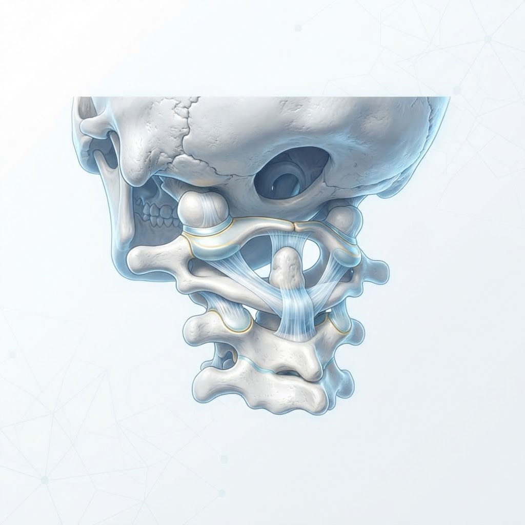
    At its core, chronic sinusitis is defined as an inflammatory condition of the paranasal sinuses—the hollow, air-filled spaces surrounding the nasal cavity—that lasts for at least twelve consecutive weeks. While an acute sinus infection often follows a common cold or allergic reaction and subsides relatively quickly, chronic sinusitis represents a persistent, recurring state of mucosal swelling and obstruction. When the lining of these passages becomes chronically inflamed, the normal drainage of mucus is blocked. This stagnation creates an environment where bacteria, viruses, or fungi can thrive, perpetuating the cycle of discomfort.

    Understanding this condition requires looking at the anatomy of the sinuses. Under healthy conditions, your sinuses produce thin, watery mucus that flows freely into the nasal cavity, acting as a natural trap for dust, pollen, and debris. Tiny hair-like structures called cilia move this mucus toward the throat, where it is harmlessly swallowed. In someone suffering from chronic sinusitis, this process is disrupted. The mucosal lining thickens, the cilia struggle to move debris, and the ostia (the small drainage openings) become constricted. This functional failure is what transforms a temporary nuisance into a long-term health challenge.

    It is helpful to distinguish between the two primary forms often discussed in the context of chronic sinusitis treatment. Some cases present with nasal polyps—soft, painless growths on the lining of your nasal passages or sinuses—while others occur without them. Both types share the common denominator of persistent inflammation, but they may require slightly different strategies for management. Many experts emphasize that chronic sinusitis is rarely the result of a single isolated factor. Instead, it is frequently a multi-faceted issue influenced by genetics, environmental exposures, immune responses, and even structural variations in the nasal anatomy, such as a deviated septum.

    Because the condition is persistent, those dealing with it often find that conventional over-the-counter medications provide, at best, a temporary reprieve. Relying solely on decongestants or steroid sprays can sometimes lead to a “rebound effect,” where symptoms worsen once the medication is stopped. Consequently, looking for natural relief for sinus infection and addressing the systemic inflammation is crucial. The goal for anyone seeking long-term sinus health is not just to clear the nasal passages in the moment, but to stabilize the mucosal lining and restore the body’s innate ability to drain and protect the sinuses. By shifting the perspective from “fighting an infection” to “cultivating a healthy environment,” individuals can move beyond the cycle of recurring flares and toward a more resilient state of sinus health.

    Common Symptoms of Persistent Sinus Issues

    Recognizing chronic sinusitis symptoms early is a major step toward preventing the frustration and fatigue associated with long-term nasal distress. While the symptoms can fluctuate in severity, the hallmark of the condition is its persistence. Unlike a standard cold, which typically peaks and then resolves within ten days, chronic sinus issues tend to hang on, often presenting as a dull, underlying discomfort that flares during specific times of the day or in response to environmental triggers.

    The most frequently reported symptom is a persistent feeling of congestion or “fullness” in the face. This pressure is often localized around the eyes, cheeks, forehead, and bridge of the nose. Many people describe it as a heavy, dragging sensation that makes concentration difficult. Because these sinuses are interconnected with the rest of the head, the pain can frequently migrate, manifesting as a dull, aching headache—particularly upon waking in the morning when mucus has accumulated overnight.

    Another key sign is persistent nasal discharge. This may not always be the thick, yellowish-green mucus associated with acute bacterial infections. In chronic cases, the discharge is often thin but constant, or sometimes clear and viscous. This leads to a persistent need to clear the throat or blow the nose, which can irritate the delicate nasal tissues further and create a cycle of physical trauma. Furthermore, chronic sinus inflammation often results in a diminished sense of smell and taste. When the nasal passages are chronically inflamed, the olfactory nerves struggle to detect subtle scents, which can impact your appetite and overall enjoyment of food.

    Fatigue is a commonly overlooked symptom. When your body is in a constant state of low-grade inflammation, it directs significant energy toward the immune response. This internal tug-of-war leaves many patients feeling perpetually tired, brain-fogged, and generally “run down.” Sleep is often disrupted as well; mouth breathing caused by congestion leads to dry throat, snoring, and poor sleep quality, compounding the feelings of fatigue during the day.

    Finally, individuals may experience a chronic cough, particularly at night, which is caused by “post-nasal drip”—a process where excess mucus drips down the back of the throat. This is a classic indicator that the sinuses are failing to drain properly into the nasal cavity. If you have been experiencing one or more of these symptoms for three months or longer, it is time to look deeper into the underlying sinus inflammation causes rather than simply treating the symptoms with temporary remedies.

    Approach Mechanism Best For
    Saline Irrigation Physically flushes out debris and moisturizes the mucosal lining. Daily maintenance and clearing of allergens/mucus.
    Steam Inhalation Adds moisture to air passages to help thin out trapped mucus. Immediate relief during acute flare-ups of dryness.
    Dietary Anti-Inflammatories Reduces systemic inflammation via antioxidants and fatty acids. Long-term systemic reduction of sinus sensitivity.
    Environmental Control Limits exposure to triggers like pollen, dust, and smoke. Prevention of seasonal or indoor allergen-induced flares.

    Identifying the Root Causes of Sinus Inflammation

    To effectively manage chronic sinusitis, one must move beyond the symptoms and investigate the underlying sinus inflammation causes. The sinuses are highly reactive cavities, and when they remain inflamed for months on end, it is usually because they are reacting to a persistent irritant, a structural blockage, or an immune system that has become hyper-vigilant. Identifying these triggers is the most important step in developing an effective strategy for lasting sinus health.

    Environmental triggers are among the most common culprits. For many, modern living environments are rife with microscopic irritants. Dust mites, mold spores, pet dander, and chemical vapors—such as those from household cleaning products or synthetic fragrances—can keep the nasal lining in a constant state of agitation. If your living space has high humidity, it can encourage mold growth; if it is too dry, the cilia cannot function properly, leading to inflammation. Determining your specific sensitivity requires careful observation. Does your congestion worsen at night (dust mites in bedding)? Does it increase during specific seasons (pollen)? Does it flare up when you are at the office (potentially poor ventilation or mold)?

    Another major category involves structural and anatomical issues. Some individuals have a deviated nasal septum, which is a physical displacement of the wall between the nasal passages. This can physically narrow the drainage pathway, making it impossible for mucus to clear, even if the person is otherwise healthy. Similarly, some people have naturally smaller sinus openings, or they may have nasal polyps—non-cancerous growths—that act as physical obstructions. These structural issues often require an evaluation to see if they are the primary driver preventing natural drainage.

    Immune system imbalances also play a critical role. In many cases, the body’s immune system mistakenly identifies harmless substances as threats, triggering an inflammatory response that manifests in the sinuses. This is often linked to sensitivities or allergies. When the immune system is constantly triggered, the resulting inflammation causes the nasal mucosa to swell, which in turn traps mucus and invites secondary bacterial or fungal colonization. This is why many people find that “sinus infections” are actually a recurring byproduct of a deeper, systemic issue with how their body handles environmental triggers.

    Finally, we cannot ignore the influence of stress and lifestyle. High levels of chronic stress trigger the release of cortisol, which can dampen the immune system’s efficiency while simultaneously increasing systemic inflammation. This creates a “perfect storm” for chronic sinus issues. When the body is stressed, it is less capable of managing the minor, everyday irritants that we all encounter. By viewing chronic sinusitis through this lens—as a signal from the body that something is out of balance—you shift the focus from “fixing” the sinuses to “harmonizing” the body’s internal state. Understanding these factors provides the foundation for creating a personalized plan for natural relief for sinus infection.

    The Connection Between Gut Health and Sinus Congestion

    In recent years, researchers have begun to map the “gut-sinus axis,” an intriguing connection between our digestive tract and our respiratory system. This concept suggests that the state of your gut microbiome—the vast community of bacteria living in your digestive system—may directly influence your sinus health. When we talk about how to clear sinuses naturally, we must look beyond the nasal passage itself and examine the core of our immune function: the gut.

    The majority of the human immune system is located within the gut. This makes sense from an evolutionary perspective, as the digestive tract is the primary point of contact between our internal systems and the external world. When the gut microbiome is in a state of balance, or symbiosis, it sends signals to the rest of the body to regulate inflammation and maintain healthy immune responses. However, when the balance of “good” bacteria is disrupted—a state known as dysbiosis—this can trigger systemic inflammation. This systemic inflammation often manifests in the weakest or most reactive parts of our bodies, which, for many, includes the sinuses.

    When your gut is struggling, your immune system may become over-reactive. This over-reactivity often manifests as an increased sensitivity to allergens and irritants. If you have an imbalanced gut, your body may be more prone to allergic responses that lead directly to sinus inflammation. Furthermore, some research suggests that the gut microbiome and the nasal microbiome communicate with each other through a chemical signaling process. An imbalance in the gut might lead to an overgrowth of certain microbes in the nasal passages, contributing to the cycle of chronic infection and congestion.

    Nutrition plays a pivotal role in this axis. Diets high in processed sugars and refined carbohydrates are known to disrupt gut flora and promote inflammation, both locally in the gut and systemically throughout the body. Conversely, diets rich in fermented foods, fiber, and diverse plant-based nutrients support a robust microbiome, which in turn helps to downregulate the body’s inflammatory response. Many individuals find that by making specific changes to their diet—such as increasing probiotic intake, reducing inflammatory triggers like dairy or gluten, and focusing on whole foods—they experience a marked reduction in their sinus symptoms.

    While it may seem counterintuitive to address a sinus issue by focusing on your digestion, this holistic approach is essential for long-term recovery. If you are struggling with chronic sinusitis, consider your diet as a core component of your treatment plan. By reducing the load of inflammatory foods and providing your gut with the nourishment it needs to host a healthy ecosystem, you are essentially “calming” your entire immune system from the inside out. This makes it significantly easier to achieve lasting sinus health and prevents the condition from becoming a permanent fixture in your life. Integrating gut-supporting habits into your daily routine is one of the most effective ways to build systemic resilience against the recurring discomfort of chronic sinus issues.

    Environmental Triggers to Avoid

    For individuals navigating the complexities of chronic sinusitis, the environment acts as a primary battlefield. When your sinus passages are already prone to inflammation, the air you breathe can either provide sanctuary or exacerbate the condition. Understanding and mitigating environmental triggers is a cornerstone of long-term sinus health management.

    Indoor Air Quality and Allergens: The home should be a haven, yet it is often a reservoir for triggers. Dust mites, mold spores, and pet dander are notorious for initiating the inflammatory cascade that keeps sinuses blocked. Dust mites thrive in soft surfaces; consequently, experts often recommend using allergen-proof covers for mattresses and pillows. Furthermore, keeping humidity levels between 30% and 50% is crucial. If the air is too dry, it irritates the mucus membranes, but if it is too moist, it promotes the growth of mold, which is a frequent culprit in sinus inflammation causes.

    Chemical Irritants: Many households rely on chemical cleaning agents, air fresheners, and scented candles. While these products make a home smell “fresh,” they release Volatile Organic Compounds (VOCs) that can be highly irritating to delicate sinus tissues. Switching to unscented, natural, or non-toxic cleaning products can significantly reduce the cumulative chemical load on your respiratory system.

    Outdoor Pollutants and Weather: Outdoor air quality is often out of your control, but your exposure to it is not. During peak pollen seasons, the microscopic particles can infiltrate the sinuses, causing swelling and mucus buildup. On high-pollen days, keeping windows closed and using a high-quality air purifier can provide much-needed relief. Additionally, sudden shifts in barometric pressure—often associated with changing weather fronts—are known to cause significant discomfort for those with chronic sinus conditions. While you cannot change the weather, being aware of these shifts allows you to pre-emptively use saline irrigation to keep the passages clear.

    Tobacco Smoke and Combustion Particles: Tobacco smoke is perhaps the most aggressive environmental enemy of the sinuses. It directly paralyzes the cilia—the tiny, hair-like structures responsible for sweeping mucus out of the sinuses. When cilia are damaged, mucus stagnates, leading to infection. Beyond direct smoking, “third-hand” smoke (residue on clothes and surfaces) and smoke from wood-burning fireplaces can also trigger sinus distress.

    Natural Ways to Reduce Sinus Pressure and Inflammation

    Managing chronic sinusitis symptoms often requires a multi-pronged approach that focuses on reducing the physical pressure within the nasal cavity. When the sinuses are blocked, the goal is to thin the mucus and encourage drainage.

    Saline Irrigation (The Neti Pot and Beyond): Saline irrigation remains one of the most effective ways to clear sinuses naturally. By flushing the nasal cavities with a sterile saline solution, you physically remove allergens, irritants, and excess mucus. It is essential, however, to use distilled, sterile, or previously boiled water, as tap water can harbor bacteria and amoebas that are dangerous when introduced into the sinuses.

    Warm Compresses and Steam Inhalation: The application of heat can soothe the facial pain associated with sinus pressure. Placing a warm, moist towel across the nose, cheeks, and forehead can help soften the mucus and improve circulation in the affected area. Similarly, steam inhalation—whether through a bowl of hot water or a dedicated facial steamer—provides direct moisture to the sinus linings. Adding a few drops of eucalyptus or peppermint oil (provided there are no allergies) can provide a cooling, decongestant effect, though these should be used sparingly as they can sometimes over-stimulate sensitive membranes.

    Facial Massage: Gentle manual manipulation of the sinus areas can encourage drainage. Using the fingertips to apply light, circular pressure around the bridge of the nose, the cheekbones, and above the eyebrows can help shift stagnant mucus toward the drainage pathways. This should be done with clean hands and light pressure; if it causes pain, discontinue immediately.

    Natural Remedy Mechanism of Action Best For
    Saline Irrigation Physical removal of mucus/debris Daily maintenance and allergy relief
    Steam Inhalation Hydration of nasal membranes Immediate relief of dry, crusty congestion
    Facial Massage Encourages lymphatic drainage Relieving localized sinus pressure and pain
    Warm Compresses Increased blood flow and vasodilation Soothing inflammation around the eyes/cheeks

    The Role of Hydration and Dietary Changes

    Dietary habits and hydration levels directly influence the consistency of the mucus in your sinuses. When you are dehydrated, your mucus becomes thick and sticky, which makes it significantly harder for your body to expel naturally. Maintaining optimal hydration is fundamental to sinus health.

    The Importance of Fluid Intake: Water is the most important tool for thinning mucus. Drinking adequate fluids throughout the day ensures that the body has the resources it needs to maintain thin, watery mucus that flows freely through the drainage ostia. Herbal teas, clear broths, and electrolyte-balanced water are excellent choices. Conversely, beverages that cause dehydration, such as excessive caffeine and alcohol, can counteract your progress and should be limited during a sinus flare-up.

    Anti-Inflammatory Nutrition: Chronic sinusitis is, at its heart, an inflammatory condition. Adopting an anti-inflammatory diet can help reduce the systemic response that contributes to sinus swelling. Focus on whole, unprocessed foods. Omega-3 fatty acids, found in fatty fish, walnuts, and flaxseeds, are known for their ability to dampen inflammation. Fruits and vegetables rich in antioxidants—such as berries, leafy greens, and citrus—support the immune system’s ability to fight off the pathogens that often accompany chronic sinus issues.

    Foods to Moderate: Certain individuals find that dairy products contribute to a feeling of thicker mucus, though this is debated in clinical literature. Keeping a food diary is a practical way to determine if dairy or other common allergens like gluten are exacerbating your personal symptoms. Reducing intake of processed sugars is also advisable, as high blood sugar levels can impair immune function and potentially feed the inflammation cycle.

    When to See a Professional for Sinus Issues

    While many natural strategies are effective for management, chronic sinusitis can sometimes escalate or reveal underlying anatomical or infectious issues that require clinical intervention. Recognizing the “red flags” is essential for protecting your long-term health.

    Duration and Severity: If you have symptoms of a sinus infection that persist for more than 10 to 12 weeks despite consistent home management, this is the clinical definition of chronic sinusitis. It is time to seek an evaluation from a primary care physician or an Ear, Nose, and Throat (ENT) specialist. They can perform nasal endoscopy to look directly into the passages and assess the level of inflammation.

    Warning Signs of Complications: Certain symptoms indicate that a sinus issue may be spreading beyond the nasal cavity. Seek immediate medical attention if you experience:

    • High fever that does not respond to over-the-counter measures.
    • Severe headache, stiff neck, or confusion (which could indicate a neurological complication).
    • Vision changes, such as double vision or swelling around the eyes.
    • Pain that is significantly worse on one side of the face compared to the other.

    Anatomical Considerations: Sometimes, the cause of chronic sinusitis is structural. A deviated septum, nasal polyps, or enlarged turbinates can physically obstruct the sinus openings, making it impossible to clear them naturally. A professional can identify these structural issues via diagnostic imaging like a CT scan. In such cases, surgical options, such as functional endoscopic sinus surgery (FESS) or balloon sinuplasty, may be necessary to widen the pathways and allow for permanent relief.

    Treatment Planning: An ENT can also help differentiate between allergic sinusitis and infectious sinusitis. If allergies are the root cause, they may suggest immunotherapy or prescription-grade nasal sprays that go beyond what is available over the counter. Do not attempt to self-diagnose or rely indefinitely on decongestant nasal sprays, as these can cause “rebound” congestion, making your symptoms worse in the long run.

    Frequently Asked Questions

    Is chronic sinusitis the same as a common cold?

    No, they are quite different. A common cold is a viral infection that typically resolves within a week or two. Chronic sinusitis is a long-term condition characterized by inflammation of the sinus linings that persists for 12 weeks or longer, regardless of treatment efforts.

    Can a sinus infection affect my vision?

    Yes, though this is a serious complication. Because the sinuses are located very close to the eye sockets, severe or spreading infection can cause eyelid swelling, redness, or pain. Any changes in vision associated with sinus pain require immediate professional medical evaluation.

    Does drinking milk make sinus congestion worse?

    While scientific evidence is mixed, many people report that dairy products cause their mucus to feel thicker or more persistent. If you suspect dairy is triggering your symptoms, try an elimination diet for two weeks to see if your sinus congestion improves.

    Are air purifiers actually effective for sinus health?

    Yes, high-quality air purifiers equipped with HEPA (High-Efficiency Particulate Air) filters are effective at removing airborne irritants such as dust, pet dander, and mold spores. By reducing the load of these allergens in your breathing environment, you can significantly lower the triggers that cause sinus inflammation.

    How long can I safely use a nasal saline rinse?

    Saline irrigation is generally considered safe for long-term, daily use. In fact, many individuals with chronic issues find that making it a part of their daily hygiene routine—much like brushing their teeth—is the best way to keep the nasal passages clear and reduce the frequency of flare-ups.

    Why does my sinus pain get worse when I fly or drive up mountains?

    This is due to changes in barometric pressure. The sinuses are air-filled cavities. When the external pressure changes rapidly, the air inside your sinuses must equalize. If the drainage pathways are blocked due to inflammation, the air cannot move, leading to significant pressure buildup and sharp, localized pain.

    Conclusion

    Chronic sinusitis is a challenging condition, but it is not one that you must simply endure. By understanding the root causes of sinus inflammation—from environmental triggers to structural and dietary factors—you can take control of your sinus health. The path to relief is rarely a single “magic bullet” but rather a combination of consistent habits, such as daily saline irrigation, a commitment to anti-inflammatory nutrition, and the proactive avoidance of irritants like tobacco smoke and harsh chemicals.

    While natural relief for sinus infections is a powerful first line of defense, it is vital to know when the limits of home care have been reached. Your sinuses are a critical part of your respiratory system; if symptoms persist beyond three months or if you experience warning signs like vision changes or severe, localized pain, do not hesitate to contact a professional. By staying observant and proactive, you can manage your chronic sinusitis effectively and regain your quality of life. Take the first step today by assessing your indoor air quality or incorporating a gentle saline rinse into your morning routine.

    By healthauthoritylife Editorial Team

    This article is for informational purposes only and is not a substitute for professional medical advice. Always consult your doctor before making changes to your diet, exercise routine, or treatment plan.
  • Gallbladder vs Biliary Dyskinesia: Understanding the Difference

    Gallbladder vs Biliary Dyskinesia: Understanding the Difference

    Key Takeaways

    • Gallbladder dyskinesia and biliary dyskinesia are often used interchangeably to describe a gallbladder that fails to contract effectively, despite the absence of physical stones.
    • The hallmark symptoms include chronic, post-meal upper abdominal pain, nausea, and bloating, which can mimic other gastrointestinal conditions like GERD or IBS.
    • Optimal digestion relies on the coordinated release of bile from the gallbladder into the small intestine to emulsify fats, a process frequently disrupted by biliary motility disorders.
    • The CCK-HIDA scan serves as a primary diagnostic tool by measuring the Ejection Fraction (EF), providing a functional look at the gallbladder rather than just a static image.
    • Functional issues are frequently missed by standard imaging like ultrasounds because these tests prioritize structural abnormalities over kinetic performance.

    For millions of individuals struggling with persistent digestive distress, the journey to a diagnosis can feel like a labyrinth. You may have experienced recurring bouts of sharp pain, nausea, and discomfort after meals, yet every conventional imaging report—from standard ultrasounds to CT scans—returns with the frustrating notation of “no stones found.” This scenario often points toward a functional rather than a structural problem within the biliary system. While the terms gallbladder dyskinesia and biliary dyskinesia are frequently discussed in clinical forums, many patients remain confused about what these conditions actually represent. Understanding these disorders requires moving beyond the “stone-focused” model of care and looking at the physiological mechanics of how your body manages bile. In this guide, the healthauthoritylife Editorial Team explores the nuances of biliary motility disorders, how they disrupt digestion, and why identifying these functional issues is the critical first step toward reclaiming your quality of life.

    Defining Gallbladder and Biliary Dyskinesia

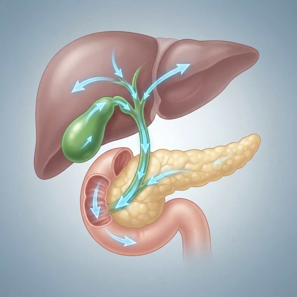
    To understand the scope of gallbladder dyskinesia, we must first distinguish it from the more commonly understood “biliary colic” caused by gallstones (cholelithiasis). In a healthy system, the gallbladder acts as a reservoir. It stores and concentrates bile produced by the liver, holding it until a meal—specifically one containing fat—triggers the release of cholecystokinin (CCK). This hormone signals the gallbladder to contract, pushing bile through the cystic duct and into the common bile duct, where it eventually enters the small intestine to aid in the breakdown of fats.

    Biliary dyskinesia is an umbrella term for conditions where this movement—or motility—is impaired. When we speak of gallbladder dyskinesia specifically, we are typically referring to a scenario where the gallbladder muscle wall, known as the muscularis, is unable to contract with sufficient force to empty its contents. It is not necessarily blocked by a physical obstruction like a stone, polyp, or sludge, but rather inhibited by a loss of coordination or muscular strength. Essentially, the “pump” is not working properly.

    Because the gallbladder is not a standalone organ but part of a complex biliary tract, the term biliary dyskinesia is often used more broadly. It encompasses issues within the entire biliary tree, including the Sphincter of Oddi—the valve that controls the flow of bile into the duodenum. If the sphincter fails to relax at the precise moment the gallbladder contracts, the resulting pressure can cause significant pain, even if the gallbladder itself is physically capable of squeezing.

    These functional disorders are fundamentally different from structural diseases. In structural cases, a surgeon can point to a gallstone and explain exactly why it is causing a blockage. In cases of gallbladder dyskinesia, the anatomy may look pristine on a high-definition scan, yet the physiological output remains deficient. This leads to a state of chronic frustration for patients who suffer from legitimate, debilitating pain that does not show up on traditional diagnostic “checklists.” Recognizing that the gallbladder is an active, kinetic organ—not just a storage sac—is the first step toward understanding why these motility disorders are so difficult to diagnose and manage.

    Primary Symptoms of Biliary Motility Issues

    The symptoms of biliary dyskinesia are notoriously difficult to distinguish from other gastrointestinal conditions, which is why it is often referred to as a “diagnosis of exclusion.” Because the biliary system shares neurological pathways with the stomach and the intestines, the distress often presents as generalized abdominal pain that can easily be mistaken for gastritis, peptic ulcers, or irritable bowel syndrome (IBS).

    The most characteristic symptom reported by patients is biliary pain—a dull, aching, or sometimes sharp, stabbing sensation located in the right upper quadrant (RUQ) of the abdomen, just beneath the rib cage. This pain frequently radiates to the center of the upper abdomen (epigastrium) or travels through to the mid-back, particularly beneath the right shoulder blade. This “referred pain” occurs because of the shared nerve innervation between the gallbladder and the phrenic nerve.

    Crucially, these symptoms are almost always linked to food intake. Unlike some conditions where pain is constant, biliary motility issues typically flare up within 30 to 60 minutes after eating, particularly meals that are high in fat or heavy in oils. Because the body is struggling to secrete enough bile to emulsify these fats, the gallbladder works overtime, contracting repeatedly against an ineffective system or failing to empty entirely, leading to a sensation of intense pressure.

    Beyond the pain, secondary symptoms often emerge as a direct result of poor fat digestion. These include:

    * Nausea and early satiety: Many individuals feel full after only a few bites of food, or they experience a persistent, low-level wave of nausea that lasts for hours after a meal.
    * Bloating and flatulence: When fat isn’t properly broken down, it remains in the gut, where it can cause fermentation and gas buildup.
    * Stool changes: Some patients report pale, clay-colored stools or, conversely, frequent diarrhea, as the lack of bile reaching the intestines alters the pH and consistency of waste.
    * Belching: Excessive burping is a common attempt by the digestive system to relieve the pressure buildup in the upper GI tract.

    It is important to note that the severity of these symptoms does not always correlate with the severity of the motility issue. Some individuals with only a minor reduction in gallbladder function report debilitating, daily pain, while others with significant “sluggishness” may only experience intermittent discomfort. This unpredictability makes it essential for patients to track their symptoms in relation to specific triggers, as this data becomes invaluable during the diagnostic process.

    How the Biliary System Functioning Impacts Digestion

    The biliary system is the silent workhorse of the digestive tract. While the stomach manages mechanical and acid-based breakdown, the liver and gallbladder are responsible for the critical chemical processing of dietary fats. When this system experiences dyskinesia, the consequences ripple through the entire digestive process, affecting everything from nutrient absorption to the balance of the gut microbiome.

    To appreciate the impact, we must look at the role of bile salts. Bile acts as a natural detergent. By breaking large fat globules into tiny droplets (emulsification), it increases the surface area for the enzyme lipase to work. If gallbladder function is impaired, the release of bile becomes mistimed or insufficient. When a fat-laden meal hits the small intestine without an adequate bile “pulse,” the fats remain largely unprocessed in the upper GI tract.

    This undigested fat creates a cascade of physiological disturbances:

    1. Malabsorption of Fat-Soluble Vitamins: Vitamins A, D, E, and K require bile for absorption. Chronic biliary issues can lead to subclinical deficiencies in these essential nutrients, even if an individual’s diet appears healthy. Over time, this can affect skin health, bone density, and immune function.
    2. Intestinal Flora Imbalance: Bile is naturally antimicrobial. It helps regulate the population of bacteria in the small intestine. When bile flow is inadequate, some experts suggest it can contribute to small intestinal bacterial overgrowth (SIBO), where bacteria from the large intestine migrate upward, leading to excessive bloating, diarrhea, and nutrient competition.
    3. Increased Gastrointestinal Transit Time: Undigested fats and lipids that travel too far into the large intestine can irritate the intestinal lining, potentially contributing to either rapid transit (diarrhea) or a slowing of the system as the gut attempts to compensate for the digestive delay.

    The following table summarizes how different approaches to assessing or managing these biliary concerns compare in a clinical context:

    Approach Primary Focus Best For
    Abdominal Ultrasound Structural Integrity Ruling out stones, polyps, or thickening
    CCK-HIDA Scan Functional Motility Quantifying ejection fraction and emptying efficiency
    Dietary Modification Symptom Management Reducing post-meal triggers and inflammation
    Endoscopic Ultrasound Detail Diagnostics Visualizing biliary sludge or small, hidden stones

    Ultimately, the biliary system’s function is about timing. It is a finely tuned synchronization between the stomach, the gallbladder, and the duodenum. When this system fails, it isn’t just about pain—it’s about the body’s fundamental inability to harvest energy from the most calorie-dense macro-nutrient in our diet. Recognizing these systemic effects is vital for anyone who suspects their digestive issues might stem from biliary motility disorders.

    The Role of CCK-HIDA Scans in Modern Diagnosis

    Because standard imaging is often blind to the inner workings of the gallbladder, the HIDA scan (Hepatobiliary Iminodiacetic Acid scan), augmented with a CCK (cholecystokinin) injection, has become the gold standard for diagnosing functional biliary disorders. Unlike an ultrasound, which provides a high-resolution snapshot of the gallbladder’s appearance, the CCK-HIDA scan acts as a “stress test” for the gallbladder’s performance.

    The process typically involves the administration of a radioactive tracer, which is taken up by the liver and excreted into the bile ducts. Once the tracer reaches the gallbladder, the patient is monitored to ensure the gallbladder fills properly. This part of the scan confirms that there is no structural blockage in the cystic duct. If the tracer can enter the gallbladder, the ducts are generally considered patent, or open.

    The diagnostic “magic” happens when the clinical team administers a synthetic form of CCK. This hormone is essentially the command that tells the gallbladder to contract. By measuring the tracer’s exit from the gallbladder after the CCK injection, clinicians can calculate the Gallbladder Ejection Fraction (GBEF). This percentage represents how much bile the gallbladder successfully squeezed out during the test.

    Generally, an Ejection Fraction above a certain threshold is considered normal, indicating that the gallbladder is pumping effectively. However, if the GBEF is significantly low, it suggests the gallbladder is underactive or “lazy,” which is the hallmark of gallbladder dyskinesia. Conversely, in rare instances, the ejection fraction may be abnormally high—sometimes referred to as hyperkinetic biliary dyskinesia—which can also cause intense, spasm-like pain.

    It is important to remember that the HIDA scan is not just about the numbers. It is also about the patient’s subjective experience during the test. For many, the injection of CCK triggers the exact same pain or nausea they feel after eating a heavy meal. When the low ejection fraction is paired with the reproduction of familiar symptoms, it provides strong evidence that the gallbladder is the source of the patient’s discomfort.

    While modern medicine relies heavily on this test, it is not without limitations. The accuracy of the result can be influenced by medications the patient may be taking, their fasting state, or the specific protocol used by the radiology department. Furthermore, the GBEF threshold for what is considered “abnormal” can vary among clinical institutions, meaning that a patient might be told their gallbladder is “borderline” in one facility and “dyskinetic” in another. Therefore, the CCK-HIDA scan should always be interpreted as one piece of a larger diagnostic puzzle, rather than a standalone arbiter of health.

    Why Conventional Tests Often Miss Functional Issues

    The frustration shared by many patients is rooted in the “normal” result they receive from their doctor after a standard diagnostic workup. A patient may present with classic symptoms of biliary disease—right-sided pain, nausea, and intolerance to fatty foods—only to have an abdominal ultrasound come back clear. In the traditional medical model, this is often interpreted as “everything looks fine.” But why does it look fine, yet feel so wrong?

    The primary reason is that conventional diagnostic tools, such as basic ultrasounds, CT scans, and MRIs, are designed primarily to detect structural pathologies. They are looking for solid, physical evidence of disease. They excel at identifying:

    * Calculi (Gallstones): Hardened deposits that occupy space.
    * Cholecystitis: Inflammation or wall thickening that signifies acute infection.
    * Masses or Tumors: Abnormal tissue growth.
    * Ductal Dilation: Physical widening caused by a blockage.

    These tests are akin to checking the structural integrity of a building; they can tell you if the foundation is cracked or if there is a wall missing, but they cannot tell you if the electricity is working or if the plumbing is flowing at the correct pressure. Functional issues like biliary dyskinesia are effectively an “electrical” or “kinetic” problem. The gallbladder may be structurally perfect—no stones, no thick walls, no sludge—but the communication between the brain, the hormones, and the muscular gallbladder wall is disrupted.

    Additionally, “biliary sludge” is another functional factor that is frequently overlooked. While sludge is not a stone, it consists of tiny, microscopic crystals of cholesterol and calcium salts that can significantly alter the viscosity of bile. Standard ultrasounds may miss small amounts of sludge, especially if the gallbladder is not imaged during a fasting state or if the equipment resolution is low. Yet, this “sludge” can act as a sandpaper-like irritant or create micro-obstructions that trigger spasms and pain, even if it does not technically fit the definition of a stone.

    Furthermore, the intermittent nature of biliary dyskinesia means that a patient can have a perfectly healthy-looking ultrasound on Tuesday, but suffer an agonizing biliary attack on Wednesday. The scan only captures a moment in time. If the gallbladder happens to be in a “rest” phase or if the trigger isn’t present during the imaging, the system will appear perfectly normal.

    Understanding these diagnostic blind spots is critical for patient advocacy. When symptoms persist despite “clean” imaging, it does not mean the pain is psychosomatic or that the symptoms are invalid. It suggests that the testing protocol has reached its limit in terms of detecting structural disease and that the investigation must pivot toward functional assessments—like the CCK-HIDA scan—to uncover the underlying motility dysfunction. Moving from a structural focus to a functional one often represents the biggest hurdle in getting the correct diagnosis for biliary motility disorders.

    Common Triggers for Biliary Dyskinesia Flare-ups

    Understanding the specific factors that provoke symptoms is a vital step in managing biliary dyskinesia. Because this condition involves an ineffective gallbladder—one that fails to contract properly in response to signals from the digestive system—it is inherently tied to how your body processes intake. When the gallbladder is sluggish, the presence of certain substances or physiological stressors can overwhelm its limited capacity, leading to the sharp, cramping abdominal pain characteristic of the disorder.

    The most common trigger is the ingestion of high-fat meals. Bile is the body’s primary emulsifier for fats; its role is to break down large fat globules into smaller, absorbable particles. When you consume a heavy, greasy, or high-fat meal, the duodenum releases cholecystokinin (CCK), a hormone that instructs the gallbladder to empty its stores. In a person with biliary dyskinesia, the gallbladder cannot respond effectively to this hormonal prompt. The pressure builds within the biliary tree, resulting in intense discomfort or pain in the upper right quadrant of the abdomen.

    Stress and nervous system dysregulation also play a significant role in biliary motility disorders. The gallbladder is governed by the autonomic nervous system, specifically the balance between the sympathetic (fight or flight) and parasympathetic (rest and digest) branches. High levels of chronic stress can shift the body into a sympathetic state, which often suppresses digestive motility. When the gallbladder is already struggling to empty, a sympathetic-dominant state can further inhibit its ability to contract, exacerbating the sluggishness.

    Furthermore, irregular eating patterns—such as skipping meals or prolonged fasting—can be surprisingly detrimental. The gallbladder acts as a reservoir; it fills with bile between meals and empties during them. If you skip meals, bile may sit in the gallbladder for an extended period, potentially becoming thick or sludgy, which complicates the emptying process when you finally do eat. Consistent, smaller, and balanced meals are generally better for maintaining biliary health than erratic or infrequent feeding schedules.

    Natural Strategies to Support Bile Flow and Motility

    Supporting gallbladder function often requires a multi-faceted approach aimed at thinning the bile and encouraging the smooth muscle of the gallbladder to function more efficiently. While these strategies are not replacements for medical intervention, many individuals find them helpful in reducing the frequency of symptom flare-ups.

    Hydration is the most foundational element in supporting bile flow. Bile is primarily composed of water. If you are chronically dehydrated, your bile can become more viscous, which increases the physical resistance the gallbladder must overcome to empty. Ensuring adequate daily fluid intake helps maintain the proper consistency of bile, making it easier for the gallbladder to push it through the cystic and common bile ducts.

    The use of “bitter” herbs and foods is a traditional strategy for supporting biliary motility. Bitter compounds found in foods like dandelion greens, arugula, radicchio, and artichokes are thought to stimulate the production of digestive juices, including bile. By incorporating these bitter elements into your meals, you may signal to the digestive system that it is time to prepare for fat metabolism, which can help prime the gallbladder for the digestive process.

    Magnesium supplementation is also frequently discussed by experts in digestive health. Magnesium plays a critical role in muscle relaxation and the regulation of smooth muscle tissue. Because the gallbladder relies on smooth muscle contractions to empty, adequate magnesium levels are essential for optimal function. Many people are deficient in this mineral, and ensuring you are meeting your requirements through magnesium-rich foods—such as pumpkin seeds, spinach, and almonds—can support overall smooth muscle health in the digestive tract.

    Additionally, mild, consistent physical activity can support digestive motility. While high-intensity exercise may be difficult during a flare-up, gentle movement like walking after a meal can aid in general digestive transit. Gravity and the movement of the abdominal muscles can assist in the digestive process, potentially taking some of the mechanical pressure off a struggling biliary system.

    Strategy Mechanism of Action Best For
    Hydration Maintains bile viscosity Daily routine, preventing sludge
    Bitter Foods Stimulates digestive enzyme/bile flow Pre-meal priming
    Magnesium-Rich Diet Supports smooth muscle function Long-term motility support
    Gentle Post-Meal Walking Encourages gastric and biliary transit Managing post-prandial fullness

    Dietary Adjustments for Managing Dyskinesia Symptoms

    When dealing with biliary dyskinesia, the goal of a dietary strategy is to reduce the mechanical workload placed on the gallbladder. Because the organ is malfunctioning, the objective is to provide the body with the nutrition it needs without requiring massive, sudden surges of bile release.

    The primary dietary shift for most individuals is moving away from high-fat, fried, and processed foods. Instead of avoiding fat entirely, which is essential for hormone regulation and fat-soluble vitamin absorption, the focus should be on healthy, easily digestible fats in moderate, consistent portions throughout the day. This “drip-feed” approach prevents the gallbladder from being overwhelmed by a sudden, large dose of dietary fat.

    Fiber is another critical component. A diet rich in soluble fiber can bind to bile acids in the digestive tract. While this helps clear excess bile, it also encourages a more efficient enterohepatic circulation. Incorporating foods like oats, apples, lentils, and chia seeds can help stabilize the digestive environment. However, fiber should be increased gradually to avoid bloating, which can exert uncomfortable pressure on the upper abdomen.

    Avoiding common food sensitivities is also vital. In some cases, biliary distress is aggravated by specific triggers such as refined sugars, dairy, or gluten. Keeping a detailed food diary is one of the most effective ways to identify personal triggers. Note the time you ate, the composition of the meal, and any symptoms that follow. Over several weeks, a pattern usually emerges, allowing you to refine your diet to suit your specific digestive tolerance.

    Finally, focus on high-quality proteins that are low in hidden fats, such as skinless poultry, white fish, or plant-based proteins like tempeh. Cooking methods also matter; opting for steaming, poaching, baking, or grilling is far better for a sensitive gallbladder than frying or heavy sautéing in oils. By simplifying your cooking methods, you reduce the total fat load per serving, which can significantly decrease the intensity of biliary symptoms.

    When to Consult a Specialist for Biliary Health

    While lifestyle changes can manage mild cases, biliary dyskinesia can progress or lead to complications that require professional medical oversight. Knowing when to escalate your concerns is critical for preventing long-term damage or acute medical crises.

    You should consult a gastroenterologist or a biliary specialist if you experience symptoms that are becoming more frequent, longer-lasting, or more intense. If you find that you can no longer eat a normal variety of foods without experiencing significant pain, it is a sign that your current management plan is no longer effective. Chronic pain, even if mild, can significantly decrease your quality of life and indicates an underlying issue that needs further diagnostic investigation, such as a HIDA scan.

    Be particularly vigilant for “red flag” symptoms. These include jaundice (yellowing of the skin or eyes), which suggests a blockage in the biliary system; high fever or chills, which could indicate an infection of the gallbladder (cholecystitis) or the bile ducts (cholangitis); and dark urine or pale, clay-colored stools. These symptoms are medical emergencies and require immediate attention at an urgent care facility or emergency room.

    Furthermore, if you have been previously diagnosed with gallstones and are now experiencing changes in your pain patterns, professional assessment is necessary. Sometimes, dyskinesia and stones can coexist, and the presence of stones increases the risk of duct blockage. A specialist can help differentiate between simple motility issues and complications that might necessitate surgical intervention, such as a cholecystectomy, to resolve the pain and prevent future risks.

    Frequently Asked Questions

    Can biliary dyskinesia lead to the formation of gallstones?

    While biliary dyskinesia is primarily a problem of movement rather than composition, the lack of effective emptying can result in bile staying in the gallbladder for too long. Over time, stagnant bile is more likely to develop into sludge and eventually into solid gallstones, making proper management of motility essential for long-term health.

    Is surgery the only way to treat biliary dyskinesia?

    Surgery, specifically the removal of the gallbladder, is often considered when conservative management fails to control symptoms. However, it is not always the first step. Many patients successfully manage their condition through dietary adjustments, stress management, and close monitoring by a gastroenterologist before considering surgical intervention.

    How does a HIDA scan accurately diagnose this condition?

    A HIDA scan uses a radioactive tracer that is absorbed by the liver and excreted into the bile. A specialist monitors the movement of this tracer into the gallbladder. By administering a CCK hormone injection, doctors can measure the gallbladder ejection fraction (GBEF). A low percentage typically confirms that the gallbladder is not contracting sufficiently, which is the hallmark of biliary dyskinesia.

    Can stress really cause gallbladder pain?

    Yes, stress impacts the autonomic nervous system, which dictates digestive function. High stress levels can induce muscle tension and alter digestive hormone release, potentially impeding the gallbladder’s ability to contract. Managing stress through breathwork, mindfulness, or therapy is a recognized, albeit indirect, way to support biliary health.

    What does it mean if I have a normal ultrasound but still have symptoms?

    An ultrasound is excellent at detecting structural issues like stones or wall thickening, but it does not measure function. You can have a perfectly healthy-looking gallbladder on an ultrasound that is, in reality, functionally “lazy” or dyskinetic. This is precisely why functional tests like the HIDA scan are necessary when structural imaging shows nothing, yet symptoms persist.

    Can children or teenagers develop biliary dyskinesia?

    While most common in adults, biliary dyskinesia can occur in younger populations. When it does occur in children or adolescents, it is often investigated by pediatric specialists who look for factors such as rapid weight changes, dietary habits, or other underlying metabolic conditions that might be disrupting the biliary tract.

    Conclusion

    Navigating the complexities of biliary health can be daunting, but distinguishing between simple gallbladder issues and biliary dyskinesia is a fundamental first step toward finding relief. Whether you are currently experiencing intermittent discomfort or are working with a specialist to manage a formal diagnosis, the key lies in understanding your body’s unique triggers and supporting your biliary system through consistent, healthy habits. By prioritizing hydration, thoughtful nutrition, and stress management, many individuals successfully mitigate their symptoms and improve their overall digestive function.

    However, never underestimate the value of professional guidance. If your symptoms interfere with your daily life or if you experience warning signs of complications, seek medical advice promptly. Armed with the right information and a proactive approach to your health, you can make informed decisions that promote long-term comfort and well-being. Take the first step today by tracking your symptoms and discussing them with a healthcare provider.

    By healthauthoritylife Editorial Team

    This article is for informational purposes only and is not a substitute for professional medical advice. Always consult your doctor before making changes to your diet, exercise routine, or treatment plan.

  • Post-Prandial Hypotension: Symptoms, Causes, and Natural Relief

    Key Takeaways

    • Post-prandial hypotension is a sudden, abnormal drop in blood pressure that occurs shortly after eating a meal.
    • Common symptoms include dizziness, lightheadedness, and fatigue, which can be particularly concerning for older adults or those with underlying metabolic conditions.
    • Digestion requires significant blood flow to the gastrointestinal tract, which can trigger systemic blood pressure drops if the cardiovascular system fails to compensate.
    • Key triggers include heavy carbohydrate intake, rapid eating, and certain underlying health conditions that affect autonomic nervous system regulation.
    • Lifestyle modifications, such as eating smaller, more frequent meals and staying hydrated, are essential for managing and finding natural relief for hypotension.

    There are few things as disorienting as finishing a satisfying meal, only to feel an immediate, wave-like sensation of faintness or profound lethargy. While many of us have joked about the “food coma,” for a significant portion of the population, this is not just a sign of a hearty lunch—it is a physiological event known as post-prandial hypotension. This condition, characterized by a measurable decrease in blood pressure shortly after eating, can range from a minor annoyance to a serious health hurdle that increases the risk of falls and fainting. Understanding the mechanics behind why your body sometimes fails to maintain stable blood pressure after you dine is the first step toward reclaiming your daily energy and safety. By examining the symptoms, biological triggers, and lifestyle influences that contribute to this phenomenon, you can begin to distinguish between a natural digestive dip and a more complex metabolic warning sign.

    What Is Post-Prandial Hypotension?

    Post-prandial hypotension is clinically defined as a drop in systolic blood pressure of 20 mmHg or more within two hours of consuming a meal. While the body has a sophisticated, finely tuned mechanism to manage blood flow during digestion, this process can occasionally go awry. In a healthy state, when you eat, your body sends a rush of blood to the stomach and small intestine to assist in the breakdown and absorption of nutrients. Simultaneously, the heart rate increases slightly, and blood vessels in other parts of the body constrict to compensate for the diversion of blood flow toward the digestive tract. This delicate balancing act maintains systemic blood pressure, ensuring that your brain and vital organs continue to receive a consistent oxygen supply.

    However, when this compensatory system falters, the result is post-prandial hypotension. It is most commonly observed in older adults, particularly those who may already have underlying conditions such as hypertension, diabetes, or autonomic nervous system dysfunction. The condition acts as a bridge between your cardiovascular health and your metabolic state. Because the digestive process is metabolically demanding, any interference in how the body handles glucose, hormones like insulin, or neurological signaling can cause that systemic blood pressure drop. It is crucial to note that this is not merely “feeling sleepy.” It is a hemodynamic event that reflects the body’s struggle to adjust to the demands of processing food.

    The prevalence of this condition suggests it is often underdiagnosed because the symptoms are frequently dismissed as simple post-meal fatigue. Many individuals unknowingly suffer from these episodes for years, attributing the sudden lightheadedness to “eating too much” or the temperature of the room. Recognizing that this is a specific medical phenomenon allows you to move away from ignoring the signals your body is sending. By identifying the root causes—which often involve the interplay between insulin spikes and vascular resistance—you can better prepare your daily routine to mitigate the impact. Whether it stems from how quickly you eat or the composition of your meal, post-prandial hypotension is a clear indicator that your body’s internal regulation systems are currently under pressure and may require targeted adjustments to maintain stable health.

    Common Symptoms of Low Blood Pressure After Eating

    Identifying the symptoms of low blood pressure after eating is the first move in self-awareness. Because these symptoms often manifest immediately after a meal, it is easy to assume they are standard digestive reactions. However, the signs of post-prandial hypotension are distinct in their neurological and physical presentation. The most frequent complaint is dizziness or a sense of lightheadedness, often described as a “floating” or “spinning” sensation that occurs as you stand up from the dining table. This occurs because the body is already struggling to maintain blood flow to the brain due to the digestive redirect; standing up adds the challenge of gravity, which the body can no longer effectively counter.

    Other common indicators include unexpected fatigue, confusion, or “brain fog.” You may find that after a lunch that previously felt manageable, you suddenly struggle to concentrate or feel a heavy, overwhelming urge to sleep. In some cases, individuals experience visual disturbances, such as blurred vision or spots in their field of vision, which is a common sign of insufficient blood flow to the ocular nerves. Furthermore, nausea is frequently reported alongside these dips in pressure, which creates a difficult cycle: you feel sick because your blood pressure is low, and your low blood pressure is worsened by the digestive distress. In more severe cases, or in individuals with fragile cardiovascular systems, one might experience chest pain or heart palpitations as the heart beats faster in a frantic attempt to compensate for the sudden drop in pressure.

    It is important to pay attention to the timing of these sensations. Are they occurring within 30 minutes of your first bite? Do they last for an hour or more? Keeping a symptom log can be an incredibly useful tool for both your own understanding and any future conversations with healthcare providers. If you notice that you are feeling unusually weak, experiencing sudden falls, or suffering from “fainting spells” (syncope) after large meals, it is vital to acknowledge these as more than just minor inconveniences. These symptoms are the body’s way of communicating that its compensatory mechanisms are overwhelmed. Recognizing these warning signs allows you to act proactively, rather than waiting for a fainting event to occur, and helps you distinguish between normal food-induced relaxation and true hemodynamic instability.

    Approach Mechanism Best For
    Meal Fractionation Eating smaller, frequent meals instead of large ones. Preventing sudden blood surges to the gut.
    Hydration Strategy Drinking water 15–30 minutes before consuming food. Increasing blood volume and pressure stability.
    Carbohydrate Control Reducing simple sugars and refined grains. Avoiding rapid insulin and vasodilation responses.
    Post-Meal Movement Engaging in light walking or standing activity. Promoting circulatory return to the heart.

    Understanding the Biological Causes

    The biological causes of post-prandial hypotension are rooted in the complex physiological dance between your nervous system, your hormones, and your vasculature. When you consume food, especially high-carbohydrate or high-sugar meals, your body initiates a rapid increase in insulin secretion. Insulin, while vital for processing glucose, acts as a mild vasodilator—meaning it signals the smooth muscles surrounding your blood vessels to relax and widen. In a system that is functioning perfectly, this widening is offset by other nerves that keep the rest of your circulation tight and efficient. However, in individuals with metabolic sensitivities or aging vasculature, this signal can lead to a systemic drop in pressure because the body fails to “tighten” those vessels fast enough to compensate for the blood now pooling in the digestive system.

    Another primary factor is the release of gastrointestinal hormones, such as glucagon-like peptide-1 (GLP-1), which are released during digestion. These hormones help manage blood sugar but also have direct effects on heart rate and vascular tone. For those with autonomic nervous system dysfunction—a condition where the nerves that control “automatic” processes like blood pressure become less responsive—the body loses its ability to increase heart rate or constrict peripheral blood vessels when blood is redirected to the stomach. This effectively leaves the “pressure” in your cardiovascular system low, resulting in those classic symptoms of dizziness and fatigue.

    Furthermore, we must consider the role of glucose metabolism. When blood glucose levels rise rapidly after eating, the body attempts to process the influx, which consumes energy and alters electrolyte balance. This process is particularly taxing for individuals with insulin resistance. The chronic state of managing high blood sugar can weaken the compensatory response of the nervous system over time. Many experts suggest that the “tug-of-war” between the digestive need for blood and the systemic need for blood pressure is at the heart of the problem. If your body is unable to effectively transition between these two states, the result is an inevitable, temporary crash. Understanding this biological disconnect is crucial, as it shifts the perspective from seeing this as a digestive issue to seeing it as a systemic cardiovascular and metabolic management challenge.

    Why Digestion Impacts Your Blood Pressure

    To understand why digestion impacts your blood pressure, you must view the cardiovascular system as a giant plumbing network with a finite amount of fluid. When you eat, your body treats the act of digestion as a high-priority task. It requires an immense amount of energy and, crucially, a massive influx of oxygenated blood. The splanchnic circulation—the blood vessels that serve the stomach, liver, and intestines—expands significantly to accommodate this need. Under normal circumstances, this expansion is manageable. However, if the volume of blood is not sufficient or if the nervous system’s feedback loops are delayed, the diversion of blood to the gut comes at the expense of systemic blood pressure.

    The intensity of this impact depends heavily on the volume and composition of the meal. A large, heavy meal necessitates a much larger diversion of blood than a small, light snack. This is why many people find their symptoms occur primarily after their largest meal of the day. Furthermore, the rate at which food reaches the small intestine plays a pivotal role. If your stomach empties rapidly, the surge of nutrients hitting the intestines triggers a faster and more aggressive demand for blood flow. This sudden, acute demand is precisely what causes the rapid drop in pressure. The body tries to counteract this by increasing the heart rate, but if the individual has any underlying cardiovascular limitations—such as stiffer arteries or a heart that cannot pump as efficiently—the compensation falls short.

    Additionally, hydration plays a silent but critical role in this dynamic. Blood volume is the bedrock of blood pressure. If you are even slightly dehydrated, your total blood volume is lower, making the internal competition for blood during digestion even more intense. When you eat, your body is effectively spreading a thinner blanket of blood over a larger area of demand. This is why individuals who are well-hydrated often report fewer and less severe episodes of post-prandial hypotension. The digestive process is, in many ways, the ultimate stress test for your cardiovascular system. It reveals how well your body can distribute resources under pressure. By respecting this biological need for blood in the gut, we can begin to implement strategies—such as slowing down the digestive process—that keep the systemic blood pressure within a healthy range, preventing the sudden, disorienting drops that characterize this condition.

    Lifestyle Factors That Worsen Hypotension

    Several lifestyle factors act as silent contributors to worsening post-prandial hypotension, often turning mild symptoms into disruptive episodes. One of the most significant culprits is the “speed” of your lifestyle. In our modern, fast-paced culture, many of us eat our meals quickly, often while distracted or in a state of high stress. When you eat rapidly, you consume more volume in a shorter period, leading to a much steeper and faster demand for blood flow to the gut. This sudden “loading” of the digestive tract forces the cardiovascular system to react with equal speed—a requirement that many individuals simply cannot meet due to age-related changes in vascular elasticity or autonomic health.

    Another major lifestyle factor is the composition of the diet. Meals high in refined carbohydrates and sugars cause the most dramatic insulin spikes. Because insulin is a vasodilator, high-sugar meals effectively “force” the body to dilate blood vessels just as it is trying to redirect blood to the gut. This creates a double-hit effect: the gut demands more blood, and the insulin-induced vasodilation ensures that blood pressure drops. Conversely, meals that are balanced with high-quality fats, lean proteins, and fiber tend to be digested more slowly and result in a more measured, stable release of insulin and nutrients. This steady digestion gives the nervous system time to calibrate, preventing the dramatic, symptom-inducing drops in blood pressure.

    Alcohol consumption is another significant factor that often goes overlooked. Alcohol is a known vasodilator and can also cause a temporary, yet significant, reduction in blood volume due to its diuretic effects. When combined with a meal, alcohol can drastically amplify the symptoms of post-prandial hypotension. Similarly, certain medications, particularly those intended to lower blood pressure, can become problematic when taken just before or after a meal. If your medication is already working to lower your blood pressure, adding the digestive “dip” on top of that can lead to a dangerous drop. Finally, a sedentary lifestyle can contribute to poor venous return—the ability of your body to pump blood from the lower extremities back up to the heart. When the circulation is already sluggish, the redirection of blood to the gut becomes much more problematic. Integrating regular, moderate activity helps maintain the efficiency of the circulatory system, ensuring that your body is better equipped to handle the metabolic demands of eating without sacrificing your systemic blood pressure stability.

    How to Prevent Dizziness After Eating

    Preventing post-prandial hypotension—the sudden drop in blood pressure following a meal—requires a proactive approach to how you structure your daily nutrition and movement. When you consume a meal, particularly one high in carbohydrates, your body directs a significant volume of blood toward the gastrointestinal tract to facilitate digestion. In individuals prone to vascular fluctuations, this shift can lead to reduced blood flow to the brain, manifesting as lightheadedness or dizziness.

    The first line of defense is modifying the size and frequency of your meals. Rather than consuming three large, calorie-dense meals that place a heavy demand on your circulatory system, transition to five or six smaller, nutrient-dense portions throughout the day. This “grazing” method reduces the intensity of the splanchnic blood flow response, keeping your blood pressure more stable over time.

    Furthermore, physical movement immediately following a meal can be a powerful tool. Gentle activity, such as a ten-to-fifteen-minute slow-paced walk, engages the skeletal muscles in your legs. These muscles act as a secondary pump for your vascular system, helping to encourage venous return and preventing blood from pooling in the digestive region. Avoid intense exercise, which might trigger an even greater demand for blood flow, but embrace light, restorative movement.

    Postural transitions are equally critical. Many people experience symptoms because they stand up abruptly immediately after finishing a meal. After eating, remain seated for at least 30 minutes. When you do stand, move slowly, rising in stages to allow your baroreceptors—the body’s internal blood pressure sensors—time to adjust to the change in gravity. If you feel even a mild sense of instability, sit back down immediately or lean against a stable surface until the sensation passes.

    Dietary Adjustments for Stable Blood Pressure

    Your dietary choices are perhaps the most influential factor in managing post-prandial hypotension. Refined carbohydrates and simple sugars are the most frequent culprits, as they cause rapid spikes in insulin and subsequent rapid changes in blood glucose and vascular tone. To stabilize your blood pressure, prioritize a diet rich in complex carbohydrates, lean proteins, and healthy fats.

    Fiber is an essential component of a heart-healthy, pressure-regulating diet. By slowing the rate of gastric emptying, high-fiber foods ensure that the nutrients from your meal enter the bloodstream gradually. This prevents the “rush” of blood to the gut that characterizes the post-meal drop. Consider incorporating legumes, leafy greens, berries, and whole grains like quinoa or oats into every meal.

    It is also beneficial to monitor the temperature of your food. Extremely hot beverages or meals can trigger vasodilation, which widens your blood vessels and makes it harder for your body to maintain stable blood pressure. Allowing your food to reach room temperature before consumption may mitigate this risk.

    Table 1: Strategic Dietary Choices for Vascular Stability

    Food Category Example Options Best for
    Complex Carbs Sweet potatoes, brown rice, lentils Sustained energy and slow digestion
    Lean Proteins Grilled chicken, wild-caught salmon, tofu Supporting muscle tone and satiety
    Healthy Fats Avocado, walnuts, olive oil Nutrient absorption and blood flow
    High-Fiber Veggies Broccoli, kale, Brussels sprouts Stabilizing post-meal glucose levels

    Hydration Strategies for Vascular Support

    Proper hydration is a fundamental pillar of vascular health. Blood volume is a primary determinant of blood pressure; when you are even mildly dehydrated, your blood volume decreases, making it significantly easier for your pressure to drop following the blood-pooling effect of a meal. However, the timing and composition of your fluid intake matter just as much as the quantity.

    Experts often recommend drinking a glass of water approximately 15 to 30 minutes before your meal. This practice increases blood volume slightly before your body begins the digestive process, providing a “buffer” for your vascular system. For some individuals, the volume of the water itself is enough to trigger a mild sympathetic nervous system response, which helps maintain blood vessel constriction and prevents the drop.

    Be mindful of the substances you mix with your water. Excessive caffeine can act as a diuretic, potentially worsening hydration status, while alcohol significantly increases vasodilation, which can exacerbate the severity of post-prandial symptoms. If you struggle with consistent hypotension, prioritize electrolyte-balanced water or herbal infusions over heavy reliance on caffeine or diuretics.

    When to See a Doctor for Post-Meal Symptoms

    While occasional lightheadedness after a large meal may be a physiological quirk, persistent or severe post-prandial hypotension can be a warning sign of underlying health issues. It is vital to consult with a healthcare professional if you notice a pattern of symptoms that interferes with your quality of life.

    You should seek medical evaluation if you experience fainting (syncope), blurred vision, chest pain, or palpitations immediately after eating. These can be indicators of more serious conditions such as autonomic nervous system dysfunction, heart rhythm irregularities, or endocrine disorders. Furthermore, if you are currently taking medications for hypertension, your doctor may need to adjust your dosage, as these drugs can sometimes interact with the natural post-meal blood flow shift, leading to excessive drops in pressure.

    A physician will typically perform a tilt-table test or monitor your blood pressure over a 24-hour period to gain a clearer picture of how your body handles post-meal transitions. Do not assume your symptoms are “normal” if they persist, even after implementing lifestyle changes.

    Natural Supplements and Herbs for Vascular Health

    Several botanical and nutritional interventions may assist in supporting vascular tone and overall circulatory health. However, these should always be integrated with caution and under guidance, as they can interact with conventional medications.

    Adaptogenic Herbs: Some individuals find that adaptogens like Ginseng or Rhodiola rosea help the body better manage stress, including the physiological stress of digestion. These herbs are believed to support the sympathetic nervous system’s responsiveness, which is essential for maintaining blood pressure during the post-meal period.

    Magnesium: A significant portion of the population does not meet daily intake requirements for magnesium. This mineral is vital for muscle relaxation and blood vessel health. Magnesium glycinate, in particular, is often noted for its bioavailability and support of the cardiovascular system.

    Ginger: Often used for digestive support, ginger may help in stabilizing the digestive process, potentially reducing the intensity of the “gut-directed” blood flow. Whether consumed as a tea or added to meals, it is a gentle way to support the gastrointestinal tract.

    Coenzyme Q10 (CoQ10): This compound is essential for cellular energy production and is found in high concentrations in the heart. While not a direct treatment for hypotension, it supports overall cardiovascular performance, ensuring the heart has the resources it needs to pump effectively when demand shifts occur.

    Frequently Asked Questions

    Can eating too much salt help with post-prandial hypotension?

    While salt increases blood pressure, it is not a recommended long-term strategy for everyone. Individuals with heart or kidney conditions should be extremely careful. Increasing salt intake should only be done under the direct supervision of a healthcare provider who can monitor your cardiovascular response.

    Is post-prandial hypotension more common in older adults?

    Yes, statistics suggest that post-prandial blood pressure drops are more prevalent among the elderly. This is largely due to natural changes in the autonomic nervous system and decreased efficiency in the body’s baroreceptor response as we age, making it harder to compensate for fluid shifts after eating.

    Should I avoid coffee after my meals?

    While caffeine can temporarily raise blood pressure, relying on it to manage post-meal drops can be counterproductive. Caffeine can cause a rapid, unsustainable spike followed by a crash, and its diuretic effects may decrease overall blood volume, ultimately leaving you more susceptible to low pressure later on.

    Does the type of meal make a difference in symptoms?

    Absolutely. Meals high in simple carbohydrates (like white bread, sugary desserts, or pasta) cause a rapid release of insulin, which can trigger a drop in blood pressure. Prioritizing complex carbohydrates, protein, and healthy fats helps slow down digestion and keeps blood pressure much more stable.

    Can lying down after a meal stop the dizziness?

    Lying down can help resolve the symptoms of dizziness because it removes the gravitational pull that makes it difficult for blood to reach the brain. However, it is better to prevent the dizziness in the first place through smaller meals and hydration, as relying on horizontal rest after every meal is not always practical.

    Are there specific medical conditions linked to these drops?

    Yes, post-prandial hypotension is frequently associated with conditions like diabetes, Parkinson’s disease, and other forms of autonomic dysfunction. If you notice these symptoms regularly, it is important to speak with your doctor to rule out these underlying health conditions.

    Conclusion

    Navigating the complexities of post-prandial hypotension requires more than just dietary changes; it requires an integrated approach to your overall metabolic and vascular well-being. By focusing on smaller, nutrient-dense meals, maintaining optimal hydration, and incorporating mindful movement, you can significantly reduce the frequency and severity of post-meal dizziness. Remember that your vascular system is a dynamic, responsive network that thrives on consistency and balance.

    While natural supplements and lifestyle adjustments are powerful tools, they should complement, not replace, a proactive relationship with your healthcare provider. If symptoms persist or escalate, seek professional guidance to ensure your cardiovascular health remains on a stable foundation. Take control of your daily habits today to enjoy a more energetic, symptom-free lifestyle.

    By healthauthoritylife Editorial Team

    This article is for informational purposes only and is not a substitute for professional medical advice. Always consult your doctor before making changes to your diet, exercise routine, or treatment plan.

  • Craniocervical Instability: Symptoms, Causes, and Natural Relief

    Craniocervical Instability: Symptoms, Causes, and Natural Relief

    Key Takeaways

    • Craniocervical instability (CCI) occurs when the ligaments connecting the skull to the spine become lax, leading to excessive motion and potential neurological compression.
    • CCI symptoms are often diverse and systemic, frequently masquerading as other conditions like fibromyalgia, dysautonomia, or chronic fatigue syndrome.
    • Ligamentous laxity, often linked to connective tissue disorders, is a primary driver of instability in the upper cervical spine.
    • Nervous system disruption is a hallmark of the condition, as the brainstem and spinal cord are highly sensitive to even minor structural misalignments.
    • While surgical intervention is sometimes necessary for severe cases, many individuals explore natural management for neck instability to improve stabilization and quality of life.

    For those navigating the complex landscape of chronic pain, unexplained neurological glitches, and persistent headaches, the term craniocervical instability (CCI) is increasingly appearing in clinical conversations. While once considered a niche structural issue, a growing body of anecdotal evidence and clinical focus suggests that many individuals suffer from upper cervical structural weaknesses without ever receiving a formal diagnosis. At its core, CCI represents a fundamental failure of the body’s “anchor point”—the junction where the skull meets the spinal column. When this area loses its structural integrity, the consequences ripple throughout the entire central nervous system, creating a challenging diagnostic puzzle for both patients and healthcare providers. By understanding the mechanical, neurological, and connective tissue components of this condition, we can better identify the path toward stability and relief.

    What is Craniocervical Instability?

    Craniocervical instability is a clinical condition characterized by excessive movement or “laxity” between the skull (the cranium) and the top two vertebrae of the neck, known as the atlas (C1) and the axis (C2). Under normal circumstances, a complex network of ligaments—specifically the alar, transverse, and apical ligaments—functions as a sophisticated tethering system. These ligaments are responsible for holding the head in a stable position while allowing for the wide range of motion the neck requires. In a state of health, this system ensures that the skull remains perfectly aligned with the spinal cord, protecting the brainstem and the vital nerves passing through the foramen magnum, which is the large opening at the base of the skull.

    When these ligaments become overstretched, torn, or developmentally loose, the “anchor” fails. The result is a mechanical instability that allows the skull to shift abnormally on the cervical spine. This is not merely a localized neck issue; it is a structural failure that creates a cascade of physiological stresses. Because the craniocervical junction is the highest point of the spine, any misalignment here acts like a tilt at the base of a tower—even a small shift at the bottom translates into significant, amplified motion at the top. This instability can cause the vertebrae to move into positions that put mechanical pressure on the spinal cord, nerve roots, and the dura mater, the protective sheath surrounding the brain and spinal cord.

    Experts generally agree that CCI is a spectrum disorder. Some individuals experience minor, sub-clinical laxity that only becomes symptomatic during physical exertion or certain head positions, while others face severe instability that threatens the integrity of their neural pathways. Unlike a standard “stiff neck,” which is often a muscular reaction to tension, CCI is rooted in the degradation of passive stabilizers—the ligaments themselves. Because these tissues have a limited blood supply compared to muscles, they often do not heal efficiently once injured. Over time, the muscles surrounding the neck, such as the trapezius and sternocleidomastoid, may go into chronic spasm as they attempt to “splint” the unstable joint. This constant state of muscular overactivity often leads to the mistaken assumption that the problem is purely muscular, when in reality, the muscles are merely compensating for the underlying skeletal instability.

    Common Symptoms of Upper Cervical Issues

    Because the craniocervical junction acts as a “bottleneck” for neurological traffic, CCI symptoms are famously broad and often systemic. Many people report that their symptoms fluctuate based on gravity—they may feel significantly worse after sitting or standing for long periods and experience some relief when lying flat. This gravity-dependent nature is a major clue that the issue is mechanical rather than strictly inflammatory or neurological. Chronic headache causes are often investigated through the lens of migraine or tension patterns, yet for those with CCI, these headaches are frequently the result of the skull putting mechanical pressure on the nerves exiting the base of the brain.

    The hallmark of CCI is often a profound, heavy feeling in the head, colloquially referred to as “bobblehead” syndrome, where the neck feels incapable of supporting the weight of the skull. This is often accompanied by cervicogenic dizziness or a sense of disequilibrium, as the proprioceptors—the sensors in the neck that tell the brain where the body is in space—become unreliable due to the joint laxity. When these sensors fire inaccurate data, the brain struggles to coordinate balance, leading to persistent lightheadedness.

    Other symptoms include auditory and visual disturbances. Patients may report tinnitus (ringing in the ears), visual blurring, or even light sensitivity. Because the vertebral arteries pass through the cervical spine, instability can also cause intermittent blood flow fluctuations, contributing to “brain fog,” cognitive processing difficulties, and fatigue. Some individuals describe an intense, burning sensation at the base of the skull or radiating pain into the shoulders and arms. Swallowing difficulties or a sensation of a lump in the throat (globus sensation) can also occur, as the soft tissues of the throat are in close proximity to the C1 and C2 vertebrae. Importantly, CCI symptoms often intensify with neck flexion (looking down), which stretches the already strained ligaments and potentially narrows the spinal canal, further compressing sensitive neural structures.

    Approach Mechanism Best For
    Postural Stabilization Strengthening deep neck flexors to support ligaments. Mild instability and prevention.
    Cervical Bracing External rigid/soft support to restrict motion. Acute flare-ups and short-term relief.
    Upper Cervical Chiropractic Gentle, precise alignment of the atlas (C1). Functional alignment improvement.
    Regenerative Therapies Injections to encourage ligamentous thickening. Chronic laxity non-responders.

    Primary Causes and Risk Factors

    Understanding CCI causes requires looking at both traumatic events and developmental vulnerabilities. For many, the trigger is a significant physical trauma, such as a motor vehicle accident (specifically whiplash), a fall, or a sports injury. During a high-velocity impact, the ligaments are stretched beyond their physiological limit, resulting in micro-tears that may never fully regain their original tension. This is why many patients can trace their symptoms back to a specific event that occurred months or even years prior to the full manifestation of their condition.

    However, trauma is not the only path to upper cervical spine health degradation. Repetitive strain also plays a critical role. In our modern era, the “text neck” phenomenon—constant forward-head posture while looking at mobile devices—places an enormous amount of mechanical leverage on the craniocervical junction. While a healthy neck can tolerate occasional flexion, holding the head in a forward, downward position for several hours a day can lead to a phenomenon known as “creep,” where the ligaments gradually stretch out over time, similar to a rubber band that has been left under tension for too long.

    Genetics and developmental anatomy are also major risk factors. Some individuals are born with anatomical variations that predispose them to instability, such as a flatter atlas or a shorter dens (the “peg” of the C2 vertebra that helps stabilize the joint). These structural variations mean that the person has less “bony” support and relies more heavily on ligamentous health. When this inherent anatomical predisposition is combined with environmental factors, the threshold for instability is significantly lowered. Furthermore, conditions that affect bone density or tissue quality can weaken the osseous and ligamentous environment of the upper neck, making it easier for minor injuries to cascade into chronic issues. Recognizing these risk factors early is essential for implementing preventative measures before symptoms become debilitating.

    The Connection Between CCI and Collagen Disorders

    One of the most significant emerging areas of research in the field of upper cervical instability is the clear association between CCI and connective tissue disorders. Collagen is the most abundant protein in the human body, acting as the “glue” that holds our tissues together. It provides the structural scaffolding for our ligaments, tendons, skin, and blood vessels. For individuals with hypermobility syndromes, the body’s collagen architecture is often less rigid or “stretchy” than that of the general population.

    When an individual has a collagen disorder, their ligaments are inherently more susceptible to laxity. In the context of the craniocervical junction, this means the natural “cables” intended to hold the head in place have a higher tendency to elongate under normal daily loads. This creates a situation where even common, everyday movements—like turning the head to look behind you or even sleeping in an awkward position—can cause the joints to shift too far. This systemic hypermobility often means that the instability is not limited to the neck; patients may also report issues with their shoulders, hips, knees, or ankles, as the same collagen structural weakness affects joints throughout the body.

    Because the ligaments in the craniocervical area are vital for neurological protection, individuals with connective tissue disorders often struggle with more severe or early-onset CCI symptoms. Managing these cases is particularly complex because simple exercise protocols that might work for a typical patient could be counterproductive for someone with hypermobility, as they may inadvertently push their joints further into a state of subluxation. Clinicians are increasingly focusing on a multidisciplinary approach for these patients, emphasizing the need for stabilizers that don’t rely solely on joint tension. Understanding that CCI can be a manifestation of a systemic connective tissue issue is life-changing for many patients, as it shifts the perspective from “my neck is weak” to “my body has a fundamental, genetic difference in how it holds itself together,” which allows for a more personalized and effective approach to natural management for neck instability.

    How CCI Affects Nervous System Function

    To understand why CCI causes such a wide variety of symptoms, we must look at the neuro-anatomy of the craniocervical junction. This region is the bridge between the brain and the body, housing the medulla oblongata, which is part of the brainstem. The brainstem is the control center for many autonomic functions, including heart rate, blood pressure, breathing, and digestion. When there is excessive motion at the C1/C2 level, it creates a “chaotic” environment for the spinal cord and the surrounding nerve roots.

    The first way CCI disrupts the nervous system is through mechanical compression. As the skull shifts, it can decrease the space available for the spinal cord, or irritate the dura mater—the sensitive, pain-sensitive lining that covers the brain and spine. Even slight irritation of the dura can trigger a massive sensory response, often resulting in widespread, non-localized pain patterns. This explains why some people with CCI experience pain that feels like it is moving around the body or why they have extreme sensitivity to touch or light.

    Secondly, the cervical spine is densely populated with proprioceptors. These are specialized sensory receptors that provide the brain with constant feedback about the head’s position. In a healthy spine, this system is quiet and efficient. In an unstable spine, the brain is bombarded with contradictory or noisy signals, leading to what is sometimes called “proprioceptive confusion.” This constant, erroneous signal traffic forces the brain to devote excessive cognitive energy to balance and stabilization, leaving fewer resources for higher-level cognitive tasks—the classic “brain fog.”

    Finally, there is the connection to the autonomic nervous system. The upper cervical region is in close proximity to the superior cervical ganglion, a major hub for the sympathetic nervous system. Chronic irritation in this area can trigger a state of “sympathetic dominance,” often referred to as a “fight or flight” response. This helps explain why many people with CCI report symptoms such as tachycardia (rapid heart rate), gastrointestinal distress, temperature regulation issues, and poor sleep quality. The nervous system is essentially locked in a defensive posture, unable to return to the calm, restorative “rest and digest” state because it is constantly receiving alarm signals from the structurally compromised neck. This neurological “noise” can be just as debilitating as the physical pain itself, making the restoration of stability a top priority for calming the entire autonomic system.

    Diagnostic Challenges and When to See a Specialist

    Diagnosing craniocervical instability (CCI) is notoriously complex. Because the craniocervical junction—the point where the skull meets the spinal column—is a region of high kinetic complexity, standard diagnostic imaging often fails to capture the nuance of instability. Many individuals with CCI struggle for years, labeled with various misdiagnoses ranging from fibromyalgia and chronic fatigue syndrome to idiopathic migraines, simply because traditional resting MRIs do not show the pathology in action.

    The primary diagnostic hurdle is that many cases of CCI are dynamic rather than static. A standard MRI is performed while the patient is lying completely still, often in a supine position. However, CCI typically manifests when the head is in motion or under the stress of gravity. Consequently, providers who specialize in upper cervical health often utilize upright or positional MRIs. These allow for imaging while the head is in various positions—flexion, extension, and rotation—to see if the brainstem or spinal cord is being compressed by the shifting of the atlas or axis vertebrae.

    Furthermore, digital motion X-ray (DMX) has become a valuable tool in assessing ligamentous laxity. By taking a continuous stream of X-ray images while the patient performs specific neck movements, clinicians can observe the vertebrae moving in real-time. This can reveal “gapping” or abnormal translation that would remain invisible in a snapshot.

    When to See a Specialist:

    You should seek a specialist—ideally a neurosurgeon or an orthopedic spine surgeon with a sub-specialty in craniocervical junction disorders—if you experience specific “red flag” symptoms. These include, but are not limited to, the feeling that your head is “too heavy” for your neck to support, frequent sensations of the head “slipping” or clicking, sudden neurological deficits such as loss of coordination, difficulty swallowing, or episodes of dizziness triggered specifically by changing the position of your head. If your neck pain does not respond to traditional physical therapy or if your symptoms worsen despite conservative care, it is time to move beyond primary care and seek out experts who understand the unique structural nature of the upper cervical spine.

    Natural Strategies for Managing Neck Stability

    While severe cases of instability may require surgical intervention, many individuals manage their symptoms through targeted, conservative strategies that prioritize stabilizing the musculature surrounding the junction. The goal is to offload the stressed ligaments by strengthening the deep neck flexors and proprioceptive muscles that hold the vertebrae in place.

    Isometrics are often considered the gold standard for initiating stability. Unlike traditional neck strengthening exercises that involve movement, isometrics involve applying resistance against the head without allowing the neck to move. This builds strength while minimizing the risk of overstretching already lax ligaments. Small, repetitive movements using a neutral spine position are also encouraged to improve proprioception—your brain’s awareness of where your head is in space.

    In addition to strengthening, managing inflammation is critical. Inflammation can soften connective tissues, further exacerbating laxity. Implementing anti-inflammatory lifestyle habits, such as identifying food sensitivities or managing systemic stress, can reduce the “foggy” sensations and muscle spasms that often accompany CCI. Many also find that cold therapy applied to the base of the skull helps manage acute flare-ups, while heat therapy applied to the upper traps can help relax the compensatory muscle guarding that occurs when the neck is trying to “hold on” to an unstable head.

    The Role of Posture and Ergonomics in Relief

    In an era defined by screen time, the “tech neck” phenomenon is a significant contributor to neck instability. For someone with underlying CCI, forward head posture acts as a lever, exponentially increasing the force applied to the craniocervical junction. For every inch the head moves forward of the shoulders, the weight of the head on the cervical spine increases significantly, placing an immense burden on the ligaments at the top of the neck.

    Optimizing ergonomics is not merely about comfort; it is a structural intervention. Screens should be at eye level to prevent the constant flexion required to look down at phones or laptops. Chairs should provide adequate lumbar support to maintain the natural curve of the lower spine, which in turn helps support the cervical spine. If the lumbar spine slumps, the thoracic spine compensates by rounding, which forces the cervical spine into a forward, precarious position.

    Ergonomic Support Table: Choosing the Right Tools

    Tool/Strategy Function Best for
    Adjustable Monitor Arm Elevates screen to eye level Preventing forward head posture
    Lumbar Support Pillow Maintains spinal curvature Reducing thoracic kyphosis
    Cervical Traction Pillow Provides gentle, passive stretch Decompressing the junction while sleeping
    Standing Desk Encourages movement variety Breaking up long periods of static posture

    Beyond the workspace, consider how you sleep. Using a pillow that supports the natural curve of the neck without pushing the head into excessive flexion is vital. Some individuals find that a “cervical roll” placed inside a pillowcase offers better support than a standard bed pillow, as it fills the gap between the neck and the mattress without altering the alignment of the jaw or skull.

    Nutritional Support for Connective Tissue Health

    Because CCI involves the compromise of ligaments and collagenous structures, nutritional support is aimed at optimizing the body’s ability to repair connective tissue. Collagen, the primary structural protein in the body, is the foundation of ligaments. While supplemental collagen is popular, it must be paired with the correct cofactors to be effectively synthesized into strong, resilient tissue.

    Vitamin C is perhaps the most critical nutrient in this process, as it is a necessary cofactor for collagen cross-linking. Without sufficient Vitamin C, the body cannot assemble collagen fibers into strong, stable structures. Additionally, nutrients like copper, manganese, and zinc play essential roles in the enzymes that stabilize these collagen fibers. Many individuals also explore the use of supplements that may support joint health, such as glucosamine, chondroitin, or omega-3 fatty acids, which help maintain the fluidity of the joints and reduce systemic inflammation.

    Hydration also plays an underrated role in spinal health. The intervertebral discs and the connective tissues surrounding the neck rely on water content for shock absorption. Dehydration can lead to a decrease in the volume of these tissues, potentially narrowing the space around the nerves and blood vessels at the craniocervical junction. Prioritizing mineral-rich hydration—often with added electrolytes—can ensure that tissues remain supple and capable of performing their protective functions.

    When Lifestyle Changes Are Not Enough

    It is important to acknowledge that there is a limit to what conservative management can achieve. If the ligaments are significantly damaged, or if the instability leads to progressive neurological symptoms—such as drop attacks, worsening dysautonomia, or severe compression of the brainstem—lifestyle changes, physical therapy, and nutritional support may be insufficient to maintain safety and quality of life.

    When conservative measures fail, medical providers may discuss interventional options. These can range from regenerative medicine injections, such as Prolotherapy or Platelet-Rich Plasma (PRP) therapy, which aim to trigger a healing response in the lax ligaments, to surgical stabilization. Surgical fusion of the craniocervical junction is typically considered a last resort. It is a major procedure that fixes the vertebrae in place, eliminating the instability but also permanently limiting the range of motion in the neck.

    The decision to move toward surgical intervention should involve a multidisciplinary team. It is essential to have an honest conversation with your medical team about the expected outcomes versus the risks, as well as the long-term implications of having a fused upper cervical spine. Never assume that “natural” methods will be enough for advanced structural failure; recognize that while lifestyle is the foundation of health, the structural reality of the spine sometimes requires surgical support to prevent permanent damage.

    Frequently Asked Questions

    Is Craniocervical Instability reversible through exercise?

    While exercise is a vital part of managing CCI, it is generally not considered “curable” in the sense that lax, overstretched ligaments rarely return to their original, taut state. However, many people achieve significant functional recovery by building a “muscular corset” around the neck that holds the vertebrae in place, effectively bypassing the need for surgical intervention.

    Can stress make CCI symptoms worse?

    Yes, stress is a major trigger for CCI symptoms. When the body is under stress, the sympathetic nervous system triggers muscle guarding and tension, particularly in the shoulders and neck. This tension pulls on the vertebrae, potentially worsening the instability of the craniocervical junction and leading to an increase in headaches and neurological discomfort.

    Why do people with CCI often have autonomic nervous system issues?

    The craniocervical junction is a high-density area for nerves that feed into the brainstem and the vagus nerve. When the vertebrae in the upper neck are unstable, they can cause micro-compression or irritation of these pathways. This often results in dysautonomia, which may manifest as heart rate fluctuations, temperature regulation issues, and digestive problems.

    Is it safe to see a chiropractor if I have suspected CCI?

    You must exercise extreme caution. Traditional high-velocity, low-amplitude (HVLA) adjustments—the “popping” or “cracking” type of manipulation—are generally contraindicated for individuals with proven or suspected craniocervical instability. The forceful nature of these adjustments could cause significant harm. Always seek out an upper cervical specialist who uses gentle, non-force techniques if you choose to pursue chiropractic care.

    How do I know if my headache is from CCI or something else?

    Headaches associated with CCI are often “cervicogenic,” meaning they originate from the neck. They typically present at the base of the skull, often referred to as “coat hanger” pain that travels up the back of the head and behind the eyes. A key differentiator is that CCI-related headaches often worsen with physical activity or changes in head position and may improve slightly when lying down.

    What is the role of the Vagus Nerve in CCI?

    The vagus nerve passes through the jugular foramen near the craniocervical junction. Because this area is prone to shifting in CCI, the vagus nerve can become physically compressed or irritated. Since the vagus nerve controls the parasympathetic nervous system (the “rest and digest” system), its irritation can lead to systemic issues including anxiety, nausea, and poor heart rate variability.

    Conclusion

    Navigating the world of craniocervical instability is a journey that requires patience, advocacy, and a deep commitment to understanding the mechanics of your own body. While the symptoms of CCI can feel overwhelming and life-altering, a comprehensive approach that combines smart ergonomics, targeted physical therapy, and nutritional support can provide a roadmap to stability and improved quality of life. By focusing on strengthening the deep supportive musculature and minimizing the mechanical stress placed on the upper cervical spine, many people find they can significantly reduce their pain and regain their daily function.

    If you suspect you are dealing with instability, start by tracking your symptoms alongside your head positions and physical activities. Seek out specialists who prioritize functional, motion-based diagnostics and never be afraid to ask for a second opinion. You are the expert on your own experience; ensure that your care team treats you as an active partner in your healing process. Take the first step today by auditing your workspace and speaking with a qualified professional about your concerns.

    By healthauthoritylife Editorial Team

    This article is for informational purposes only and is not a substitute for professional medical advice. Always consult your doctor before making changes to your diet, exercise routine, or treatment plan.

  • Intermittent Fasting for Beginners: A Simple Step-by-Step Guide

    Intermittent Fasting for Beginners: A Simple Step-by-Step Guide

    Key Takeaways

    • Intermittent fasting is an eating pattern that cycles between periods of eating and fasting, rather than a diet focused on food restriction.
    • The core mechanism of fasting involves metabolic switching, where the body shifts from burning glucose to burning stored fat for energy.
    • Multiple fasting schedules exist, ranging from daily time-restricted eating to weekly periodic fasting methods.
    • Evidence suggests that consistent fasting may improve insulin sensitivity, support cellular repair, and aid in long-term weight management.
    • Staying hydrated with zero-calorie beverages is essential for successful fasting and maintaining metabolic health throughout the day.

    In an era of constant snacking and late-night meals, the concept of deliberate abstinence from food may seem counterintuitive. However, the practice of intermittent fasting has moved from the fringes of alternative health to the forefront of mainstream nutrition science. Far more than a passing trend, this approach to eating recognizes that the when of your meals is just as important as the what. By providing your digestive system with a necessary period of rest, you allow your body to tap into deeper metabolic processes that are often suppressed by a constant influx of calories. This comprehensive intermittent fasting guide is designed to help you navigate this transition, whether you are looking to simplify your daily routine, improve your metabolic health, or find a sustainable approach to weight management.

    What Is Intermittent Fasting and How Does It Work?

    At its most fundamental level, intermittent fasting is an eating protocol that involves voluntary abstinence from food for specific periods. Unlike traditional “dieting,” which requires you to meticulously track calories, points, or specific macronutrients, intermittent fasting is primarily a timing-based strategy. It organizes your day around a specific eating window and a designated fasting window. While the rules of the eating window vary based on your personal philosophy—some prefer low-carb, others Mediterranean—the primary focus remains on the window itself.

    The science behind how to start intermittent fasting lies in the concept of metabolic switching. When we eat, our bodies prioritize the use of glucose—derived from carbohydrates—as the primary fuel source. This process is supported by insulin, a hormone that regulates blood sugar levels. When insulin levels are high, the body is generally in a “storage” mode, focusing on building energy reserves rather than breaking them down. This is an efficient system for an active person, but in the modern environment of abundant, constant food access, our insulin levels rarely get the chance to drop low enough to signal the body to burn fat.

    When you fast, your insulin levels begin to decline significantly. After several hours without caloric intake, your body completes the digestion process and shifts its focus. As glucose stores in the liver, known as glycogen, become depleted, the body searches for an alternative fuel source. It begins to mobilize stored body fat, converting it into ketones. This metabolic switch is the primary driver behind fasting for weight loss. By teaching your body to be “metabolically flexible,” you become better at switching between burning sugar and burning fat, which can lead to more stable energy levels, reduced cravings, and a more efficient metabolism over time.

    Beyond the simple caloric deficit often created by shortening the eating window, fasting encourages a form of cellular “spring cleaning.” During extended fasts, the body initiates a process called autophagy. This is a vital maintenance mechanism where cells begin to break down and recycle damaged proteins and organelles. Think of it as a cellular recycling center that clears out the “junk” that has accumulated over time, allowing for more efficient cell function. This is why many advocates suggest that intermittent fasting is not just about weight, but about longevity and cellular health. By working with your body’s natural biological rhythms—rather than against them—you can foster a state of internal balance that is difficult to achieve when the digestive system is constantly active.

    Common Methods of Intermittent Fasting Explained

    One of the reasons intermittent fasting has become so popular is its inherent flexibility. Because there is no “one size fits all” approach, you can choose a method that aligns with your professional schedule, social life, and personal comfort levels. Understanding the most common methods is essential for anyone looking to incorporate this practice into their daily life.

    Method Description Best for
    16/8 Method 16 hours of fasting followed by an 8-hour eating window. Beginners and consistent routines.
    5:2 Diet Eat normally for 5 days, restrict calories to 500-600 on 2 days. People who prefer less daily structure.
    Eat-Stop-Eat A full 24-hour fast once or twice per week. Experienced fasters seeking efficiency.
    OMAD (One Meal A Day) Fasting for 23 hours and consuming all calories in one hour. Advanced users with high discipline.

    The 16/8 method is widely considered the most accessible entry point for newcomers. It is simple to implement: you might skip breakfast and have your first meal at noon, finishing your final meal by 8:00 PM. This schedule fits well within the social structure of many work environments and allows for a substantial portion of the fasting window to occur while you sleep, which makes the hunger signals much more manageable for the average person. It is often the “gold standard” for those asking how to start intermittent fasting because it requires minimal lifestyle disruption.

    For those who struggle with the idea of a restrictive daily schedule, the 5:2 method offers a different approach. Instead of focusing on the clock, you focus on the calendar. By eating a normal, healthy diet for five days of the week and limiting intake to a very low calorie count—typically around 500 to 600 calories—on the other two, you achieve a significant caloric deficit without the daily intensity of time-restricted feeding. This can be easier for people with fluctuating social lives or variable work shifts, though some may find the “low calorie” days challenging to sustain.

    The Eat-Stop-Eat method and the OMAD protocol represent the more advanced end of the spectrum. These methods involve longer periods of abstinence, ranging from 24 hours to 23 hours. While these methods are often praised for their efficiency in fat loss and potential for deep autophagy, they require a higher degree of mental fortitude and body awareness. They are generally not recommended for individuals who have a history of disordered eating or those who are just beginning their health journey. The key to choosing a method is honesty; start with the approach that feels sustainable for your life, rather than the one that seems most “effective” on paper. Consistency is far more powerful than intensity when it comes to long-term health outcomes.

    Health Benefits Backed by Scientific Research

    The interest in fasting extends far beyond weight loss, encompassing a wide range of markers related to metabolic health tips. Research into these mechanisms is ongoing, but many experts agree that the systemic effects of periodic fasting provide significant physiological advantages. When you move away from the “eat every three hours” mentality, you give your endocrine system a break, specifically regarding insulin production. This reduction in circulating insulin is a cornerstone of improved metabolic health, as chronic high insulin levels are often associated with weight gain and increased resistance to the hormone’s signaling, which can complicate long-term health management.

    One of the most frequently observed benefits of fasting is improved blood sugar control. By reducing the frequency of insulin spikes, the body becomes more efficient at regulating glucose levels. For many individuals, this stabilization of blood sugar is directly linked to more consistent energy levels throughout the day. Many people who adopt an intermittent fasting schedule report an end to the “afternoon slump,” a common side effect of blood sugar dips following high-carbohydrate meals. Furthermore, this stable glucose regulation can reduce the frequency and intensity of hunger pangs, as the body learns to rely on stored fat when external fuel is unavailable.

    In addition to metabolic benefits, fasting is often lauded for its potential to support cardiovascular health. While human studies are still evolving, some evidence suggests that fasting can help improve markers such as blood pressure, resting heart rate, and lipid profiles. By lowering inflammation—a common driver of various chronic conditions—fasting may help protect the lining of the blood vessels and reduce the overall burden on the heart. It is important to remember that these benefits are often cumulative; they occur over time as the body adapts to the new rhythm of intake and rest.

    There is also compelling, though emerging, research regarding the impact of fasting on brain health. Some studies indicate that the production of brain-derived neurotrophic factor (BDNF) may increase during periods of fasting. BDNF is a protein that plays a crucial role in the survival of existing neurons and the growth of new ones. It is often described as “fertilizer for the brain,” supporting cognitive function, memory, and emotional regulation. By potentially enhancing neuroplasticity, intermittent fasting may provide protective effects that extend well into the aging process. While it is not a cure-all, the cumulative impact of improved insulin sensitivity, lower inflammation, and enhanced cognitive support positions intermittent fasting as a multi-faceted tool for anyone looking to optimize their long-term health and wellness.

    How to Choose the Right Fasting Schedule for Your Lifestyle

    Choosing the right schedule is less about following a rigid set of rules and more about understanding your personal rhythm. If you attempt to force a schedule that clashes with your natural hunger cues or your social obligations, the likelihood of long-term adherence drops significantly. Before you begin, take a few days to track your current eating patterns. When do you feel most hungry? When do you find yourself snacking mindlessly? Identifying these triggers will help you choose a window that minimizes discomfort and maximizes the likelihood of success.

    If you are a morning person who enjoys a protein-rich breakfast, an 8:00 AM to 4:00 PM eating window might be perfect for you. This allows you to front-load your nutrition, supporting your energy levels during the part of the day when you are most active. Conversely, if you are a social eater who values evening dinners with family or friends, a noon to 8:00 PM window might be more practical. The goal is to build the fasting schedule around your existing lifestyle, rather than trying to build your life around the fasting schedule. If you find that a 16-hour fast is too difficult to start, don’t feel discouraged. You can start with a 12-hour fast—essentially just closing the kitchen after dinner and skipping a late-night snack—and gradually increase the duration by 30 to 60 minutes each week until you reach your goal.

    Your work environment also plays a role in your choice of schedule. High-stress jobs that require deep focus during specific hours may make it difficult to ignore hunger in the early stages of fasting. In these cases, choosing a schedule that aligns your fasting window with your most intense work hours can be a double-edged sword: you might benefit from the increased mental clarity that some report during fasting, but you might also find the physical sensation of hunger to be a distraction. Experimenting with your window over a weekend or a period of lighter work can provide you with the data you need to make a long-term decision.

    Finally, listen to your body’s feedback loop. If you find yourself consistently feeling lightheaded, irritable, or unable to focus, it may be an indication that your fasting window is too aggressive for your current physiology. There is no medal for suffering. The most effective intermittent fasting schedule is the one you can stick to consistently for months, not just for a few days. Be prepared to adjust. If you hit a plateau in your weight loss goals or find your energy flagging, it is perfectly acceptable to shift your window or reconsider the composition of the food you eat during your active hours. Your metabolic health is a dynamic system, and your fasting practice should be as well.

    What Can You Drink During Your Fasting Window?

    Hydration is the single most critical factor in successful fasting. When you are not consuming food, your body may lose water more quickly because you are no longer getting the fluid content typically found in meals. Furthermore, hunger is often confused with thirst; by maintaining proper hydration, you can often mitigate the physiological “empty” feeling that arises during the first few days of adjusting to a new schedule. However, not all beverages are fasting-friendly. To keep your metabolism in that sought-after “fasted” state, you must ensure that your intake during the fasting window does not trigger an insulin response.

    Plain water is, and always will be, the gold standard. Whether it is tap, filtered, or mineral water, it contains zero calories and zero additives, making it the safest choice for keeping your digestive system at rest. If you find the lack of flavor challenging, carbonated water (sparkling or seltzer) is an excellent alternative. Just ensure that it is plain; many flavored sparkling waters contain “natural flavors” that may include traces of sugars or sweeteners which can potentially break a fast for the most sensitive individuals. If you do use sparkling water, check the label to ensure there is no hidden calorie count or added sweetener.

    Black coffee and unsweetened tea are widely considered acceptable additions during a fast. Both are essentially calorie-free and can provide a slight boost in alertness and metabolism. Coffee contains antioxidants and caffeine, which may even help suppress appetite temporarily, making it easier to push through the final hours of a fast. Similarly, green tea and black tea are packed with beneficial compounds. However, the key word here is “unsweetened.” Once you add milk, cream, honey, or artificial sweeteners, you change the chemical profile of the beverage. Even a small splash of dairy or a dollop of creamer contains enough calories and macronutrients to trigger an insulin spike, effectively ending your fasting state.

    Be particularly cautious with artificial sweeteners. While they are technically zero-calorie, some studies suggest that the intense sweetness can trick the brain and the gut into preparing for sugar, potentially triggering an insulin response and increasing cravings for real sugar later in the day. If your goal is to achieve the deepest levels of metabolic repair, the cleanest approach is to stick to water, black coffee, and plain, unsweetened tea. Avoid energy drinks, “zero-sugar” sodas, and sports drinks during your fasting hours, as these are often loaded with chemical additives that may hinder your progress. By keeping your fasting window “clean,” you ensure that your body remains focused on the primary objective: internal restoration and fat utilization.

    Managing Hunger and Cravings During the Initial Adjustment

    When you first transition into an intermittent fasting schedule, your body may protest. For years, most of us have been conditioned to eat on a rigid schedule of three meals plus snacks. When you remove those traditional eating windows, your hunger hormones—specifically ghrelin—can spike at the times you typically consumed food. Understanding that this hunger is a physiological habit rather than a sign of starvation is your most powerful tool during the first week.

    The sensation of hunger is often wave-like. It rises in intensity for 15 to 20 minutes and then naturally subsides as your body switches to burning stored energy. During these waves, the most effective strategy is to stay occupied. Engage in tasks that require mental focus, such as a work project, organizing a space, or taking a brisk walk. Movement, in particular, has been shown to temporarily suppress appetite by shifting blood flow and releasing neurotransmitters that can provide a mild boost in energy.

    Hydration is your best defense against false hunger signals. Often, the brain misinterprets mild dehydration as a signal for food. Keep a large bottle of water or sparkling water nearby. Herbal teas like peppermint, ginger, or rooibos are excellent choices because they provide flavor and warmth without breaking your fast. Black coffee is also widely accepted during fasting windows, as the caffeine can help dull the appetite, provided you do not add sweeteners or cream.

    If you find that your cravings are becoming unbearable, it may be time to evaluate your “feeding window” meals. Are you consuming enough protein and healthy fats? High-satiety foods, such as avocado, eggs, nuts, and lean meats, take longer to digest and keep your blood sugar stable. Conversely, if your last meal was high in refined carbohydrates, you are likely setting yourself up for a blood sugar crash the following morning, which manifests as intense cravings. Adjusting your macronutrient balance often solves the hunger issue within a few days.

    Common Mistakes Beginners Make and How to Avoid Them

    Embarking on a fasting journey requires strategy. Many people approach it with a “more is better” mentality, which can lead to burnout or metabolic stress. Avoiding these common pitfalls will ensure your journey is sustainable.

    • The “Eat Anything” Trap: Just because you are fasting does not mean you can ignore the nutritional quality of your food. Relying on ultra-processed foods during your eating window can lead to nutrient deficiencies and sluggishness. Focus on whole, nutrient-dense foods to support your metabolic health.
    • Neglecting Electrolytes: As your body sheds excess water during the early stages of fasting, you lose essential minerals like sodium, potassium, and magnesium. This can lead to headaches and fatigue. Incorporating a pinch of high-quality sea salt into your water can make a significant difference.
    • Over-Exercising While Fasted: While fasted workouts can be beneficial for some, doing high-intensity interval training while your body is still adapting to fasting can lead to unnecessary cortisol spikes. Start with low-intensity activities like walking or yoga until your body has fully adapted.
    • Extreme Caloric Restriction: You should not starve yourself. Your eating window must provide enough energy to fuel your basal metabolic rate. If you feel dizzy, irritable, or perpetually weak, you may be cutting your calories too low.
    • Inconsistent Scheduling: Frequent changes to your fasting window can disrupt your circadian rhythm. Try to keep your start and end times relatively consistent to allow your body to anticipate when it will receive fuel.

    To help you decide which approach might suit your lifestyle best, consider the following table regarding popular fasting styles.

    Fasting Style Structure Best For
    16:8 Method 16 hours fasting, 8 hours eating Beginners and social eaters
    12:12 Method 12 hours fasting, 12 hours eating Those with metabolic sensitivities
    Circadian Rhythm Aligning fasting with sunset Improving sleep and hormonal balance
    Eat-Stop-Eat 24-hour fast, once or twice a week Advanced fasters looking for a reset

    Signs That Intermittent Fasting Might Not Be Right for You

    While intermittent fasting offers numerous potential benefits for metabolic health and weight management, it is not a universal solution. Certain physiological and psychological conditions necessitate a more traditional approach to eating. It is vital to listen to your body and recognize when a method is no longer serving your health goals.

    Individuals with a history of disordered eating, such as anorexia or bulimia, should generally avoid intermittent fasting. The rigid structure of tracking hours and restricting food can exacerbate unhealthy preoccupations with calories and body image. In these instances, fostering a flexible and intuitive relationship with food is significantly more important than following a specific schedule.

    Furthermore, if you are pregnant, nursing, or trying to conceive, your nutritional needs are significantly higher. Consistent, regular energy intake is required to support both you and your baby’s development. Fasting can place unnecessary stress on the body during these phases. Similarly, those with chronic illnesses, particularly type 1 diabetes or conditions requiring medication that must be taken with food, should strictly avoid fasting unless directly supervised by a primary care physician.

    Persistent signs that you should stop or adjust your approach include: chronic fatigue, severe brain fog that does not dissipate, irregular menstrual cycles in women, hair loss, or poor sleep quality. If you find yourself obsessed with the clock and feeling anxious about eating windows, the psychological burden likely outweighs any potential metabolic gain. Health is holistic; if a dietary practice creates misery or prevents you from participating in social or physical activities, it is likely not the right fit for your current chapter of life.

    How to Prepare Your First Healthy Meal to Break Your Fast

    How you break your fast is just as important as the fast itself. After hours of resting your digestive system, your body is highly sensitive to the nutrients you introduce. A meal that is too heavy, refined, or sugary can cause an immediate spike in insulin, leading to lethargy and further cravings.

    Start with something gentle. Bone broth is the gold standard for breaking a fast because it is rich in amino acids like glycine and collagen, which soothe the gut lining and provide easy-to-absorb minerals. Follow this with a small serving of protein and healthy fats. For example, a “break-fast” plate could include a soft-boiled egg, a quarter of an avocado, and a handful of leafy greens with a splash of olive oil. The fats and protein signal to your hormones that the fast is over without overwhelming your digestive system.

    Avoid starting with large portions of heavy grains or sugary fruits. Your goal is to keep blood sugar stable. If you consume a massive meal right away, you may experience “post-prandial somnolence”—that heavy, sleepy feeling that occurs when blood flow is diverted almost entirely to the digestive tract. Eating slowly is essential; chew your food thoroughly to aid digestion and give your satiety signals time to reach your brain, which typically takes about 20 minutes.

    By prioritizing fiber, quality proteins, and healthy fats in your first meal, you prime your metabolism for the hours of activity ahead. This thoughtful approach ensures that you feel energized rather than depleted, helping you maintain consistency throughout your eating window and making your next fast much easier to manage.

    Frequently Asked Questions

    Can I drink water, coffee, or tea during my fasting window?

    Yes, water, black coffee, and unsweetened tea are generally permitted during your fasting window. These beverages have negligible caloric impact and do not trigger a significant insulin response. Avoid adding creamers, sugar, or artificial sweeteners, as these can negate the metabolic benefits of the fast.

    Will intermittent fasting cause muscle loss?

    If you consume adequate protein and engage in resistance training during your eating window, you can typically maintain or even build muscle while fasting. The risk of muscle loss usually occurs only when people consume an extremely low-calorie diet while neglecting protein intake over an extended period.

    How long does it take to see results?

    Results vary significantly based on your starting point, your consistency, and your overall lifestyle. Some people notice improved energy levels and less bloating within the first two weeks, while noticeable changes in body composition may take several weeks or months of consistent practice.

    Is it okay to change my eating window day-to-day?

    While some flexibility is acceptable, keeping a consistent window is generally better for your internal clock. Consistency helps your body regulate appetite hormones more effectively, making it easier to stick to your routine in the long run.

    Can I fast if I have a high-stress job or lifestyle?

    Fasting is a form of physiological stress. If your life is already highly stressful, consider a more moderate approach, such as the 12:12 method. Always ensure you are not overworking your body to the point of exhaustion; prioritize recovery and adequate sleep alongside your fasting routine.

    What if I accidentally eat something during my fasting window?

    Do not be discouraged. One “broken” fast will not undo your progress. Simply acknowledge it, forgive yourself, and resume your fasting schedule at your next available opportunity. The success of intermittent fasting lies in the long-term habit, not in being perfect every single day.

    Conclusion

    Intermittent fasting is more than just a diet trend; it is a powerful lifestyle tool that can help you regain control over your metabolic health, energy levels, and relationship with food. By understanding the mechanics of your hunger, avoiding common mistakes, and listening to your body’s unique signals, you can turn fasting into a sustainable practice that fits seamlessly into your life. Remember that health is a journey of small, consistent improvements. Start slow, stay hydrated, and focus on nourishing your body with high-quality, whole foods during your eating windows.

    Are you ready to take the next step toward a healthier version of yourself? Start by tracking your current eating patterns this week and identify a manageable 12-hour window where you can commit to closing the kitchen. Small shifts lead to significant results—begin your intermittent fasting journey today and experience the difference for yourself.

    By healthauthoritylife Editorial Team

    This article is for informational purposes only and is not a substitute for professional medical advice. Always consult your doctor before making changes to your diet, exercise routine, or treatment plan.

  • Pseudogout: Symptoms, Causes, and Natural Relief Strategies

    Pseudogout: Symptoms, Causes, and Natural Relief Strategies

    Key Takeaways

    • Pseudogout is a form of arthritis caused by the deposition of calcium pyrophosphate crystals in joint tissues.
    • While symptoms mimic gout, the root causes and crystal compositions are fundamentally different.
    • Age, genetics, and metabolic conditions are primary contributors to the development of these deposits.
    • Identifying a pseudogout flare up early is essential for effective joint inflammation treatment and long-term joint health.
    • While medical intervention is often necessary, natural relief for pseudogout focuses on lifestyle adjustments and inflammatory control.

    If you have ever experienced sudden, intense joint pain, swelling, and warmth, you may have immediately suspected an injury or a typical gout attack. However, when these symptoms persist or appear in joints less commonly associated with metabolic conditions, the culprit might be something entirely different: pseudogout. Often misunderstood and frequently misdiagnosed, this form of arthritis affects thousands of individuals, particularly as they navigate the aging process. Understanding the mechanics of your joints and the nature of crystal deposition is the first step toward reclaiming your mobility. In this comprehensive guide, the healthauthoritylife Editorial Team explores the intricacies of this condition, from the underlying causes of pseudogout to practical, evidence-informed natural relief strategies.

    What Is Pseudogout and How Does It Affect Joints?

    Pseudogout, medically referred to as Calcium Pyrophosphate Deposition (CPPD) disease, is a crystalline-induced arthritis that occurs when microscopic mineral crystals settle into the joint spaces. Unlike many other forms of arthritis that result from wear-and-tear or autoimmune dysfunction, pseudogout is specifically a metabolic-mechanical issue. The condition occurs when calcium pyrophosphate crystals form within the cartilage, the synovial fluid, or the protective tissues surrounding a joint. When these crystals shed into the joint space, the immune system perceives them as foreign invaders, triggering a rapid and painful inflammatory response.

    The joints most frequently affected by this condition are the knees, though it is not uncommon for the wrists, ankles, shoulders, and hips to be impacted. Because these crystals deposit into the cartilage itself—a process known as chondrocalcinosis—the joint environment becomes perpetually irritated. This explains why pseudogout can exist as a chronic, low-grade discomfort or manifest as a sharp, acute pseudogout flare up management nightmare. When a flare occurs, the body’s inflammatory cascade causes the joint to become red, tender, and significantly restricted in its range of motion.

    Over time, the chronic presence of these crystals can lead to a form of secondary osteoarthritis. The persistent inflammation damages the cartilage, which weakens the joint structure and contributes to long-term degeneration. Understanding the pathology of CPPD is critical for effective joint inflammation treatment. Unlike standard arthritis, which might be managed solely through physical therapy, pseudogout requires a nuanced approach that addresses both the structural irritation caused by the crystals and the systemic inflammation they induce. Patients often report that the pain feels distinct from traditional joint stiffness, often describing it as a burning sensation or a deep, throbbing pressure that does not respond to standard rest.

    The severity of how pseudogout affects your life depends largely on the frequency of flares. While some individuals may experience one or two episodes in their lifetime, others suffer from recurring bouts that can lead to significant disability if left unchecked. By recognizing the early signs of joint irritation and understanding how to modify your environment and nutrition to reduce inflammation, you can mitigate the impact these crystals have on your day-to-day activities. The goal is to calm the immune response and create an internal environment where crystal formation is inhibited, rather than encouraged.

    Common Symptoms and Warning Signs of Pseudogout

    Identifying the symptoms of this condition requires a keen awareness of your body’s signals, especially since they can easily be mistaken for other orthopedic issues. The most common pseudogout symptoms include sudden, intense joint pain that typically peaks within 12 to 24 hours. The affected joint will often appear red, swollen, and warm to the touch. This presentation is quite similar to an infection or a standard gout attack, which is why clinical evaluation is so important for those experiencing their first flare.

    One of the hallmark signs of pseudogout is the involvement of larger joints. While traditional gout has a strong affinity for the base of the big toe, pseudogout is most commonly found in the knee. If you notice swelling in your knee that isn’t accompanied by a recent sports injury or accident, it is worth investigating as a potential metabolic event. Another warning sign is the persistence of the pain; pseudogout flares can last for several days to several weeks, significantly impacting your ability to walk, bend, or perform routine tasks. You might also notice a decreased range of motion in the days leading up to a full-blown flare, as the buildup of crystals begins to interfere with the smooth articulation of the joint surfaces.

    It is important to note that some individuals may have the presence of crystals—visible on imaging—without feeling any symptoms at all. This “asymptomatic chondrocalcinosis” is surprisingly common, especially among older populations. However, for those who do experience symptoms, the flares are often triggered by stressful events for the body, such as recent surgery, sudden illness, or extreme dehydration. Because the immune system is hyper-sensitive during these times, it is more likely to react to the presence of calcium pyrophosphate crystals.

    If you are experiencing symptoms, pay close attention to the timing. Did the pain come on suddenly overnight? Is there a fever associated with the joint pain? While fever can sometimes accompany severe pseudogout attacks due to the intensity of the systemic inflammation, it can also indicate a septic joint (an infection), which is a medical emergency. Always observe the joint for signs of systemic reaction, such as chills or fatigue, which often accompany a sharp flare. Documentation of your symptoms—when they started, how long they lasted, and what joint was affected—can provide invaluable data for health practitioners when determining your specific path toward relief.

    Key Differences Between Pseudogout and Gout

    While the names sound similar and the experience of pain is often indistinguishable to the sufferer, gout and pseudogout are distinct conditions defined by different chemical compositions. Gout is caused by the accumulation of monosodium urate crystals, which are typically the result of high uric acid levels in the blood, often linked to diet, alcohol consumption, and metabolic syndrome. In contrast, pseudogout involves the deposition of calcium pyrophosphate crystals. These are related to the body’s mineral balance and aging, rather than the breakdown of purines found in food.

    Understanding these differences is crucial for selecting the right treatment plan. For instance, the dietary changes that successfully manage gout—such as reducing purine-rich foods—are generally ineffective for managing the causes of pseudogout. Similarly, the medications used to lower uric acid levels will have no effect on the calcium-based crystal deposition seen in pseudogout. Below is a comparison to help clarify the distinctions between these two inflammatory conditions.

    Feature Gout Pseudogout Best For
    Crystal Type Monosodium Urate Calcium Pyrophosphate Targeted Diagnosis
    Typical Joint Big Toe, Mid-foot Knees, Wrists Symptom Localization
    Dietary Influence High (Purines) Low (Mineral Balance) Lifestyle Management
    Primary Driver High Uric Acid Aging/Genetic Factors Root Cause Analysis

    When clinicians diagnose these conditions, they often look for these specific differences. A definitive diagnosis is typically made through an analysis of the synovial fluid drawn from the affected joint. Under a polarized light microscope, these crystals appear different: urate crystals are typically needle-shaped and have negative birefringence, while calcium pyrophosphate crystals are often rhomboid-shaped and exhibit positive birefringence. This microscopic confirmation is the gold standard for distinguishing the two, as clinical symptoms alone can lead to inaccurate assumptions.

    The management approaches also diverge significantly. Gout is often managed by focusing on lowering systemic uric acid, whereas pseudogout management is frequently centered on symptom suppression during a flare and supporting joint integrity over the long term. Because pseudogout is associated with underlying issues like hyperparathyroidism or iron overload, a diagnosis of pseudogout often prompts a broader investigation into a patient’s overall metabolic health. Never assume that a joint flare is simply “gout” without proper diagnostic verification, as the treatment strategy for one will not necessarily alleviate the suffering caused by the other.

    What Causes Calcium Pyrophosphate Crystal Formation?

    The formation of calcium pyrophosphate crystals is a complex biochemical process that is not yet fully understood by the scientific community. However, experts generally agree that it involves an imbalance in the chemical environment within the joint space. Specifically, it centers on the concentration of inorganic pyrophosphate. Normally, pyrophosphate levels are kept in a delicate balance by specific enzymes that break down these molecules into phosphates. When this enzyme activity is insufficient, or when pyrophosphate production is abnormally high, the substance binds with calcium in the cartilage and synovial fluid to form the notorious crystals.

    Several factors appear to contribute to this buildup. One significant factor is the aging of cartilage. As we grow older, the biochemical composition of our joints shifts, making them more hospitable to crystal deposits. This is why pseudogout is remarkably rare in younger adults but becomes increasingly prevalent after the age of 60. There is also a strong genetic component to consider; many individuals who suffer from recurrent pseudogout have a family history of the condition, suggesting that some people may have a hereditary predisposition to the enzymatic imbalances that lead to crystal formation.

    Beyond genetics and age, certain metabolic conditions can act as catalysts for these deposits. Thyroid imbalances, particularly hyperparathyroidism, can significantly alter calcium metabolism in the body, which directly affects the joint environment. Similarly, conditions that cause an excess of iron or magnesium imbalances can create the conditions necessary for crystals to precipitate. Because these metabolic triggers are systemic, treating pseudogout often involves looking at blood chemistry, not just the symptomatic joint. Maintaining a stable metabolic environment through balanced nutrition and proper hydration is a fundamental strategy for those looking to prevent further crystal deposition.

    It is also worth noting that damage to a joint from a previous injury can serve as a “seeding” ground for these crystals. Once the architecture of the cartilage is compromised, it becomes easier for mineral deposits to accumulate. This creates a vicious cycle: an injury leads to crystal formation, the crystals induce inflammation, and the resulting inflammation further damages the cartilage, which in turn provides more surface area for additional crystals to settle. Understanding this mechanism is essential for anyone who has suffered a past injury to a major weight-bearing joint, as early intervention and joint-supporting care are vital to preventing the secondary development of CPPD.

    Risk Factors That Increase Your Chances of Pseudogout

    While the exact trigger for a flare can sometimes be elusive, researchers have identified several concrete risk factors that significantly increase the likelihood of developing pseudogout. First and foremost is age. The probability of having calcium pyrophosphate crystals in your joints increases dramatically as you move into your seventh and eighth decades of life. This is, in many ways, an inevitable side effect of the aging of joint tissues, though not every older person will develop the inflammatory symptoms associated with these deposits.

    Another major risk factor is the presence of underlying metabolic diseases. Hyperparathyroidism, a condition where the parathyroid glands produce too much parathyroid hormone, is a frequent partner to pseudogout. This hormone regulates calcium levels; when it is out of balance, it can lead to hypercalcemia, creating an environment where crystals are more likely to form. Similarly, hemochromatosis—a condition of iron overload—has been closely linked to CPPD. When too much iron accumulates in the body, it can damage cartilage and disrupt the enzymatic processes that prevent mineral deposition. Hypomagnesemia (low magnesium levels) is another critical factor; magnesium plays a vital role in preventing the formation of pyrophosphate crystals, and a deficiency can essentially leave the door open for these deposits to grow.

    Genetics also play a significant, if less controllable, role. Families often share the same enzymatic deficiencies or metabolic tendencies. If you have immediate family members who have been diagnosed with pseudogout or early-onset osteoarthritis, your risk profile is elevated. This doesn’t mean you are destined for the same fate, but it does mean you should be more proactive about monitoring your joint health and addressing inflammation early. Furthermore, recent trauma or surgery to a joint is a major risk factor. Many patients report their first major pseudogout attack in the weeks following knee surgery or a severe sprain, as the body’s inflammatory response to the injury can inadvertently “knock loose” existing crystals into the joint space.

    Finally, lifestyle choices and systemic health status cannot be ignored. Dehydration is a surprisingly common trigger for crystal concentration. When the body is dehydrated, the synovial fluid can become more concentrated, potentially facilitating the aggregation of minerals. Additionally, chronic systemic inflammation—often caused by poor diet, high stress, or lack of sleep—can keep the immune system on high alert. When the immune system is already in a state of heightened reactivity, it is far more likely to identify and attack the presence of these crystals in the joint, turning a dormant deposit into a painful, acute flare. By managing these risk factors through a combination of regular medical checkups and proactive lifestyle management, you can gain better control over your joint health and significantly reduce the frequency of painful episodes.

    How Doctors Diagnose Pseudogout and Joint Inflammation

    Diagnosing pseudogout can be a complex process because its presentation often mirrors other inflammatory conditions, most notably rheumatoid arthritis and septic arthritis. Because there is no single blood test that definitively confirms the presence of calcium pyrophosphate crystals, clinicians rely on a combination of physical assessments, medical history analysis, and diagnostic imaging to rule out other potential triggers of joint inflammation.

    The gold standard for diagnosing pseudogout involves arthrocentesis, or joint aspiration. During this procedure, a healthcare professional uses a sterile needle to draw a small amount of synovial fluid—the lubricating substance within your joint space—to be analyzed in a laboratory. Under a polarized light microscope, a technician can identify the specific shape and birefringence of calcium pyrophosphate crystals. These crystals appear as short, rod-like, or rhomboid structures, which distinguish them from the needle-like urate crystals associated with typical gout.

    Beyond fluid analysis, imaging plays a critical role in confirming a diagnosis. An X-ray is often the first line of defense, as it can reveal chondrocalcinosis—a telltale sign where calcium deposits show up as thin, white lines or patches within the joint cartilage. While X-rays are excellent for identifying long-term structural changes, ultrasound is increasingly used to detect active inflammation and smaller, early-stage crystal deposits that might not be visible on standard radiographs. In cases where the diagnosis remains elusive, MRI or CT scans may be ordered to visualize the soft tissues and confirm that the pain is truly originating from crystal deposition rather than a ligament injury or systemic autoimmune flare.

    Blood work is typically requested, not to diagnose the pseudogout itself, but to screen for metabolic disorders that might be causing the crystal buildup. Physicians often check levels of calcium, magnesium, iron, and thyroid hormones, as chronic imbalances in these areas can predispose individuals to the accumulation of calcium pyrophosphate crystals. By systematically eliminating other conditions through this multi-modal approach, healthcare providers can provide a more accurate diagnosis and develop a targeted treatment plan.

    Natural Anti-Inflammatory Strategies for Flare-Up Relief

    When a pseudogout flare-up occurs, the immediate goal is to calm the immune system’s overreaction to the crystals within the joint. While medical interventions like non-steroidal anti-inflammatory drugs (NSAIDs) are common, many individuals find success incorporating natural relief strategies to help manage symptoms and promote a swifter recovery.

    The most fundamental natural relief strategy is the consistent use of cryotherapy. During an acute flare, the affected joint is typically hot, red, and swollen. Applying a cold pack or an ice compress wrapped in a thin towel for 15 to 20 minutes several times a day can help constrict blood vessels and reduce localized swelling. It is important to avoid placing ice directly on the skin to prevent tissue damage, but the cooling effect remains one of the most effective ways to dampen the inflammatory response and provide temporary numbing of the affected area.

    Following the acute phase—once the extreme heat has dissipated—gentle movement becomes essential. Prolonged immobilization can lead to joint stiffness and muscle atrophy, making it harder to return to normal function later. When the sharp, stabbing pain subsides, low-impact, non-weight-bearing exercises such as swimming, stationary cycling, or gentle range-of-motion stretching can stimulate synovial fluid circulation, which helps distribute nutrients to the cartilage and may assist in the long-term health of the joint.

    Furthermore, botanical support, specifically through the use of turmeric and ginger, has gained popularity for its potential to support systemic inflammation management. Curcumin, the active compound in turmeric, is often highlighted for its ability to modulate inflammatory pathways. Pairing this with black pepper or healthy fats can increase its bioavailability. Gingerol, found in fresh ginger, is similarly valued for its soothing properties on the digestive tract and its potential to reduce the intensity of inflammatory markers. While these are not cures for crystal deposition, they serve as valuable tools to support the body’s innate ability to handle stress and irritation during a flare.

    Strategy Mechanism of Relief Best for
    Cryotherapy Vasoconstriction and nerve numbing Immediate, hot, swollen joint flare-ups
    Turmeric/Curcumin Natural inflammatory pathway modulation Long-term daily systemic support
    Hydration Joint lubrication and toxin transport Preventing stiffness and crystal concentration
    Epsom Salt Baths Magnesium absorption and muscle relaxation Reducing secondary muscle tension near joints
    Low-Impact Mobility Increased synovial fluid movement Post-acute phase recovery

    Dietary Adjustments to Support Joint and Bone Health

    While diet does not cause pseudogout in the same direct way it might trigger regular gout—which is often tied to high-purine foods—adopting a nutrient-dense anti-inflammatory diet is essential for supporting overall bone and joint health. The focus should shift toward minimizing processed sugars and inflammatory fats, which can exacerbate systemic inflammation and make joints more susceptible to discomfort.

    Incorporating a variety of omega-3 fatty acids is a primary strategy. Cold-water fish like salmon, mackerel, and sardines, as well as plant-based sources like walnuts, flaxseeds, and chia seeds, provide essential fatty acids that help regulate the body’s inflammatory response. Many individuals find that reducing the intake of omega-6 rich vegetable oils, which are common in processed snacks and fast foods, and balancing them with higher omega-3 intake, helps reduce the “background noise” of systemic inflammation.

    Magnesium-rich foods are also vital. Because magnesium plays a role in regulating calcium metabolism, ensuring an adequate intake of dark leafy greens, pumpkin seeds, almonds, and legumes may help maintain a healthier balance in the body. When calcium and magnesium levels are optimized, the potential for aberrant crystal formation may be better managed by the body’s homeostatic mechanisms.

    Equally important is staying properly hydrated. Water acts as the medium for nutrient transport throughout the joints. Dehydration can lead to increased concentration of minerals in synovial fluid, potentially making it easier for crystals to precipitate. Maintaining consistent hydration throughout the day supports the viscosity of joint fluids, which is crucial for reducing friction between cartilaginous surfaces. By prioritizing whole, unprocessed foods and maintaining high water intake, you provide your joints with the structural support they need to maintain resilience against inflammatory triggers.

    When to Seek Medical Attention for Joint Pain

    While natural relief strategies can be helpful, pseudogout can cause significant, debilitating pain, and it is vital to recognize when the situation requires professional medical intervention. Joint pain is a non-specific symptom, and in some cases, it can signal an underlying condition that necessitates urgent treatment.

    You should prioritize a visit to a healthcare provider if you experience a fever or chills alongside your joint pain. These symptoms can be indicative of septic arthritis—a bacterial infection within the joint space that is considered a medical emergency. Septic arthritis requires immediate antibiotic intervention to prevent permanent joint destruction or systemic infection, and it cannot be managed with natural remedies alone.

    Seek medical care if the pain is so intense that you are unable to bear any weight on the joint or if you experience significant redness, heat, and swelling that prevents you from moving the joint at all. Persistent, unexplained pain that does not show signs of improvement after 48 to 72 hours of conservative management should also be evaluated. A doctor can rule out more severe underlying triggers, such as hyperparathyroidism or hemochromatosis, and determine if prescription medication—such as low-dose colchicine, corticosteroids, or other targeted anti-inflammatory drugs—is necessary to stop the flare-up and preserve the structural integrity of your joint.

    Long-Term Lifestyle Habits to Prevent Future Attacks

    Preventing future pseudogout attacks requires a proactive approach to your overall physical health. Because the condition is often linked to underlying metabolic factors, keeping your internal environment stable is the best defense against recurring crystal deposits.

    First, weight management is critical. Excess body weight places mechanical stress on weight-bearing joints like the knees and ankles. This stress can contribute to cartilage wear and tear, which creates the perfect environment for calcium pyrophosphate crystals to settle into the damaged tissue. A sustainable, moderate-intensity exercise routine that builds the musculature around the joints can provide extra support, reducing the direct pressure on the cartilage during daily activities.

    Second, manage any chronic health conditions diligently. Since conditions like thyroid disorders, high blood sugar, and chronic iron overload are linked to a higher risk of crystal deposition, annual physicals and blood panels are essential. If you have been diagnosed with an endocrine or metabolic issue, strictly following your medical management plan is one of the most effective ways to reduce the risk of secondary pseudogout flare-ups.

    Finally, practice “joint hygiene” throughout your day. Avoid repetitive strain injuries by using proper ergonomics at work and while exercising. If you engage in heavy labor or high-impact athletics, ensure you have proper footwear and implement regular rest days to allow your tissues to recover. By combining these lifestyle habits—maintaining a healthy weight, monitoring systemic health, and practicing careful joint usage—you can significantly improve your quality of life and minimize the frequency of painful episodes.

    Frequently Asked Questions

    Is pseudogout the same as regular gout?

    No, they are different conditions caused by different types of crystals. Gout is caused by uric acid crystals, which often result from high-purine diets, whereas pseudogout is caused by calcium pyrophosphate crystals, which are typically associated with aging and metabolic imbalances.

    Can pseudogout affect multiple joints at once?

    While pseudogout most commonly presents in a single joint, such as the knee, it can certainly affect multiple joints simultaneously. This is known as polyarticular pseudogout, which can mimic the symptoms of rheumatoid arthritis and may require more comprehensive medical management.

    Are there specific foods I should avoid to prevent pseudogout?

    Unlike regular gout, there is no specific “pseudogout diet” that eliminates triggers like purines. However, it is generally recommended to avoid highly processed foods, excess sugars, and inflammatory fats, as these can contribute to systemic inflammation that makes joints more susceptible to flare-ups.

    Can pseudogout lead to permanent joint damage?

    Over time, repeated inflammatory attacks and the continued presence of crystals can cause damage to the cartilage, leading to a form of arthritis known as pyrophosphate arthropathy. This can result in chronic pain, stiffness, and reduced range of motion, which is why effective long-term management is important.

    Is surgery ever required for pseudogout?

    Surgery is rarely the primary treatment for pseudogout. However, if chronic inflammation has led to severe cartilage degradation or bone damage, orthopedic procedures such as joint replacement might be considered to restore function and relieve chronic pain.

    How long does a typical pseudogout flare-up last?

    A pseudogout flare-up can last anywhere from a few days to several weeks, depending on the severity of the inflammation and the effectiveness of the treatment plan. With early intervention and proper care, many people can shorten the duration of the pain significantly.

    Conclusion

    Navigating the challenges of pseudogout requires a multifaceted approach that balances medical diagnosis with proactive lifestyle choices. While the pain associated with a flare-up can be sudden and intense, understanding the underlying mechanism—the deposition of calcium pyrophosphate crystals—is the first step toward reclaiming control over your joint health. By combining professional diagnostic insights, smart anti-inflammatory strategies, and a diet that supports overall metabolic resilience, you can effectively manage symptoms and work toward preventing future episodes. Remember that your joints are the pillars of your mobility; by treating them with consistent care and paying attention to early warning signs, you are investing in a more active and pain-free future. Start small by adjusting your hydration, incorporating low-impact movement, and discussing any persistent pain with your primary care provider today.

    By healthauthoritylife Editorial Team

    This article is for informational purposes only and is not a substitute for professional medical advice. Always consult your doctor before making changes to your diet, exercise routine, or treatment plan.
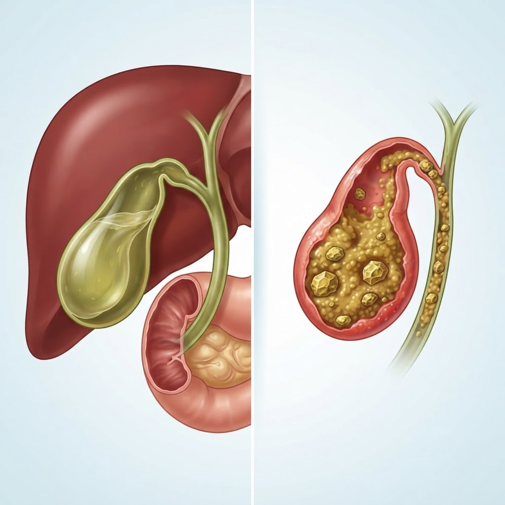
  • Gastroparesis: Symptoms, Causes, and Natural Relief Strategies

    Gastroparesis: Symptoms, Causes, and Natural Relief Strategies

    Key Takeaways

    • Gastroparesis is a chronic condition characterized by slow stomach emptying, often resulting in significant digestive discomfort and nutritional challenges.
    • The vagus nerve serves as the primary communication bridge between the brain and the digestive system, playing a critical role in controlling gastric motility.
    • Common symptoms like persistent nausea, vomiting, and early satiety are often mismanaged as general indigestion without formal diagnostic testing.
    • Managing the condition typically requires a multi-faceted approach, including specialized dietary modifications, lifestyle adjustments, and, when necessary, medical intervention.
    • Identifying the underlying cause—whether it is diabetes, post-surgical complications, or idiopathic factors—is essential for developing an effective long-term management plan.

    Living with a digestive system that seems to work against you can be an incredibly isolating and taxing experience. For those navigating the complexities of gastroparesis, every meal becomes a calculated risk, and the simple act of eating can turn into a source of anxiety rather than nourishment. This condition, defined by abnormally slow stomach emptying, affects the body’s ability to move food from the stomach into the small intestine efficiently. While the medical community continues to explore the nuances of this disorder, many individuals find that understanding the physiological roots—and learning how to support their body’s natural rhythms—is the first step toward reclaiming their quality of life. Whether you are dealing with a new diagnosis or searching for answers to persistent, unexplained digestive distress, this comprehensive guide aims to shed light on the mechanisms of gastroparesis and provide actionable strategies for your journey toward wellness.

    What Is Gastroparesis and Why Does It Happen?

    At its core, gastroparesis—a term derived from “gastro” (stomach) and “paresis” (paralysis/weakness)—is a condition where the muscles in the stomach wall do not function with the normal strength or coordination required to break down food and move it into the small intestine. In a healthy digestive system, the stomach acts like a powerful mixer, grinding solid food into smaller, semi-liquid particles known as chyme. Once this process is complete, the stomach empties its contents into the small intestine, where nutrient absorption occurs. In individuals with gastroparesis, this mechanical process is significantly hindered or slowed, leading to food remaining in the stomach for an extended duration.

    When food sits in the stomach for too long, several physiological issues arise. Primarily, the food may begin to ferment, which causes bloating, abdominal discomfort, and the development of hard masses of undigested food called bezoars. Furthermore, because the stomach is not emptying as intended, the body often struggles to absorb the necessary vitamins, minerals, and calories required for daily energy. This can lead to unexplained weight loss, fatigue, and a host of secondary complications related to malnutrition.

    The “why” behind gastroparesis is complex because it is rarely a standalone disease. Instead, it is usually a symptom or complication of an underlying systemic issue. The stomach muscles are controlled by a series of complex nerve signals and hormones. When these systems are disrupted—often due to damage to the stomach lining, the nerves governing digestion, or even hormonal imbalances—the motility of the stomach is compromised. Experts generally agree that the condition is most frequently linked to endocrine disorders, such as diabetes, where high blood sugar levels can damage the nerves that control the digestive tract over time. However, it can also manifest following viral infections, specific abdominal surgeries, or in cases where no clear cause can be identified, which is clinically referred to as idiopathic gastroparesis.

    Understanding that your body is attempting to digest food despite compromised signaling can help shift the perspective from one of frustration to one of management. It is not necessarily a failure of willpower or diet in the conventional sense; rather, it is a physiological slowing of a mechanical process. By recognizing that the stomach needs assistance with the “grinding” phase, patients and caregivers can begin to implement strategies—such as texture modification and meal timing—that reduce the burden on the gastric muscles. In the following sections, we will delve into the symptoms, triggers, and the essential role of the nervous system in ensuring this vital process remains as efficient as possible.

    Common Symptoms of Delayed Gastric Emptying

    Because the stomach is the initial processing center for everything we consume, the symptoms of slow stomach emptying can be systemic, affecting everything from energy levels to general comfort. Recognizing these gastroparesis symptoms early is crucial, as they are frequently mistaken for standard acid reflux, food poisoning, or irritable bowel syndrome (IBS). Because the condition involves the stomach being unable to empty, the most classic sign is an intense sense of fullness after consuming only a small amount of food. This is often termed “early satiety,” and it can occur even if the individual has not eaten for several hours.

    Nausea is perhaps the most prevalent and distressing symptom reported by those living with this condition. Because food remains in the stomach, the stomach feels “overloaded,” sending signals to the brain that trigger the nausea reflex. For many, this nausea is constant, but it often intensifies after a meal or when consuming high-fiber or high-fat foods that naturally take longer to digest. In more severe cases, this nausea culminates in vomiting. Interestingly, the vomit may consist of food that was eaten many hours, or even days, prior, which is a significant clinical indicator that the stomach has not emptied as it should.

    Abdominal pain and bloating are also hallmark complaints. As food sits in the stomach, it may cause a sensation of pressure in the upper abdomen. This can sometimes be confused with heart-related issues, making it important to have a professional medical evaluation to rule out other cardiac or thoracic concerns. Because the stomach is struggling to move contents, the gut microbiome may also be affected, leading to excessive gas, belching, and visible distension of the abdomen. This bloating can be uncomfortable and often prevents individuals from wearing standard-fitting clothing, further impacting their daily life.

    Another often-overlooked symptom is a significant fluctuation in blood sugar levels, particularly in diabetic patients. When digestion is unpredictable, the release of glucose from food into the bloodstream becomes erratic. One hour you may experience hypoglycemia because the food hasn’t reached the intestines yet, and the next hour you may experience hyperglycemia because the stomach finally empties a large bolus of nutrients at once. Finally, it is important to note the impact on nutritional status. Chronic symptoms lead to an avoidance of food, resulting in unintentional weight loss, anemia due to poor iron absorption, and electrolyte imbalances. If you find yourself frequently feeling “full” after three bites, experiencing nausea that doesn’t resolve with standard antacids, or noticing weight loss despite an appetite, it is essential to track these patterns and discuss them with a healthcare professional to determine if a motility study is warranted.

    Symptom Profile Typical Manifestation Best For
    Early Satiety Feeling full after only a few bites of food. Identifying volume limitations.
    Post-Prandial Nausea Persistent queasiness occurring shortly after meals. Tracking digestive triggers.
    Vomiting Undigested Food Ejection of food eaten many hours earlier. Measuring emptying severity.
    Blood Sugar Instability Erratic glucose levels in diabetic patients. Managing metabolic balance.

    Primary Causes and Risk Factors for Gastroparesis

    While the symptom profile of gastroparesis is fairly consistent, the underlying triggers are incredibly diverse. Understanding the etiology of your specific case is important, as it often dictates the potential success of various treatment strategies. Perhaps the most recognized cause is diabetes mellitus. High blood glucose levels over a prolonged period can cause damage to the autonomic nervous system, which includes the nerves that regulate stomach contractions. This is known as diabetic gastroparesis, and it remains one of the most frequently diagnosed forms of the condition. For those with diabetes, maintaining strict blood glucose control is often the primary goal to prevent further nerve damage and improve gastric function.

    Beyond diabetes, another major category of causes involves post-surgical complications. Surgeries that involve the stomach, esophagus, or the upper small intestine—such as bariatric procedures, fundoplication, or esophagectomies—can inadvertently injure the vagus nerve or disrupt the delicate muscular structures required for normal emptying. Even if a surgery is performed perfectly, the post-operative healing phase or the formation of scar tissue can sometimes lead to impaired gastric motility. It is a known complication that requires careful post-surgical monitoring.

    Viral infections represent a more transient yet impactful cause of gastroparesis. Many patients report that their digestive symptoms began immediately following a significant viral illness, such as the flu or other gastrointestinal infections. In these instances, the virus may cause a temporary inflammation of the nerves controlling the stomach, a condition often called “post-viral gastroparesis.” While some individuals may regain normal function as the inflammation subsides, for others, the damage may persist, requiring long-term dietary and medicinal management.

    There are also connective tissue disorders, such as scleroderma or lupus, which can contribute to the development of gastroparesis by causing scarring or dysfunction of the stomach muscle tissue itself. Additionally, certain medications—including some opioids, antidepressants, and blood pressure-lowering drugs—can slow down the gastrointestinal tract as a known side effect. Finally, there is a large group of cases labeled as “idiopathic,” meaning that after thorough testing, no clear anatomical, hormonal, or infectious cause can be identified. In these cases, it is hypothesized that there may be subtle, underlying micro-damage to the nervous system or a primary disorder of the stomach’s pacemaker cells, known as the Interstitial Cells of Cajal. By identifying the root cause, a patient can work with their clinical team to discontinue potentially problematic medications or address systemic diseases like diabetes with greater precision, which is often the foundation for any successful long-term recovery strategy.

    The Connection Between Vagus Nerve Function and Digestion

    To truly understand why the stomach fails to empty, one must understand the conductor of the digestive orchestra: the vagus nerve. The vagus nerve is the longest and most complex of the cranial nerves, wandering from the brainstem down into the abdomen and branching out to virtually all the major organ systems. In the context of digestion, the vagus nerve acts as a high-speed communication highway. It carries signals from the brain to the stomach, telling the muscles when to contract and when to relax. Without these precise electrical commands, the mechanical process of digestion simply cannot occur with the necessary rhythm and force.

    When the vagus nerve is damaged or functionally suppressed, the stomach muscles effectively go “off-duty.” This is why vagus nerve health is such a hot topic in the wellness community. The nerve is a key component of the parasympathetic nervous system, which is often called the “rest and digest” state. In our fast-paced, high-stress modern environment, many people spend a disproportionate amount of time in the sympathetic “fight or flight” mode. When the body is constantly stressed, the parasympathetic nervous system is dialed down, which can directly inhibit the vagus nerve’s ability to stimulate digestive motility. This is a primary reason why stress is often considered an exacerbating factor for those already dealing with sluggish stomach emptying.

    Natural remedies for gastroparesis frequently focus on “vagus nerve stimulation.” This is not just a theoretical concept; researchers have found that by encouraging parasympathetic tone, one can sometimes help optimize the signals sent to the gut. Techniques such as deep, rhythmic diaphragmatic breathing, cold exposure (like cold-water face splashes), and gentle physical activity have been suggested to help “tone” the vagus nerve. The idea is to shift the body away from the stress-induced state and into a state of relaxed digestion before and during meals.

    Furthermore, the vagus nerve also regulates the production of stomach acid and digestive enzymes. If the nerve signals are weak, not only is the motility affected, but the chemical breakdown of food is also compromised. This creates a double-edged sword: food is moving too slowly, AND it is not being properly broken down into the molecules the body needs. This is why many experts emphasize the importance of “mindful eating” in the management of gastric motility issues. By eating slowly, chewing thoroughly, and ensuring the environment is calm, one can theoretically leverage the body’s natural parasympathetic pathways to send the strongest possible signals down the vagus nerve to the stomach. While this does not act as a cure-all, especially for those with severe nerve damage, it provides a practical way for patients to actively support their digestive health and potentially reduce the severity of their symptoms on a day-to-day basis.

    Diagnostic Tests to Identify Gastroparesis

    Because the symptoms of gastroparesis can overlap with many other gastrointestinal conditions—including peptic ulcer disease, gallbladder issues, or even gastric outlet obstructions caused by tumors—formal diagnosis is an essential prerequisite to starting any treatment plan. The goal of diagnostic testing is two-fold: first, to confirm that the stomach is indeed emptying too slowly, and second, to rule out any mechanical blockage or structural abnormality that might be mimicking the condition.

    The gold standard for diagnosis is the gastric emptying scintigraphy study. During this procedure, the patient consumes a standardized meal, usually consisting of eggs or an egg substitute mixed with a tiny amount of a radioactive tracer, such as technetium-99m. Because the tracer is not absorbed by the body, it serves as a way to visualize the food within the stomach. After eating, the patient is placed under a gamma camera at specific intervals—typically at one, two, and four hours—to measure how much of the meal remains in the stomach. If a significant percentage of the meal is still present after four hours, the diagnosis of gastroparesis is confirmed. This test provides a quantitative measurement that helps doctors categorize the severity of the delay.

    Before ordering a emptying study, however, doctors will typically perform an upper endoscopy (EGD). In this procedure, a flexible tube with a camera is passed down the esophagus into the stomach. This allows the gastroenterologist to physically inspect the lining of the stomach and the pyloric sphincter (the valve that allows food to exit the stomach). It is vital for ruling out physical obstructions like polyps, tumors, or severe scar tissue from past ulcers that could be preventing food from passing through, rather than a failure of the stomach muscles themselves.

    In some cases, additional tests may be utilized depending on the clinical suspicion. For example, if there is a concern regarding an underlying systemic disease, blood tests may be ordered to check for diabetes (hemoglobin A1c), thyroid function, or autoimmune markers that might point to conditions like scleroderma. In very complex or persistent cases, a doctor might also order an ultrasound or a CT scan of the abdomen to get a detailed view of the surrounding organs, such as the pancreas and gallbladder, to ensure they are not impacting gastric function. It is important for patients to understand that the diagnostic process can take time. Because symptoms can vary day-to-day, the clinical picture is often built over several visits. However, having a definitive diagnosis is empowering, as it allows the patient and their health team to transition from “trial and error” to a targeted, evidence-based management strategy that addresses the specific nature of their delayed gastric emptying.

    Nutritional Strategies: Best Foods for Gastroparesis

    Managing a gastroparesis diet requires a complete shift in how you approach meal composition. Because the stomach is struggling with slow stomach emptying, the primary goal is to minimize the mechanical work required by the digestive system. This means moving toward smaller, more frequent meals rather than large, heavy ones. When the stomach is overloaded, food sits for extended periods, leading to fermentation, bloating, and nausea.

    The best foods for gastroparesis are those that are low in fat and low in fiber. While fiber is usually celebrated in nutritional science, it can act like a clog in a slow-moving drain for someone with gastroparesis. Fiber remains in the stomach longer than other nutrients, which can exacerbate symptoms of obstruction or fullness. Similarly, fat slows down gastric emptying even further, so it must be kept to a minimum.

    Prioritize nutrient-dense, easily digestible foods:

    • Pureed Soups: Blending vegetables removes the fibrous structure, making it easier for the stomach to pass the liquid through the pylorus.
    • Well-Cooked Proteins: Focus on lean sources like white fish, poultry, or eggs. Ensure these are cooked thoroughly until they are soft and easy to break down.
    • Refined Grains: While whole grains are typically healthier, white rice, white pasta, or sourdough bread are often better tolerated by individuals with gastroparesis because they lack the hard-to-digest bran and germ components.
    • Smoothies and Shakes: Liquid calories are your best friend. By pulverizing fruits and a scoop of protein powder into a liquid base (like almond or soy milk), you bypass the initial grinding phase of digestion.
    • Cooked Fruits and Vegetables: Raw vegetables can be incredibly difficult to process. Steaming, roasting, or boiling vegetables until they are soft significantly reduces the strain on your stomach muscles.

    Foods and Habits to Avoid for Better Motility

    If you are experiencing symptoms of slow stomach emptying, certain “trigger foods” can turn a manageable day into a painful one. Identifying these is a matter of trial and error, but the following categories are generally considered problematic by those managing gastrointestinal motility issues.

    High-fat foods are the most significant culprits. Fats take the longest to empty from the stomach because the body releases hormones in response to fat that specifically signal the stomach to slow down. This is an evolutionary survival mechanism, but for someone with gastroparesis, it is detrimental. Avoid fried foods, high-fat dairy, marbled meats, and even heavy oils or butter.

    Hard-to-digest fibrous foods act as physical barriers. Foods like raw broccoli, cauliflower, Brussels sprouts, beans, legumes, and nuts can remain in the stomach for hours or even days. Additionally, carbonated beverages should be avoided; the gas trapped in soda or seltzer can expand inside a stomach that is already struggling, leading to severe discomfort and pressure.

    Habits are just as important as food choices. Drinking fluids during meals can cause the stomach to reach its maximum capacity prematurely, leading to early satiety. Instead, try to drink fluids between meals, keeping at least a 30-minute window before and after eating to allow for better digestion.

    Food Category Examples to Avoid Best For
    High-Fiber Produce Raw broccoli, celery, skin of apples, berries with seeds Short-term symptom avoidance
    High-Fat Proteins Ribeye steak, bacon, fried chicken, salmon skin Reducing stomach-slowdown signals
    Whole Grains Brown rice, quinoa, high-fiber cereals Minimizing gastric transit time
    Gas-Forming Foods Beans, lentils, onions, cabbage, carbonated soda Preventing bloating and pressure

    Natural Supplements to Support Stomach Emptying

    While diet is the cornerstone of management, certain natural supplements may assist in supporting the vagus nerve and improving digestive flow. It is important to note that these should be used as supportive tools rather than replacements for clinical treatment.

    Ginger: Ginger is perhaps the most widely recognized natural remedy for digestive motility. Gingerols, the active compounds in ginger, have been shown to help stimulate gastric contractions. Consuming ginger tea or ginger extract before meals may help “wake up” the stomach. Many individuals find that ginger helps reduce the nausea associated with delayed emptying.

    Peppermint: Peppermint oil has antispasmodic properties. While it is often used for irritable bowel syndrome (IBS), it can also help relax the stomach muscles, which may relieve the sensation of tightness and cramping that often accompanies slow stomach emptying. Use caution, however, as peppermint can sometimes relax the lower esophageal sphincter, which might trigger heartburn.

    Prokinetics and Enzymes: Digestive enzymes, specifically those that assist in protein and carbohydrate breakdown, can reduce the amount of time food needs to spend in the stomach. Some people find success with supplemental pancreatic enzymes. Furthermore, some botanical supplements that target the vagus nerve—such as those containing bitters or certain herbal stimulants—are sometimes used to encourage the digestive tract to move food more effectively.

    Always exercise caution when introducing supplements. The stomach in a patient with gastroparesis is hypersensitive; even a small pill can be irritating. Always trial one supplement at a time to monitor for reactions.

    Lifestyle Adjustments to Improve Digestive Flow

    Beyond what you put into your body, the physical way you navigate your day significantly impacts the vagus nerve and overall digestive motility. The vagus nerve acts as the primary communication highway between your brain and your gut. When you are in a state of chronic stress, the “fight or flight” (sympathetic) nervous system takes over, which naturally inhibits digestion. To improve motility, you must encourage “rest and digest” (parasympathetic) states.

    Practice Mindful Eating: Eating while distracted or stressed prevents the brain from properly signaling the stomach to prepare for food. Take time to chew your food thoroughly—until it is almost liquid—before swallowing. This reduces the mechanical load on the stomach.

    Gravity-Assisted Digestion: Do not lie down immediately after eating. Gravity helps keep food moving downward through the digestive tract. Aim to remain upright for at least two hours after your last meal. If you struggle with night-time symptoms, consider elevating the head of your bed.

    Vagus Nerve Stimulation: Simple, non-invasive techniques can help tone the vagus nerve. Activities like deep, diaphragmatic breathing, gargling water, or humming can stimulate the nerves in the throat and chest, which may have a positive downstream effect on the stomach’s ability to contract.

    Regular, Gentle Movement: While strenuous exercise is often out of the question during a flare-up, gentle, low-impact movement like walking can help stimulate motility. Avoid intense bouncing or high-impact activities that can increase nausea.

    When to Seek Professional Medical Help for Digestion

    Gastroparesis is a serious medical condition that requires professional oversight. While lifestyle and dietary changes are helpful, they are not a substitute for medical diagnostics and management. You should seek medical attention promptly if you experience “red flag” symptoms.

    These signs include, but are not limited to: inability to keep down liquids for more than 24 hours, signs of severe dehydration (dark urine, dizziness, extreme lethargy), unexplained and rapid weight loss, or persistent abdominal pain that feels sharp rather than dull or crampy. Furthermore, if you are diabetic, gastroparesis can make glycemic control extremely difficult; if your blood sugar levels are erratic, you must work closely with your endocrinologist to manage the interplay between your medication and your digestive speed.

    A doctor can provide motility testing, such as a gastric emptying study, which is the gold standard for diagnosis. They can also screen for underlying issues like mechanical blockages, severe electrolyte imbalances, or nerve damage. Do not attempt to self-manage severe, persistent symptoms without a formal evaluation.

    Frequently Asked Questions

    Is gastroparesis a lifelong condition?

    For some, gastroparesis is a chronic, lifelong condition, especially if it is linked to diabetes or nerve damage. However, for others, it may be temporary, triggered by viral infections, surgery, or medication side effects. Management focuses on minimizing symptoms and improving quality of life, and in some cases, the underlying cause can be addressed to restore normal motility.

    Can stress make gastroparesis symptoms worse?

    Yes, significantly. Stress activates the sympathetic nervous system, which directs blood flow away from the gut and inhibits digestive motility. Because the vagus nerve, which controls stomach movement, is highly sensitive to stress, emotional or physical pressure can lead to increased nausea, bloating, and slower gastric emptying.

    Should I avoid all fiber if I have slow stomach emptying?

    Most experts recommend a low-fiber diet for gastroparesis because fiber adds bulk and stays in the stomach much longer than other nutrients. While you should avoid high-fiber foods like raw vegetables and whole grains, you may still be able to tolerate small amounts of cooked, soft fiber depending on your individual tolerance. It is best to work with a dietitian to find your specific threshold.

    Why do I feel full after only a few bites of food?

    This phenomenon, known as early satiety, occurs because your stomach is not emptying properly. When previous meals are still sitting in the stomach, there is no physical space for new food. Even a small amount of food can trigger the stomach’s stretch receptors, signaling to the brain that you are “full,” even if you haven’t received adequate nutrition.

    Is it safe to exercise with gastroparesis?

    Light, gentle exercise like walking is often recommended as it can help stimulate the digestive system and improve blood flow. However, you should avoid high-intensity workouts or activities that involve jumping or straining, as these can increase nausea and physical distress. Always listen to your body and avoid exercise immediately after eating.

    Can diabetes-related gastroparesis be reversed?

    Reversing diabetic gastroparesis is challenging, but strict glycemic control is the most important factor in preventing further nerve damage. By keeping blood sugar levels within a healthy range, some people may prevent the condition from worsening. While full reversal is rare, diligent management can lead to significant improvements in symptom frequency and intensity.

    Conclusion

    Gastroparesis is a complex, often debilitating condition that demands a comprehensive approach to health. By understanding the mechanical limitations of your stomach and the role of the vagus nerve, you can begin to implement targeted dietary and lifestyle changes that provide genuine relief. From prioritizing liquid nutrition and low-fat, low-fiber meals to embracing stress-reduction techniques that calm the nervous system, you have the power to influence your digestive flow significantly.

    Remember, this journey is not about finding a single “cure,” but rather about building a lifestyle that minimizes discomfort and supports your body’s specific needs. Be patient with yourself, keep a food journal to track your triggers, and maintain open communication with your healthcare team. You do not have to struggle in silence; take charge of your digestive wellness today by making one small change—such as swapping a heavy meal for a nutrient-dense soup—and observing how your body responds.

    By healthauthoritylife Editorial Team

    This article is for informational purposes only and is not a substitute for professional medical advice. Always consult your doctor before making changes to your diet, exercise routine, or treatment plan.

  • Sorbitol Intolerance: Symptoms, Causes, and Dietary Tips

    Sorbitol Intolerance: Symptoms, Causes, and Dietary Tips

    Key Takeaways

    • Sorbitol intolerance occurs when the small intestine fails to absorb this sugar alcohol effectively, leading to bacterial fermentation in the colon.
    • Common symptoms of sorbitol malabsorption include significant abdominal bloating, gas, cramping, and osmotic diarrhea.
    • Sorbitol is frequently hidden in “sugar-free” processed products, often labeled as sweeteners or humectants.
    • Managing gut health often requires identifying and strictly limiting high-sorbitol fruits like apples, pears, and stone fruits.
    • Adopting a low sorbitol diet is a primary strategy for those seeking relief from chronic digestive discomfort.

    For many individuals, the journey toward better digestive health begins with understanding how specific carbohydrates interact with the gut microbiome. While fiber and natural sugars are often touted as healthy staples, certain compounds—specifically sugar alcohols—can become significant barriers to comfort for those with a sensitive digestive system. Sorbitol, a polyol commonly found in both nature and synthetic sweeteners, is a primary offender in this category. For those living with sorbitol intolerance, even small amounts of this sugar alcohol can trigger a cascade of internal distress, turning a seemingly healthy snack into a source of painful bloating and irritability. In this guide, the healthauthoritylife Editorial Team explores the mechanics of sorbitol malabsorption, how to navigate the grocery aisles to identify hidden triggers, and the essential steps toward reclaiming your digestive well-being through intentional dietary management.

    What Is Sorbitol and Why Does It Cause Digestive Distress?

    Sorbitol is a type of sugar alcohol, scientifically classified as a polyol. It occurs naturally in a variety of fruits and vegetables, and it is also manufactured industrially for use as a food additive. In the food industry, sorbitol is prized for its ability to retain moisture, prevent crystallization in sugary confections, and provide a sweet taste without the caloric impact of table sugar. Because of these versatile properties, it appears as a common ingredient in everything from chewing gum and protein bars to sugar-free chocolates and pharmaceutical syrups.

    The core of the problem lies in the human body’s capacity to process this compound. In a healthy digestive tract, sorbitol is meant to be absorbed in the small intestine through a process of diffusion. However, the mechanism of this absorption is notably inefficient compared to other sugars like glucose or fructose. For the average person, consuming small amounts of sorbitol may pass through the gut with minimal disruption. But for those with sorbitol intolerance, the small intestine simply lacks the necessary efficiency to move these molecules into the bloodstream at a sufficient rate.

    When sorbitol remains unabsorbed in the small intestine, it travels further down the gastrointestinal tract, eventually reaching the large intestine, or colon. This is where the biological conflict intensifies. The colon is densely populated with trillions of bacteria that constitute our gut microbiome. These bacteria view undigested carbohydrates, such as sorbitol, as a readily available fuel source. Through a process known as fermentation, the bacteria begin to break down the sorbitol. This process is a natural part of gut ecology, but when an excessive amount of undigested sugar alcohol is present, the fermentation happens too rapidly and in too high a volume.

    The metabolic byproduct of this bacterial fermentation is the production of gases, specifically hydrogen, carbon dioxide, and occasionally methane. Furthermore, sorbitol is osmotic, meaning it acts like a magnet for water. It draws fluid into the intestinal lumen, which can lead to significant changes in stool consistency and frequency. This combination—rapid gas production and an increase in fluid volume—creates a “perfect storm” for digestive distress. Understanding that this is a mechanical and chemical process, rather than an allergy in the traditional immunological sense, is the first step toward effective gut health management. By recognizing that your body is effectively saying “no” to this specific molecule, you can begin the process of adjusting your intake to prioritize comfort and long-term digestive stability.

    Common Symptoms of Sorbitol Malabsorption

    Symptoms of sorbitol intolerance are often delayed, appearing anywhere from thirty minutes to several hours after consumption. Because these symptoms mirror those of other conditions like irritable bowel syndrome (IBS) or food sensitivities, individuals often struggle to connect their diet to their discomfort. However, there is a distinct clinical profile associated with sorbitol malabsorption that the healthauthoritylife Editorial Team suggests keeping a close eye on.

    Abdominal bloating is arguably the most frequently reported symptom. Many people describe it as a feeling of fullness or “tightness” in the midsection, often worsening throughout the day as more fermentable substances are processed. This bloating is the direct physical result of gas buildup pressing against the intestinal walls. Alongside this, sharp, localized abdominal cramping is common. These cramps occur as the intestines stretch and contract in an effort to move gas bubbles and fluid through the system. For many, these pains are intense enough to cause significant distraction and distress, often leading to a cycle of anxiety regarding future meals.

    Increased flatulence is another telltale sign. Because the fermentation process in the colon produces a high volume of gas, individuals with intolerance often experience chronic, excessive gas that may be accompanied by unpleasant odors. While gas is a normal byproduct of digestion, a consistent, uncomfortable increase that correlates with specific food choices—like consuming sugar-free candy or certain fruits—is a strong indicator of malabsorption.

    In addition to gas and pain, sorbitol’s osmotic properties often trigger digestive urgency or diarrhea. Because the unabsorbed sorbitol pulls water into the colon, the resulting stool is typically soft, loose, or even liquid. This can be particularly jarring for those who are accustomed to normal digestive function. Some individuals also report a sense of incomplete evacuation, where the digestive system feels “sluggish” despite the watery nature of the stool. It is important to note that the severity of these symptoms is highly dose-dependent. A small amount might cause minor rumbling, while a large serving of a sorbitol-heavy protein bar might result in acute, debilitating discomfort. Keeping a detailed food diary can help you identify your personal threshold, as some people can tolerate trace amounts, while others may find themselves highly reactive to even minute quantities of sugar alcohols.

    Symptom Type Physical Manifestation Best For
    Osmotic Reaction Loose, frequent stools and urgency Immediate hydration management
    Fermentation Build-up Sharp, lingering bloating and distension Long-term dietary elimination
    Enteric Nervous Response Cramping and abdominal tenderness Lifestyle and stress reduction

    How Sorbitol Triggers Bloating and Gas in the Gut

    To understand why sugar alcohols and bloating go hand-in-hand, one must look at the specific physical environment of the human gut. The gastrointestinal tract relies on a delicate balance of fluid dynamics and bacterial activity to maintain homeostasis. When you ingest a normal carbohydrate—such as starches from rice or glucose from fruit—your body uses specialized enzymes and transporters to absorb these nutrients into the bloodstream before they ever reach the colon. The system is designed to be highly efficient, leaving only minimal remnants for the colonic flora to process.

    Sorbitol disrupts this efficiency. Because humans lack a specific transporter that handles polyols with high speed and capacity, a significant portion of the sorbitol you ingest remains in the lumen of the small intestine. As this sugar alcohol makes its way toward the large intestine, it brings its chemical properties with it. First, there is the osmotic effect. Water, which is usually reabsorbed in the intestines, is held back or pulled into the tract to dilute the concentration of sorbitol. This creates a more liquid environment in the colon, which inherently alters the transit time of waste.

    Once the sorbitol enters the colon, the bacteria encounter a high-energy substrate. The microbiome is essentially an ecosystem of diverse microorganisms, many of which thrive on these types of sugars. They utilize the sorbitol for energy, but this metabolic process releases gases as a byproduct. In a healthy gut, this gas is absorbed into the bloodstream or passed through the digestive system at a rate that is mostly unnoticeable. However, because sorbitol fermentation can be quite rapid, the volume of gas produced often overwhelms the body’s ability to move it along. This creates trapped pockets of air, which cause the walls of the intestines to expand—this is the physical sensation of bloating.

    Furthermore, the increased distension caused by the gas can trigger the gut’s nervous system. The intestines are lined with an intricate network of nerves known as the enteric nervous system, often referred to as the “second brain.” When the gut wall is stretched, these nerves send signals of discomfort or pain to the brain. In individuals with heightened visceral sensitivity, this physical stretching is perceived as much more painful than it might be for others. This is why sorbitol intolerance is a frequent subject in the discussion of functional gut health management; the substance acts as both a chemical irritant and a mechanical trigger for pain. Recognizing this dual nature is vital, as it emphasizes that the goal of a low sorbitol diet is not just to reduce the frequency of gas, but to protect the integrity and comfort of the entire digestive system.

    Identifying Hidden Sorbitol in Processed Foods

    Navigating the grocery store for someone with sorbitol intolerance can feel like a minefield. While fresh produce is easy to track, processed foods often contain sorbitol under a variety of names or as part of a complex ingredient list. Manufacturers frequently use sorbitol not just for sweetness, but as a humectant—a substance that keeps food moist and prevents it from drying out. This makes it an incredibly popular additive in products that rely on shelf stability, such as baked goods, nutrition bars, and even some chewing gums and mints.

    The first step in identifying these triggers is to become a dedicated label reader. Look for the word “sorbitol” explicitly, but also be aware that it may appear as “E420” in international markets or on certain imported goods. Furthermore, sorbitol is often found in products labeled as “sugar-free,” “diabetic-friendly,” or “keto-friendly.” If a package boasts that it is “reduced sugar,” check the ingredients list to see what replaced the sugar. If you see sugar alcohols listed, there is a high likelihood that sorbitol is among them, or that another polyol like xylitol or mannitol is present, which may also cause issues for those with sensitive guts.

    It is not just sweet treats you need to watch out for. Sorbitol and other sugar alcohols are common in many “health-conscious” products. Protein bars are a major culprit; the texture of these bars often requires humectants to remain chewy and appealing. Additionally, check the labels of liquid medications, cough syrups, and mouthwashes. Many of these products use sorbitol to improve the flavor and mouthfeel of the liquid. Even some brands of toothpaste or specialized dietary supplements may contain trace amounts that, when combined throughout a single day, can cross the threshold of what your gut can handle.

    To master the art of the low sorbitol diet, create a “blacklist” of common food categories that frequently harbor these ingredients. Sugar-free chewing gum is arguably the most common source of concentrated exposure. Because people often chew gum throughout the day, the cumulative dose of sorbitol can lead to chronic, low-grade digestive issues that seem impossible to pin down. When shopping, prioritize whole, single-ingredient foods whenever possible. By shifting your reliance away from ultra-processed snacks and towards items prepared from scratch, you significantly reduce the risk of accidental exposure to hidden sweeteners. If you must buy a packaged product, look for items that use natural sugar alternatives like stevia or monk fruit, which are typically easier on the gut, though it is always wise to assess your own tolerance even with these alternatives.

    Which Natural Fruits and Vegetables Are High in Sorbitol?

    While processed foods are the most common source of extreme sorbitol intake, it is equally important for those managing their gut health to understand the natural sources of this compound. Many fruits and vegetables contain sorbitol as a naturally occurring sugar. For someone with significant sorbitol malabsorption, eating a large portion of a high-sorbitol fruit can trigger the same symptoms as eating a handful of sugar-free mints. Understanding the hierarchy of sorbitol content in whole foods is essential for building a diet that supports rather than hinders your digestion.

    Fruits in the Rosaceae family are the most notorious offenders. This group includes apples, pears, peaches, nectarines, apricots, and plums. These fruits are high in sorbitol, and many also contain high levels of fructose, which can further complicate digestive issues for those who struggle with sugar absorption in general. When you eat a fresh pear or a crisp apple, you are getting a concentrated dose of sorbitol. For some individuals, eating just one of these fruits is enough to trigger a bloating response. Dried fruits, such as prunes, dates, and dried apricots, are even more concentrated; since the water has been removed, the density of the sugar alcohol per bite is significantly higher than that of the fresh counterpart.

    Berries generally have a lower sorbitol content, which is why they are often recommended as better alternatives. Strawberries, blueberries, and raspberries tend to be much more tolerable for those with sensitivity. However, this is not a universal rule, and individual tolerance can vary. Some people may be able to handle moderate portions of blackberries, while others may still feel the effects of the minor amount of polyols present. The key is to test your own limits through a systematic process, starting with smaller portions and observing how your body responds over a 24-hour period.

    Vegetables are generally safer than fruits, but there are still notable exceptions. Mushrooms, while technically a fungus, are often grouped with vegetables in culinary contexts and are surprisingly high in polyols, including mannitol, which behaves similarly to sorbitol in the gut. Asparagus, cauliflower, and sweet potatoes may also cause mild issues for those who are particularly sensitive to fermentable carbohydrates. If you are struggling with chronic bloating, try a temporary elimination of these high-sorbitol fruits and vegetables, replacing them with lower-polyol options like leafy greens, cucumber, zucchini, or citrus fruits like oranges and lemons. By identifying these natural triggers, you can curate a meal plan that provides the nutrients you need without the digestive distress that accompanies the fermentation of high-sorbitol plant matter. Always remember that nutritional needs are individual, and what triggers one person may be perfectly acceptable for another; monitoring your symptoms with a diary remains the most reliable tool in your gut health arsenal.

    Diagnosing Sorbitol Intolerance: Testing and Elimination

    Identifying whether your digestive distress stems from sorbitol malabsorption requires a methodical approach. Because the symptoms of sorbitol intolerance—such as bloating, flatulence, and diarrhea—are common to many gastrointestinal disorders, it is essential to distinguish them from conditions like Celiac disease, lactose intolerance, or Irritable Bowel Syndrome (IBS). The diagnostic process typically begins with a thorough medical history, followed by specialized testing or a supervised elimination strategy.

    The gold standard for identifying carbohydrate malabsorption is the hydrogen breath test. During this procedure, you consume a measured dose of sorbitol after fasting. If your small intestine fails to adequately absorb the sugar alcohol, it passes into the large intestine, where gut bacteria ferment it. This fermentation process releases hydrogen gas, which is absorbed into the bloodstream and eventually exhaled through the lungs. A clinical technician monitors your breath samples at specific intervals; a significant rise in hydrogen levels confirms that the sorbitol is not being processed correctly in the upper digestive tract.

    If clinical testing is unavailable or inconclusive, an elimination and reintroduction diet serves as a practical alternative for gut health management. This involves removing all high-sorbitol foods from your diet for two to four weeks. During this period, you must become an expert at reading ingredient labels, as sorbitol is a frequent additive in “sugar-free” products. Once your symptoms subside, you systematically reintroduce sorbitol in controlled amounts to determine your individual threshold. Maintaining a detailed food diary during this phase is crucial, as it allows you to correlate specific intake levels with the severity of your digestive response.

    The Connection Between Sorbitol and SIBO

    There is a significant intersection between sorbitol malabsorption and Small Intestinal Bacterial Overgrowth (SIBO). In a healthy digestive system, the majority of bacterial activity is concentrated in the large intestine. In SIBO, however, an excessive amount of bacteria migrates into the small intestine. When you consume foods containing sorbitol—which is normally intended to be absorbed before reaching the colon—these bacteria “get to it first.”

    Because these bacteria are present in the wrong location, their fermentation of sorbitol occurs much earlier in the digestive process. This leads to immediate and often intense symptoms, including rapid-onset bloating, distention, and abdominal pain. For those suffering from chronic digestive health issues, sorbitol acts as a fuel source that exacerbates the overgrowth. If you find that your sensitivity to sugar alcohols has increased suddenly, it is worth discussing the possibility of SIBO with a gastroenterologist. Managing sorbitol intolerance in the context of SIBO often requires a dual approach: treating the bacterial overgrowth while simultaneously restricting fermentable carbohydrates to provide symptomatic relief.

    Dietary Approach Mechanism of Action Best for
    Strict Elimination Removes all sorbitol triggers to allow gut inflammation to subside. Initial diagnosis and severe symptom flares.
    Low-FODMAP Diet Limits a broad range of fermentable sugars, including sorbitol. Long-term management of IBS and general bloating.
    Sorbitol Threshold Testing Systematic reintroduction to identify safe serving sizes. Those aiming to diversify their diet after initial healing.
    SIBO-Specific Protocol Combines anti-bacterial support with carbohydrate restriction. Individuals with confirmed small intestinal overgrowth.

    Low-Sorbitol Diet Strategies for Long-Term Relief

    Achieving long-term relief from sorbitol intolerance is less about total deprivation and more about building a sustainable, personalized eating pattern. The goal is to minimize the osmotic load on your bowels while maintaining a nutrient-dense diet. Many people find success by categorizing foods based on their sorbitol content and prioritizing “safe” alternatives.

    When shopping, prioritize whole, unprocessed foods. Fresh vegetables such as spinach, kale, and carrots, and proteins like lean meat, fish, and eggs are naturally free of sorbitol and pose little risk to your digestive tract. When it comes to fruits, focus on varieties with low sorbitol concentrations, such as bananas, blueberries, and citrus fruits. It is equally important to avoid processed “diet” foods, which frequently contain hidden sorbitol used as a bulk sweetener. Look for alternative labels like “xylitol-free” or “sucralose-free” if you find that sugar alcohols in general trigger your system, though always check the full ingredient list.

    Implementing a “low-sorbitol” strategy also involves mindful preparation. If you are dining out, inquire about sugar-free sauces, dressings, or chewing gums that may contain artificial sweeteners. Over time, you will learn to spot “red flag” ingredients like hydrogenated starch hydrolysates or sorbitol syrups, which are commonly hidden in protein bars and electrolyte supplements. By gradually diversifying your nutrient intake while avoiding these specific triggers, you provide your gut with the stability it needs to function optimally.

    Lifestyle Habits to Support Sensitive Digestion

    Beyond dietary choices, lifestyle factors play a major role in how your body handles fermentable carbohydrates. Chronic stress, for instance, can alter gut motility. When your digestive system is “sluggish” due to stress, food sits longer in the gut, providing more time for fermentation to occur, which makes even small amounts of sorbitol feel more problematic. Integrating stress-reduction techniques such as diaphragmatic breathing, yoga, or consistent sleep schedules can improve overall gut motility and symptom tolerance.

    Physical activity is another pillar of gut health management. Moderate exercise, such as walking after meals, encourages rhythmic contractions of the digestive tract. This helps prevent gas from becoming trapped and reduces the sensation of bloating. Additionally, hydration is key; drinking adequate water supports the digestion process and helps maintain the health of the mucosal lining in your intestines. Avoiding late-night snacking is also beneficial, as it allows your digestive system a period of “rest and repair” during sleep, ensuring that the next day’s meals are processed efficiently.

    Finally, consider the pace of your eating. Digestion begins in the mouth, and failing to chew food thoroughly forces the stomach and intestines to work significantly harder. By eating slowly and mindfully, you reduce the amount of air swallowed (which reduces gas) and prime your enzymes to break down nutrients more effectively. Small lifestyle adjustments often compound, creating an environment where your digestive system is more resilient and less prone to reacting violently to minor dietary indiscretions.

    Frequently Asked Questions

    Is sorbitol intolerance the same as an allergy?

    No, they are distinct. An allergy involves an immune system reaction to a protein, whereas sorbitol intolerance is a digestive inability to absorb a specific carbohydrate. The symptoms may overlap in terms of abdominal pain, but they occur through entirely different biological pathways.

    Can I ever eat sugar-free products again if I have sorbitol intolerance?

    Many people find that they can tolerate small amounts of sorbitol after their gut health has stabilized. However, it is vital to test your personal threshold gradually. Some individuals discover that other sugar alcohols, like stevia or erythritol, are much better tolerated than sorbitol.

    Do symptoms of sorbitol malabsorption always happen immediately?

    Symptoms typically appear within a few hours of consumption, as the undigested sugar alcohol reaches the large intestine. However, the exact timing depends on your individual transit time and what else you consumed during the meal, which can buffer or accelerate the process.

    Are there natural sources of sorbitol I should be worried about?

    Yes, sorbitol occurs naturally in certain fruits, such as stone fruits (peaches, plums, cherries) and apples. While these are healthy, they can be triggers for those with significant malabsorption. You do not necessarily need to avoid them entirely, but keeping serving sizes small is recommended.

    Could my sorbitol intolerance be a sign of a larger health issue?

    While often a primary digestive issue, chronic malabsorption can sometimes be a symptom of conditions like SIBO, Celiac disease, or inflammatory bowel conditions. If you experience unintentional weight loss, anemia, or blood in your stool, you should seek professional medical evaluation immediately.

    Does drinking more water help with sorbitol-induced bloating?

    Staying hydrated is generally beneficial for gut transit. However, simply increasing water intake will not neutralize the fermentation process happening in your colon. Water helps maintain normal bowel function, but the primary way to manage symptoms remains identifying and reducing your intake of sorbitol-heavy foods.

    Conclusion

    Navigating sorbitol intolerance requires a transition from reactive symptom management to a proactive, informed lifestyle. By understanding how your digestive tract processes sugar alcohols, recognizing the common triggers, and applying the dietary strategies outlined here, you can regain control over your gut health. Remember that your body is unique; the key to long-term relief lies in patience, observation, and the willingness to adjust your habits based on your personal response. Do not settle for persistent discomfort; start by tracking your intake today, consult with a healthcare professional to rule out underlying conditions, and move toward a digestive experience that feels stable and comfortable. Take the first step toward better gut health by keeping a detailed food diary for the next seven days to identify your own specific triggers.

    By healthauthoritylife Editorial Team

    This article is for informational purposes only and is not a substitute for professional medical advice. Always consult your doctor before making changes to your diet, exercise routine, or treatment plan.

  • Gallbladder Sludge vs Gallstones: Differences & Treatment

    Gallbladder Sludge vs Gallstones: Differences & Treatment

    Key Takeaways

    • Gallbladder sludge is a collection of microscopic particles, while gallstones are hardened, pebble-like formations within the gallbladder.
    • Both conditions arise from imbalances in bile composition, often linked to diet, rapid weight loss, or metabolic factors.
    • Understanding the difference between sludge and stones is critical, as sludge is frequently a precursor to the development of larger stones.
    • Biliary sludge symptoms often mirror those of gallstones, including upper abdominal discomfort and digestive distress after high-fat meals.
    • Prioritizing natural gallbladder health through hydration and a balanced diet can help support proper bile flow and reduce the risk of biliary issues.

    When you feel an unexpected, sharp ache in your upper abdomen after a heavy meal, your gallbladder—a small, pear-shaped organ tucked beneath your liver—might be to blame. Many people assume any pain in this region implies the presence of gallstones, but the reality is more nuanced. The gallbladder plays a central role in your digestive process, acting as a reservoir for bile, a fluid essential for breaking down fats. However, when the delicate chemical balance of this fluid is disrupted, the body can develop either gallbladder sludge or gallstones. Distinguishing between these two conditions is more than a clinical exercise; it is the first step toward managing your digestive comfort and preventing more serious complications. In this guide, the healthauthoritylife Editorial Team explores the intricate relationship between gallbladder sludge vs gallstones, how they form, and the foundational steps for maintaining optimal gallbladder function.

    Understanding the Biliary System and Bile Production

    To grasp the mechanisms behind gallbladder health, one must first appreciate the biliary system—a sophisticated network comprising the liver, gallbladder, and bile ducts. The liver acts as the body’s primary chemical factory, continuously producing bile, a complex fluid consisting of cholesterol, bile salts, bilirubin, lecithin, and water. This golden-yellow liquid is not merely a waste product; it is a critical digestive tool. Once produced by the liver, bile is channeled through a series of ducts. During periods between meals, when the body does not immediately require assistance with fat digestion, this bile is diverted to the gallbladder.

    The gallbladder’s primary duty is to store, concentrate, and release bile. Upon arrival, the gallbladder absorbs water and electrolytes from the bile, effectively making it more potent and concentrated. Think of it as a biological reservoir that saves its resources for when you consume a meal containing fats. When you eat, the hormone cholecystokinin (CCK) is released in response to dietary fats. This hormone signals the gallbladder to contract, pushing the concentrated bile through the common bile duct and into the small intestine, specifically the duodenum. Once in the small intestine, the bile performs an essential function: it emulsifies fats, breaking them into smaller droplets that digestive enzymes can then process into nutrients your body can absorb.

    The efficiency of this system relies entirely on the solubility of the substances within the bile. Under normal circumstances, cholesterol remains dissolved in the bile thanks to the balancing effects of bile salts and lecithin. However, if the liver produces an excess of cholesterol, or if the gallbladder fails to concentrate or empty the bile effectively, the environment within the gallbladder changes. This disruption can lead to the precipitation of solids. If the bile remains stagnant, as it might during periods of fasting, rapid weight loss, or dehydration, the fluid loses its ability to keep cholesterol in a liquid state. Over time, these microscopic solids accumulate, beginning the transition from a healthy, free-flowing fluid to a condition characterized by sludge or stones. Maintaining this system requires a consistent flow, which is why experts often emphasize that nutrition and hydration are not just abstract concepts for general health, but specific requirements for the biliary tract to function without obstruction.

    What Is Gallbladder Sludge and Why Does It Form?

    Gallbladder sludge, formally known as biliary sludge, is an accumulation of particulate matter that precipitates out of the bile. Unlike the rigid, solid structures we associate with stones, sludge is more of a viscous, gel-like mixture. Imagine the consistency of thick, muddy water or fine sediment at the bottom of a container. It is a heterogeneous combination of mucus, cholesterol crystals, calcium salts, and other debris that has settled in the gallbladder. While it is not yet a stone, its presence indicates that the bile is supersaturated, and the gallbladder is struggling to flush its contents effectively.

    The formation of sludge is often a reactive process. It frequently occurs when bile remains in the gallbladder for too long, a state known as biliary stasis. When the gallbladder does not contract fully or frequently enough, the bile becomes stagnant. As the liquid component continues to be absorbed, the remaining substances become overly concentrated, causing particles to fall out of solution. Several lifestyle and physiological triggers contribute to this formation. Rapid weight loss is a classic example; when the body breaks down fat stores at an accelerated rate, the liver secretes extra cholesterol into the bile, quickly overwhelming the natural balance and leading to sludge accumulation.

    Pregnancy is another well-documented catalyst. Hormonal shifts can lead to a decrease in gallbladder motility, meaning the organ does not empty as vigorously as it should. Similarly, prolonged fasting or the use of certain medications that affect bile composition can trap bile in the gallbladder, encouraging the “settling” process. It is important to note that biliary sludge symptoms can be quite subtle. Many individuals may have sludge for years without experiencing a single bout of pain. However, in others, this thick sediment can act as a precursor, providing a surface upon which more solidified materials can attach and grow. Because sludge is essentially the “pre-stone” phase, managing it early is essential. Addressing the underlying dietary habits—such as ensuring adequate intake of fiber and healthy fats, which stimulate regular gallbladder contractions—can often help the body clear this sediment naturally before it has the opportunity to evolve into something more obstructive.

    Gallstones: Causes and Types of Biliary Calculi

    While sludge is a semi-fluid sediment, gallstones (medically termed cholelithiasis) are distinct, solid concretions that form within the gallbladder. Gallbladder stone formation is a complex geochemical process that occurs when the bile’s delicate equilibrium is tipped toward crystallization. When cholesterol concentrations are excessively high—or when there is a deficiency in the bile salts that keep that cholesterol dissolved—the molecules begin to clump together. These microscopic clumps act as a “nidus,” or foundation, upon which more materials layer themselves over time, eventually growing into a pebble-like stone.

    Gallstones are generally categorized by their primary composition, which often provides clues about the underlying metabolic environment of the patient. The most common variety, accounting for the vast majority of cases in developed nations, is the cholesterol stone. These are typically yellow-green in color and are composed primarily of hardened cholesterol. Their formation is closely linked to diets high in refined sugars and saturated fats, as well as obesity and sedentary lifestyles. When the gallbladder fails to empty properly, these cholesterol crystals have the time they need to aggregate into larger, solid masses.

    The second major category involves pigment stones. These are smaller, darker, and harder, composed primarily of bilirubin, a pigment produced by the breakdown of red blood cells. Pigment stones are less common than cholesterol stones and are often associated with underlying medical conditions, such as chronic liver disease or blood disorders where red blood cells are destroyed too rapidly (hemolysis). Regardless of the type, once a stone forms, it occupies space within the gallbladder. While many people live with “silent” gallstones that never cause an issue, the danger arises when a stone migrates out of the gallbladder and becomes lodged in a duct. This blockage prevents bile from exiting, causing significant pressure, inflammation, and pain. Unlike sludge, which can sometimes be cleared through improved dietary flow, stones are structural objects that rarely dissolve on their own. Their persistence often requires a deeper investigation into one’s metabolic health, as they are frequently a sign of systemic imbalances rather than just a local gallbladder issue.

    Key Differences Between Sludge and Gallstones

    When comparing gallbladder sludge vs gallstones, it is helpful to visualize the difference between a liquid-dense sediment and a solid, structural object. While they share the same biological “neighborhood” and are often caused by similar imbalances in bile chemistry, their physical properties determine how they interact with the biliary system. Sludge acts more like a thick fluid; because of its semi-liquid nature, it can sometimes be “washed out” of the gallbladder when the organ contracts vigorously. Stones, conversely, are firm, calcified, or cholesterol-hardened objects that maintain their shape and size, making them much more likely to become physically trapped within the narrow neck of the gallbladder or the bile ducts.

    Another primary difference lies in their visibility and diagnostic identification. Sludge is notorious for being difficult to pinpoint; because it lacks the sharp density of a stone, it may not show up clearly on traditional X-rays or even some lower-resolution ultrasounds. It often appears as a shifting, low-level echo within the gallbladder. Gallstones are typically much easier to identify, as they reflect sound waves strongly during an ultrasound, appearing as bright, white objects often followed by an “acoustic shadow.” This diagnostic distinction is important because the clinical approach to managing them often differs based on the maturity and severity of the condition.

    The following table illustrates the core differences between these two conditions:

    Feature Gallbladder Sludge Gallstones Best For
    Consistency Viscous, gel-like, liquid sediment Hard, pebble-like, solid mass Identifying Physical State
    Permanence Potentially reversible with lifestyle Generally permanent structural issue Setting Treatment Expectations
    Diagnostic Clarity Often subtle; hard to visualize Clear, high-contrast visibility Choosing Diagnostic Tools
    Obstruction Risk Moderate; can cause intermittent blockage High; likely to cause acute blockage Assessing Emergency Potential

    Understanding these differences is vital for informed self-advocacy. If you are diagnosed with sludge, it is often viewed as a “yellow light”—a warning sign that your gallbladder is not emptying properly and your bile composition is off-balance. It serves as an opportunity to intervene with diet and lifestyle modifications. If you are diagnosed with stones, the reality of the situation changes, as you are dealing with a permanent obstruction that requires closer monitoring and, in many cases, medical consultation regarding surgical options or more intensive management strategies. Recognizing which condition you are facing allows you to better collaborate with your healthcare provider to decide on the most appropriate, effective course of action for your unique digestive health profile.

    Common Symptoms Shared by Sludge and Gallstones

    Despite the physical differences between sludge and stones, the way they present clinically is often remarkably similar. Because both conditions interfere with the normal flow of bile, they produce symptoms primarily triggered by the gallbladder’s attempt to contract against resistance. Biliary sludge symptoms and gallstone symptoms both fall under the umbrella of “biliary colic,” which is the medical term for the intense, cramping pain that occurs when the gallbladder struggles to move bile through a partially or fully obstructed duct.

    The most hallmark symptom is a recurring pain in the upper right quadrant of the abdomen, often radiating toward the right shoulder or back. This pain is frequently described as a dull ache that suddenly transitions into a sharp, intense sensation. Many individuals notice that this discomfort is tightly linked to meal timing. Specifically, high-fat or greasy meals act as a major trigger, as they command the gallbladder to contract forcefully to release bile. If the duct is even partially obstructed by sludge or a stone, the resulting pressure is perceived as a sharp, painful cramp. The pain can last anywhere from thirty minutes to several hours, and it typically peaks and then gradually subsides as the gallbladder relaxes.

    Beyond acute pain, both conditions are frequently associated with vague, non-specific digestive distress that is easy to misidentify as simple indigestion or heartburn. Common complaints include bloating, excessive gas, belching, and a feeling of fullness in the abdomen shortly after eating. Some individuals report a persistent sense of nausea, particularly after fatty or fried foods. Because these symptoms mirror common digestive issues like GERD or gastritis, many people delay seeking a professional assessment, often blaming “bad food” for their discomfort. However, if these symptoms occur with regularity—especially following specific dietary choices—it is important to consider the gallbladder as a potential culprit. While these symptoms are shared, the frequency and intensity can vary; sludge may cause intermittent, milder bouts of indigestion, while stones are more likely to result in severe, emergency-level pain episodes. Regardless of the severity, the presence of these symptoms serves as a clear message from your biliary system that it is not functioning optimally and that an investigation into your bile health is warranted.

    Diagnostic Methods for Identifying Gallbladder Issues

    Because the clinical presentation of gallbladder sludge and gallstones often overlaps, medical professionals rely on diagnostic imaging and laboratory tests to distinguish between the two and assess the severity of the condition. Identifying the physical nature of the gallbladder content is critical, as treatment pathways for “thickened bile” versus “hardened crystals” can differ significantly.

    The primary diagnostic tool for assessing the biliary system is the transabdominal ultrasound. This non-invasive procedure uses sound waves to create real-time images of the gallbladder. Ultrasound is exceptionally effective at identifying gallstones, which appear as bright, echogenic foci that typically cast an acoustic shadow. Gallbladder sludge, conversely, typically appears as low-level echoes that settle in the dependent portion of the gallbladder and do not cast a shadow. Because sludge is mobile, clinicians may ask the patient to change positions during the ultrasound to observe if the material shifts, which confirms its semi-liquid, viscous nature.

    When ultrasound results are inconclusive, or if there is a suspicion of complications like bile duct obstruction, physicians may order a Hepatobiliary Iminodiacetic Acid (HIDA) scan. This nuclear medicine test involves injecting a radioactive tracer that is excreted by the liver into the bile. A camera tracks the flow of this tracer into the gallbladder and the small intestine. A HIDA scan provides a quantitative measurement known as the gallbladder ejection fraction. A low ejection fraction may indicate that the gallbladder is not contracting effectively, which is a common precursor to both sludge accumulation and stone formation.

    Blood tests are also vital for ruling out related complications, such as inflammation or infection. While blood work cannot visualize the gallbladder itself, elevated levels of liver enzymes, bilirubin, or white blood cells can suggest that sludge or stones have moved out of the gallbladder and are causing a blockage in the common bile duct or triggering acute cholecystitis. In complex cases, Magnetic Resonance Cholangiopancreatography (MRCP) may be utilized. This specialized MRI provides high-resolution imaging of the bile ducts to look for small stones or obstructions that might be missed by standard ultrasound imaging.

    Risk Factors That Promote Gallbladder Congestion

    Understanding why some individuals develop gallbladder sludge while others remain asymptomatic requires looking at the lifestyle and physiological factors that inhibit bile flow, a condition often referred to as biliary stasis. Bile is produced in the liver and stored in the gallbladder, where it is concentrated and released in response to dietary fat. When this cycle is disrupted, the gallbladder becomes a site for congestion.

    Rapid weight loss is one of the most significant risk factors for both sludge and stone formation. When a person adopts an ultra-low-calorie diet, the gallbladder is rarely stimulated to contract. As a result, bile remains in the gallbladder for extended periods, becomes overly concentrated, and eventually precipitates into sludge or small crystals. Similarly, pregnancy acts as a major risk factor due to hormonal fluctuations. Elevated levels of estrogen increase the cholesterol content of bile, while progesterone relaxes the gallbladder smooth muscle, slowing down its emptying process.

    Metabolic conditions also play a foundational role in gallbladder health. Insulin resistance, obesity, and type 2 diabetes are strongly linked to abnormal bile composition. In these states, the liver often secretes bile that is hyper-saturated with cholesterol, which serves as the “raw material” for stone formation. Additionally, medications can influence gallbladder motility. Certain drugs, such as ceftriaxone, octreotide, or hormone replacement therapies, are known to interfere with normal gallbladder function, occasionally leading to “pseudolithiasis,” a condition where sludge forms as a side effect of the medication.

    Age and genetics also contribute to the risk profile. As metabolism slows with age, the gallbladder’s ability to contract effectively may decrease. Furthermore, individuals with a family history of gallbladder disease may have an inherited predisposition to produce bile with an unfavorable chemical balance, making them more susceptible to stagnation even if their lifestyle habits are relatively balanced.

    Factor Mechanism of Impact Best For
    Rapid Weight Loss Lack of gallbladder stimulation Understanding caloric pacing
    High-Estrogen States Increased biliary cholesterol Hormonal health awareness
    Insulin Resistance Altered liver metabolism Metabolic disease prevention
    Low-Fat Diets Failure to trigger bile release Balanced macro-planning

    Dietary Strategies to Improve Bile Flow

    The gallbladder is essentially a “use it or lose it” organ. When it comes to natural gallbladder health, the frequency and composition of your meals are paramount. The goal is to stimulate regular, complete emptying of the gallbladder to prevent bile from sitting long enough to form sludge or stones.

    Instead of avoiding fat entirely, which is a common mistake that can actually worsen stagnation, focus on consuming high-quality, moderate amounts of healthy fats at regular intervals. Healthy fats like avocado, olive oil, raw nuts, and seeds act as a “trigger” for the gallbladder to release stored bile into the small intestine. By incorporating a small amount of healthy fat into each meal, you ensure the gallbladder contracts several times throughout the day, preventing the bile from becoming sluggish.

    Fiber is another cornerstone of a gallbladder-friendly diet. Soluble fiber, found in oats, beans, lentils, and apples, helps bind to bile acids in the digestive tract and encourages their excretion. This process forces the liver to use more cholesterol to produce fresh bile, which can improve the overall quality and flow of the bile being stored in the gallbladder. Conversely, a diet high in processed sugars and refined carbohydrates is associated with increased biliary sludge risk, as these foods promote insulin spikes and hepatic cholesterol synthesis.

    Hydration cannot be overlooked. Bile is largely composed of water; when you are chronically dehydrated, the bile becomes thicker and more viscous, making it much harder for the gallbladder to empty it effectively. Aiming for consistent water intake throughout the day supports proper bile viscosity. Furthermore, including bitter greens—such as arugula, dandelion greens, endive, and radicchio—before a meal can stimulate digestive juices, including bile, effectively priming the gallbladder for the food to follow.

    Natural Herbs and Supplements for Gallbladder Support

    Beyond dietary shifts, certain herbs and nutritional supplements have been traditionally used to support gallbladder health by promoting “choleresis”—the production of bile—and “cholekinesis”—the movement of bile. These natural aids are best used to support a healthy lifestyle rather than as a substitute for professional medical intervention.

    Milk Thistle (Silybum marianum) is perhaps the most well-known liver and gallbladder support herb. It contains silymarin, a compound that supports healthy liver function and the production of bile. By improving the liver’s ability to process and secrete high-quality bile, milk thistle helps ensure that the gallbladder is receiving the raw material it needs to function correctly.

    Dandelion root and artichoke leaf are also widely utilized as “bitter herbs.” These act as cholagogues, substances that promote the discharge of bile from the system. Artichoke leaf extract, in particular, is frequently researched for its potential to stimulate bile flow, which can be beneficial for individuals dealing with mild indigestion or a feeling of heaviness after fatty meals. Another common supplement is taurine. Taurine is a key amino acid that is essential for the formation of bile salts. Without adequate taurine, bile can become less effective at breaking down fats, potentially contributing to the stagnation that leads to sludge formation.

    Beet root, either as a supplement or in fresh juice form, is another favorite in natural wellness circles. Beets contain betaine, which has been shown in some studies to help thin bile, allowing it to flow more freely through the biliary tree. While these supplements can be helpful, it is important to note that if you have an active stone obstructing the duct, aggressive stimulation of the gallbladder can be painful. Always consult with a healthcare professional to ensure these herbs are appropriate for your specific condition.

    When to Seek Medical Attention for Gallbladder Pain

    While dietary and lifestyle adjustments can manage sub-clinical gallbladder congestion, there are clear warning signs that indicate a medical emergency. Ignoring these symptoms can lead to dangerous complications such as gallstone pancreatitis, cholangitis (infection of the bile ducts), or a gallbladder rupture.

    The most common sign of a serious issue is biliary colic—intense, steady pain in the upper right quadrant of the abdomen or the center of the stomach, often radiating to the right shoulder blade or back. While this pain can occur after a fatty meal, it should not persist for more than a few hours. If the pain is severe, constant, or lasts longer than five hours, you should seek immediate care.

    You must also seek prompt medical attention if you experience signs of infection or obstruction, which include:

    • Fever and Chills: This often indicates that the gallbladder is inflamed or that there is an infection in the biliary tree.
    • Jaundice: Yellowing of the skin or the whites of the eyes suggests that a stone or significant sludge is blocking the common bile duct, preventing bile from reaching the intestine and causing it to back up into the bloodstream.
    • Persistent Nausea and Vomiting: If you are unable to keep down fluids, it could be a sign of a high-grade obstruction.
    • Clay-colored stools or dark, tea-colored urine: These are classic indicators that bile flow is obstructed, preventing the pigments from reaching the digestive tract.

    Do not wait for symptoms to resolve on their own if they are accompanied by these red flags. Emergency rooms are equipped to provide rapid ultrasound imaging and stabilize patients who are suffering from acute biliary distress.

    Frequently Asked Questions

    Can gallbladder sludge be reversed naturally?

    Many health experts suggest that biliary sludge is reversible, especially when caught early. Because sludge is essentially “thickened” bile, it can often be thinned out by improving liver health, ensuring adequate hydration, eating a diet that supports regular gallbladder contractions, and potentially utilizing supportive herbs. However, it requires a commitment to long-term dietary changes and regular medical monitoring to ensure that the sludge does not progress into hardened gallstones.

    What is the main difference between gallbladder sludge and gallstones?

    The primary difference lies in the physical state of the material. Gallbladder sludge consists of a mixture of cholesterol crystals, calcium salts, and other debris suspended in a viscous, mucous-like bile that has not yet solidified into a rock-like structure. Gallstones, however, are hardened, pebble-like deposits that have formed when these crystals aggregate and solidify. While sludge can be a precursor to stones, they have different ultrasound signatures and different clinical implications.

    Are there foods I should avoid if I have gallbladder sludge?

    Individuals with gallbladder issues are typically advised to avoid highly processed foods, deep-fried items, and trans fats, as these can put extra strain on the liver and gallbladder. High-sugar foods and refined carbohydrates can also promote the secretion of cholesterol-rich bile, which may worsen sludge. Additionally, some people find that heavy dairy products or specific allergenic foods can trigger discomfort, though dietary triggers can be highly individualized.

    Does gallbladder sludge always turn into gallstones?

    Not necessarily. While sludge is considered a risk factor for stone formation, it is also a dynamic condition. In some cases, the sludge may clear up on its own if the patient adjusts their diet, improves hydration, and corrects the underlying gallbladder motility issues. In other instances, the sludge may persist for a long time without ever forming stones. However, because it can be an intermittent precursor to stones, it should not be ignored.

    Is surgery always required for gallbladder sludge?

    Surgery is rarely the first-line treatment for uncomplicated gallbladder sludge. It is usually reserved for cases where the sludge is causing significant symptoms, such as recurrent pain, or if it leads to complications like inflammation (cholecystitis) or blockage of the bile ducts. If a patient is asymptomatic, doctors often recommend a “watch and wait” approach, focusing on dietary lifestyle modifications to prevent the condition from worsening.

    Can stress affect my gallbladder health?

    Yes, chronic stress can negatively impact the digestive system, including the gallbladder. When the body is in a “fight or flight” state, digestion is slowed down as the body prioritizes other systems. This can inhibit proper bile release and gallbladder emptying. Furthermore, stress can lead to poor dietary choices, which further exacerbates the cycle of biliary stasis. Mindful eating and stress-reduction techniques can be beneficial parts of a holistic approach to gallbladder wellness.

    Conclusion

    Understanding the distinction between gallbladder sludge and gallstones is the first step toward reclaiming your digestive health. While sludge is often a reversible warning sign of biliary stasis, gallstones represent a more concrete, solidified concern that may require more invasive intervention. By recognizing the risk factors, embracing a diet that encourages healthy bile flow, and utilizing natural support strategies, you can significantly reduce the likelihood of gallbladder complications.

    However, knowledge is only half the battle. Your gallbladder is a vital component of your digestive system, and its health is inextricably linked to your liver, your metabolism, and your daily habits. If you suspect you are experiencing symptoms of gallbladder congestion, do not hesitate to schedule an appointment with your healthcare provider for a proper diagnosis. Taking proactive steps today is the most effective way to protect your long-term wellness.

    Take Action: Start by keeping a simple food diary to track how your body reacts to different types of fats and fiber. If you notice post-meal discomfort, speak with a registered dietitian or your primary care physician to discuss how you can optimize your gallbladder function through targeted nutrition.

    By healthauthoritylife Editorial Team

    This article is for informational purposes only and is not a substitute for professional medical advice. Always consult your doctor before making changes to your diet, exercise routine, or treatment plan.