
⚕️ Written by Dr. Sarah Mitchell, MD, MPH  •  📋 Evidence-Based Articles  •  🔍 Medically Reviewed

⚠️ Not a substitute for professional medical advice

Author: HealthAuthorityLife Editorial Team

  • MCAS and Gut Health: Why Your Microbiome Triggers Symptoms

    MCAS and Gut Health: Why Your Microbiome Triggers Symptoms

    Key Takeaways

    • The gut houses a significant portion of the body’s mast cells, creating a direct communication pathway between your microbiome and your immune system.
    • Gut dysbiosis often leads to an accumulation of histamine-producing bacteria, which can directly trigger systemic mast cell activation syndrome symptoms.
    • Intestinal permeability, or “leaky gut,” allows bacterial endotoxins to enter the bloodstream, constantly keeping mast cells in a heightened state of reactivity.
    • Small Intestinal Bacterial Overgrowth (SIBO) frequently co-occurs with MCAS, creating a vicious cycle of inflammation and histamine intolerance.
    • Addressing gut health is a cornerstone of healing MCAS naturally, as calming the digestive tract often leads to a reduction in overall systemic flares.

    For those navigating the complex landscape of Mast Cell Activation Syndrome (MCAS), the journey toward relief is rarely straightforward. You may have spent years chasing disparate symptoms—brain fog, digestive distress, hives, and tachycardia—only to realize they are all tied to a singular, hyper-responsive immune mechanism. While many patients focus heavily on antihistamines or dietary restrictions, the most overlooked piece of the puzzle is often the digestive system. The state of your gut microbiome is not just a secondary concern; it is frequently the primary driver of systemic reactivity. When the delicate balance of the gut is disrupted, it communicates directly with mast cells throughout the body, turning your internal ecosystem into a source of chronic inflammation. Understanding this bidirectional relationship is essential for those looking for meaningful, long-term ways to manage symptoms and regain their quality of life.

    The Connection Between Mast Cell Activation and the Gut

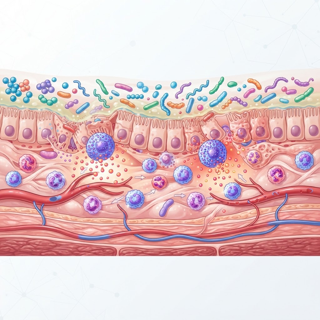
    The human gastrointestinal tract is essentially the largest immune organ in the body. Within the mucosal lining of the gut resides a massive concentration of mast cells—the specialized white blood cells responsible for releasing histamine, cytokines, and other inflammatory mediators in response to perceived threats. In a healthy individual, these cells act as vigilant sentinels, protecting against pathogens. However, for those struggling with mast cell activation syndrome, these sentinels become “trigger-happy.” When we consider the relationship between MCAS and gut health, we must view the gut not as a simple tube, but as a complex signaling center that dictates the behavior of the immune system globally.

    Mast cells in the gut are positioned near nerve endings, blood vessels, and the epithelial barrier. Because they are in constant contact with the contents of your food and the bacteria living in your microbiome, they are uniquely sensitive to the environment inside your digestive tract. When the gut environment is imbalanced, mast cells can become sensitized. This sensitization is why many people with MCAS experience “gut-brain” or “gut-systemic” flares shortly after eating or even when experiencing digestive stress. The mast cells release their contents into the surrounding tissue, which then enters the bloodstream, potentially triggering symptoms in the skin, heart, or brain.

    This localized activation often explains why patients report symptoms that seem unrelated to digestion. A shift in the gut’s chemical environment can prompt a mast cell degranulation event that manifests as a sudden drop in blood pressure or an onset of neurological symptoms. Furthermore, the gut-associated lymphoid tissue (GALT) works in tandem with these mast cells. When the microbiome is in a state of dysbiosis, the chemical signals sent to the GALT are essentially “danger signals.” The immune system interprets these signals as a reason to stay in a state of high alert. Consequently, the threshold for mast cell degranulation lowers significantly. Essentially, your gut health determines the “background noise” of your immune system. If your gut is constantly sending inflammatory signals due to poor digestion or dysbiosis, your mast cells are effectively waiting for the smallest provocation—like a high-histamine food—to erupt into a full-blown systemic flare.

    Healing this connection requires a fundamental shift in perspective. Rather than treating mast cells as the “enemy” that must be suppressed, we look at the gut as the environment that governs their behavior. By optimizing digestion, reducing irritation, and balancing the microbial population, we are essentially telling the mast cells that it is safe to return to a baseline state of calm. This approach moves the goalpost from mere symptom management to addressing the root cause, which is a vital distinction for anyone attempting to heal MCAS naturally. The gut is the command center, and stabilizing the environment within it is the most effective way to restore calm to the mast cells stationed throughout your entire body.

    How Gut Dysbiosis Triggers Systemic Mast Cell Release

    Dysbiosis, the term used to describe an imbalance in the types and quantities of bacteria residing in the digestive tract, is a major contributor to the pathology of MCAS. In a balanced microbiome, beneficial bacteria help regulate immune function and break down dietary histamine. However, when unfavorable bacteria or fungi (like Candida) proliferate, the internal chemistry shifts toward a pro-inflammatory state. This process is deeply linked to the concept of dysbiosis and histamine; when the microbial composition of the gut is skewed, the production of histamine increases, while the ability to neutralize it decreases.

    Many common strains of bacteria associated with dysbiosis are capable of decarboxylating the amino acid histidine into histamine. If these bacteria colonize the gut in high numbers, they create an endogenous source of histamine that is constantly being absorbed through the intestinal wall. For an individual with MCAS, this is akin to being on a high-histamine diet even if you are eating “low-histamine” foods. This excess histamine bypasses the usual breakdown process in the gut and enters systemic circulation, essentially flooding the body and overwhelming the DAO (diamine oxidase) enzymes that are meant to keep histamine levels in check.

    Beyond histamine production, dysbiotic bacteria produce various endotoxins and metabolites that act as direct chemical irritants to the intestinal wall. Lipopolysaccharides (LPS), which are found in the cell walls of certain gram-negative bacteria, are potent stimulators of mast cells. When LPS levels rise in the gut, they trigger immune receptors on the mast cells to degranulate. This creates a feedback loop: the dysbiosis triggers the mast cells, and the mast cell mediators (like TNF-alpha) cause further inflammation, which in turn alters the gut environment to be even more favorable to the dysbiotic bacteria. Breaking this cycle is difficult because the mast cells become hypersensitive, reacting to even minor changes in the microbiome.

    Furthermore, dysbiosis affects the pH levels and the integrity of the mucosal layer that protects the gut lining. A healthy microbiome produces short-chain fatty acids (SCFAs), such as butyrate, which nourish the cells of the gut lining and regulate immune response. Dysbiosis often leads to a deficiency in these protective SCFAs. Without this protective barrier, mast cells are left exposed to undigested food particles and bacterial antigens. This constant exposure keeps the mast cells in a perpetual state of “fight or flight,” leading to the chronic nature of mast cell activation syndrome symptoms. The clinical approach, therefore, must focus on rebalancing the flora through targeted nutrition, probiotics, or antimicrobial therapy if necessary, guided by the principle of “start low and go slow” to avoid triggering a massive mast cell release during the die-off phase.

    Approach Primary Mechanism Best For
    Low-Histamine Diet Reduces external histamine load Acute symptom management and identifying triggers
    Probiotic Support Restores commensal bacterial balance Long-term gut-immune modulation and homeostasis
    Digestive Enzyme Therapy Enhances nutrient breakdown Reducing fermentation and bacterial overgrowth
    Mucosal Repair Protocols Strengthens the intestinal barrier Reducing systemic absorption of triggers

    The Role of Intestinal Permeability in MCAS Flares

    Intestinal permeability, frequently referred to as “leaky gut,” is perhaps the most significant bridge between a malfunctioning digestive system and systemic mast cell activation. Under normal physiological conditions, the lining of the small intestine acts as a selective filter. It allows for the absorption of nutrients while keeping larger, potentially harmful molecules—such as undigested proteins, bacterial toxins, and inflammatory debris—firmly inside the intestinal lumen. In those with MCAS, this barrier is often compromised, meaning that the barrier’s “tight junctions” have become loose or damaged.

    When the gut lining is permeable, the substances mentioned above cross into the bloodstream. This is a critical event for mast cell patients. Because mast cells are distributed throughout the sub-epithelial space, they are the first line of defense when these foreign particles breach the barrier. The moment these intruders cross the lining, mast cells identify them as antigens and begin a degranulation process to neutralize the threat. This process is inherently inflammatory. While it is a natural defense response, it becomes pathological when the barrier is consistently leaky, keeping the mast cells in a chronic state of activation.

    The relationship between intestinal permeability and MCAS is cyclical and self-perpetuating. The inflammatory mediators released by mast cells—such as histamine, tryptase, and prostaglandins—have been shown to increase the permeability of the gut lining even further. This creates a damaging loop: a flare causes the gut to become leakier, which allows more triggers to enter the blood, which then causes more mast cells to degranulate, resulting in even more systemic inflammation. This is why individuals with MCAS often describe their condition as a “snowball effect.” Once a flare begins, it can feel like their entire system is turning against them, regardless of what they do or eat.

    Addressing the root cause of the permeability is essential for anyone seeking to heal MCAS naturally. This involves more than just “avoiding foods.” It requires a deliberate, methodical approach to cooling the fire in the gut. Experts generally agree that using gut-supportive nutrients, such as L-glutamine, zinc carnosine, and soothing mucosal protectants like deglycyrrhizinated licorice (DGL) or slippery elm, can help to reinforce the barrier integrity. However, it is imperative to ensure that these supplements themselves are well-tolerated, as the highly sensitive nature of mast cells means that even some “health” supplements can trigger a reaction if they are derived from histamine-containing sources or fillers that the body reacts to. By reducing the overall burden of inflammatory triggers and slowly strengthening the intestinal fence, patients can eventually lower the reactivity of their mast cells and experience fewer systemic flares.

    Identifying Common Digestive Triggers for MCAS Patients

    For those managing MCAS, the dinner table is often viewed as a minefield. Digestive triggers are rarely universal, but there are certain patterns that frequently emerge in the clinical experience of the MCAS community. The primary challenge is that symptoms are often delayed; you might eat a trigger in the morning and experience a neurological, respiratory, or dermatological flare hours later, making it difficult to pinpoint the culprit. Understanding these categories of triggers is a vital step in mapping your unique sensitivity profile.

    The most commonly discussed category is, of course, high-histamine foods. These include fermented items like sauerkraut and kimchi, aged cheeses, smoked or cured meats, and bone broth that has been cooked for long periods. While these are packed with nutrients, the slow-cooked or fermented processes result in a high buildup of histamine that the gut of an MCAS patient is ill-equipped to handle. However, it is equally important to identify “histamine liberators”—foods that are not inherently high in histamine but trigger the body’s own mast cells to dump their contents. Common offenders in this group include citrus fruits, tomatoes, egg whites, and certain food additives like artificial dyes and preservatives.

    Another frequently ignored category is the “hidden” chemical triggers. Many people with MCAS react not just to the food itself, but to the pesticides, herbicides, and waxes on non-organic produce. These chemicals can irritate the delicate lining of the gut and cause a low-grade inflammatory response that serves as a substrate for mast cell activation. Switching to a strictly organic diet is often the first, most impactful recommendation for those trying to gain a baseline of control. Furthermore, food sensitivities that are not strictly histamine-related, such as reactions to lectins or oxalates, can cause localized gut inflammation. When the gut is inflamed, the mast cells near the intestinal lining stay on high alert, even if the food being eaten is technically low-histamine.

    Lastly, we must consider the temperature and texture of food. Cold foods or extremely hot foods can, in some sensitive individuals, stimulate the vagus nerve and prompt a mast cell response. Similarly, eating while under stress is a massive, albeit overlooked, trigger. The enteric nervous system is tied to the central nervous system; when you eat in a state of “fight or flight,” the digestive process is compromised, leading to incomplete breakdown of food and increased potential for fermentation in the gut. Fermentation products, such as aldehydes and alcohols, are common triggers for mast cell degranulation. Creating a “safe” environment for eating—one that is calm, mindful, and focused—can be just as effective as dietary restriction for many patients. Keeping a detailed food and symptom diary is the most effective tool for identifying your specific triggers, as it allows you to see the patterns that your body is trying to communicate.

    Why SIBO and MCAS Often Occur Together

    The intersection of Small Intestinal Bacterial Overgrowth (SIBO) and MCAS is a frequent and challenging finding in clinical practice. SIBO is defined by an excessive growth of bacteria in the small intestine, where bacterial counts should typically be relatively low. Because the small intestine is where the majority of nutrient absorption takes place, the presence of a massive bacterial colony creates a state of chaos. The bacteria consume the nutrients before the host can, but more importantly for the MCAS patient, they produce vast quantities of gases and metabolic byproducts that disrupt the gut’s normal function.

    The link between these two conditions is often a matter of motility. In a healthy gut, the “migrating motor complex” (MMC)—a series of rhythmic contractions—acts like a “housekeeper,” sweeping undigested food and bacteria through the digestive tract. In many MCAS patients, the motility is slowed. This may be due to mast cell mediators directly affecting the smooth muscle of the gut or to underlying autonomic nervous system dysfunction (often related to conditions like POTS, which co-occurs frequently with MCAS). When motility is sluggish, bacteria have the opportunity to migrate from the large intestine into the small intestine, where they set up shop and multiply.

    Once SIBO is established, it acts as a permanent, systemic trigger for mast cell activation. The bacteria in the small intestine do not just ferment food; they create a toxic environment that keeps the immune system in a state of hyper-arousal. The chronic inflammation resulting from SIBO damages the gut lining, leading to the intestinal permeability discussed earlier. This sets up a perfect storm for MCAS, as the inflammatory mediators released by the overgrowth act as a continuous source of stimulation for the mast cells. This explains why many MCAS patients find that their condition is “treatment-resistant”—as long as the SIBO remains, the “danger signal” is constantly being broadcasted to the immune system.

    Addressing this co-occurrence requires a balanced approach. Treating SIBO can be particularly daunting for an MCAS patient, as antimicrobial protocols (whether herbal or pharmaceutical) can cause a large-scale die-off of bacteria. This die-off releases high amounts of bacterial toxins (LPS) and histamine into the system, which can trigger a severe MCAS flare if the patient is not stabilized first. Therefore, the common clinical strategy is to focus on mast cell stabilization and gut soothing *before* attacking the bacterial overgrowth. By “priming the terrain,” you can minimize the risk of a severe reaction during the treatment phase. This often involves the use of specialized diets (like low-FODMAP, specifically adapted for MCAS) and prokinetics to improve motility, ensuring that the “housekeeper” is working correctly before you begin the process of clearing the overgrowth. Understanding this relationship is a major breakthrough for many patients, as it shifts the focus from managing constant, unexplained flares to targeting the underlying bacterial population that is fueling the fire.

    Testing Methods to Evaluate Your Gut Microbiome Health

    When investigating the link between MCAS gut health and systemic inflammation, conventional testing often falls short. While standard stool tests can identify clear-cut parasites or major infections, they frequently miss the subtle imbalances characterizing dysbiosis and histamine intolerance. For the patient with mast cell activation syndrome symptoms, a more granular look at the gut environment is essential to create an effective healing protocol.

    The most comprehensive approach often involves functional stool analysis, which goes beyond simple cultures. These tests often utilize PCR (polymerase chain reaction) technology to quantify the relative abundance of commensal bacteria. By analyzing the diversity index of your microbiome, practitioners can determine if there is an overgrowth of Proteobacteria—a phylum often associated with gut inflammation—or a deficiency in beneficial butyrate-producing species that help maintain the integrity of the gut lining.

    Organic Acids Testing (OAT) is another vital diagnostic tool. By measuring metabolic byproducts in the urine, OAT can identify fungal overgrowth (like Candida) or bacterial metabolites that act as neurotoxins. Since many MCAS patients struggle with cognitive fog and systemic sensitivity, identifying these markers is crucial. If your OAT results show elevated markers for clostridia or yeast, it provides a clear roadmap for targeted antimicrobial support, which must be approached with caution in those with mast cell sensitivity.

    Finally, assessing intestinal permeability is central to managing mast cell stability. While there is no single “gold standard” test that is universally accepted, tests measuring Zonulin levels or the lactulose/mannitol ratio are often utilized. Elevated Zonulin suggests that the “tight junctions” of your gut barrier are loose, potentially allowing undigested food particles and bacterial endotoxins to enter the bloodstream, thereby triggering an immediate mast cell degranulation response.

    Dietary Strategies to Reduce Gut Inflammation and Histamine

    Diet is perhaps the most immediate lever you can pull to influence mast cell activity. However, because MCAS is highly individual, a “one-size-fits-all” approach rarely succeeds. The primary objective is to lower the total histamine load while simultaneously reducing the mechanical and chemical stressors on the intestinal mucosa.

    A low-histamine diet involves more than just removing aged cheese and fermented foods. It requires a meticulous focus on food freshness. Histamine levels in proteins—such as chicken, fish, and beef—skyrocket as the meat ages after slaughter. Buying fresh, flash-frozen meat and cooking it immediately is a cornerstone of this strategy. Leftovers are often a hidden trap for the MCAS patient, as even a 24-hour stay in the refrigerator can lead to the proliferation of histamine-producing bacteria in the food.

    Beyond histamine-specific foods, addressing intestinal permeability often requires a temporary reduction in inflammatory triggers. Gluten, cow’s dairy, and refined sugars are frequent offenders that increase gut permeability. By removing these, you allow the gut lining a chance to repair itself, which is vital for preventing the “leaky gut” cycle that drives mast cell activation.

    Dietary Strategy Mechanism of Action Best for
    Low-Histamine Diet Reduces dietary triggers of degranulation General histamine intolerance
    Anti-Inflammatory Elimination Repairs the intestinal mucosal barrier Suspected gut dysbiosis
    Low-FODMAP (Modified) Limits fermentable carbohydrates Small Intestinal Bacterial Overgrowth (SIBO)
    GAPS/Introduction Diet Starves pathogens, nourishes villi Severe digestive distress

    Probiotic Considerations for Sensitive Mast Cell Patients

    In the general population, probiotics are touted as the cure-all for gut health. However, for those with MCAS, introducing the wrong strains can exacerbate symptoms significantly. Many common probiotic strains, such as Lactobacillus casei, Lactobacillus bulgaricus, and Streptococcus thermophilus, are histamine-producers. Consuming these can rapidly undo the progress made through diet and environmental control.

    Instead, look for histamine-neutral or histamine-degrading strains. Strains such as Bifidobacterium infantis, Bifidobacterium longum, and Lactobacillus plantarum have been observed in some cases to support gut barrier function without the production of biogenic amines. Some practitioners also suggest incorporating Lactobacillus rhamnosus, which may help modulate the immune response, provided the patient does not have a specific intolerance to that strain.

    When starting any probiotic protocol, the rule of “start low and go slow” is absolute. A common mistake is jumping to a full dose. Many sensitive patients begin by taking a fraction of a capsule or even just a pinch of the probiotic powder mixed into water, slowly titrating upward over several weeks. If you experience an increase in flushing, itching, or digestive bloating, it is a clear sign to pause, reset, and reassess the formulation.

    Natural Approaches to Balancing the Gut-Immune Axis

    Balancing the gut-immune axis requires a dual-pronged strategy: soothing the existing mast cell reactivity while gently modulating the microbial ecosystem. Nature provides several compounds that can assist in this stabilization process, often referred to as natural mast cell stabilizers.

    Quercetin, a potent bioflavonoid found in red onions and apples, is widely recognized for its ability to inhibit the release of histamine from mast cells. When paired with gut-healing agents like L-glutamine and deglycyrrhizinated licorice (DGL), it can help close the gaps in the intestinal wall. L-glutamine, in particular, acts as the primary fuel source for the cells lining the small intestine, providing the building blocks necessary to reinforce the physical barrier against bacterial toxins.

    Botanical interventions should be approached with professional guidance. Many essential oils and herbal extracts used to combat dysbiosis (like oregano oil or berberine) are potent. While they can effectively reduce bacterial overgrowths, they can also cause a “die-off” reaction, which releases a surge of endotoxins and histamine into the system. Often, practitioners recommend starting with gentler, systemic immune modulators—such as vitamin C or zinc—to raise the threshold for mast cell degranulation before attempting aggressive microbial eradication.

    Lifestyle Changes to Support Long-Term Mast Cell Stability

    Lifestyle factors are the “hidden” drivers of MCAS. Even with a perfect diet, high levels of chronic stress can force the body into a state of perpetual mast cell activation. The gut and the brain are physically connected via the vagus nerve, and this nerve is highly sensitive to the chemicals released during the “fight or flight” response.

    Vagus nerve stimulation is a powerful, non-pharmacological way to modulate gut inflammation. Simple practices like deep diaphragmatic breathing, cold-water exposure (such as splashing the face with ice-cold water), and humming or singing can tone the vagus nerve, signaling the body to switch from a sympathetic (stress) state to a parasympathetic (rest and digest) state. When the body is in “rest and digest” mode, the gut wall is better protected, and the likelihood of inappropriate mast cell triggering decreases significantly.

    Sleep hygiene is equally vital. During deep, restorative sleep, the body conducts its most intensive repair work on the intestinal mucosa. Chronic sleep deprivation, conversely, is associated with increased intestinal permeability and elevated inflammatory markers. Aiming for a consistent circadian rhythm—waking and sleeping at the same time every day—helps stabilize the hormonal environment, which in turn provides a more stable baseline for immune system function.

    Frequently Asked Questions

    Can MCAS cause digestive issues if I don’t have allergies?

    Yes, absolutely. Even without classic IgE-mediated food allergies, MCAS can cause significant gastrointestinal distress. When mast cells in the gut lining activate, they release mediators like histamine, prostaglandins, and leukotrienes. These substances alter gut motility, increase secretion, and cause inflammation, leading to symptoms like diarrhea, cramping, bloating, and abdominal pain that often mimic Irritable Bowel Syndrome.

    Is it possible to heal a leaky gut while having MCAS?

    Healing the gut in the presence of MCAS is possible but requires a delicate approach. Because the gut is the primary site of mast cell activity, success depends on reducing the systemic inflammatory load. By implementing a low-histamine diet, supporting the microbiome with safe, non-histamine-producing probiotics, and utilizing nutrients that support the intestinal wall, many individuals see a reduction in permeability and a concurrent improvement in their overall symptom profile.

    Should I avoid all probiotics if I have mast cell activation syndrome?

    Not necessarily, but you must be extremely selective. Many traditional probiotic strains increase histamine levels. However, certain strains, such as specific bifidobacteria, may actually support immune balance. The key is to avoid fermented foods and multi-strain probiotics until you have tested your individual tolerance to single-strain supplements under the guidance of a practitioner.

    How does stress impact MCAS symptoms in the gut?

    The gut-brain axis is highly active in MCAS. Stress triggers the release of Corticotropin-Releasing Hormone (CRH), which directly stimulates mast cells to degranulate. This creates a vicious cycle: stress causes mast cells to release histamine, which increases gut inflammation and permeability, leading to further systemic sensitivity and renewed stress on the body. Managing the nervous system is a required component of long-term healing.

    Why do my symptoms get worse after eating leftovers?

    Leftover food is a major trigger for many MCAS patients because histamine levels accumulate as food undergoes microbial decomposition. Even when refrigerated, most leftovers experience a significant increase in histamine content within 24 hours. For those with a reduced capacity to break down histamine (due to DAO enzyme deficiency or dysbiosis), this surge causes immediate “flare” symptoms after eating.

    Are there specific supplements that help with histamine degradation?

    Many patients benefit from DAO (diamine oxidase) enzyme supplements taken just before meals. DAO is the primary enzyme responsible for breaking down ingested histamine. While it does not address the root cause of MCAS or gut dysbiosis, it can provide significant symptomatic relief by reducing the load of histamine that enters the bloodstream after consumption of food, allowing the body more time to regulate its internal processes.

    Conclusion

    Navigating the intersection of MCAS and gut health is undoubtedly a complex challenge, but it is also one of the most rewarding journeys a patient can take toward regaining their quality of life. By recognizing that your microbiome is not just a collection of bacteria, but a dynamic immune organ, you can begin to shift your focus from symptom suppression to foundational healing. From refining your dietary choices to managing the nervous system through vagus nerve stimulation, every small step contributes to a more stable immune environment.

    The path to recovery is rarely linear, and it requires patience, consistency, and a willingness to listen to your body’s unique signals. We encourage you to start with one or two changes mentioned in this guide—such as focusing on fresh food preparation or introducing a histamine-neutral probiotic—and observe how your system responds. Remember, you have the power to create a more resilient gut-immune axis. Stay informed, stay steady, and prioritize your long-term stability above short-term quick fixes. Take control of your wellness journey today by partnering with a knowledgeable healthcare provider to tailor these strategies to your specific medical history.

    By healthauthoritylife Editorial Team

    This article is for informational purposes only and is not a substitute for professional medical advice. Always consult your doctor before making changes to your diet, exercise routine, or treatment plan.
  • Craniocervical Instability: Symptoms, Causes, and Natural Relief

    Craniocervical Instability: Symptoms, Causes, and Natural Relief

    Key Takeaways

    • Craniocervical instability (CCI) occurs when the ligaments supporting the skull and upper spine become too lax to maintain structural integrity.
    • While chronic neck pain is a hallmark sign, CCI symptoms often manifest neurologically, affecting vision, balance, and cognitive function.
    • There is a well-documented clinical connection between EDS and CCI due to systemic collagen defects.
    • Ligament laxity can stem from connective tissue disorders, physical trauma, or inflammatory processes that degrade tissue stability.
    • Diagnosing CCI is notoriously complex, often requiring dynamic imaging rather than standard static X-rays or MRIs.

    For those living with persistent, unexplained neurological struggles and persistent upper cervical discomfort, the journey toward a diagnosis can feel like a labyrinth. Craniocervical instability, often referred to as CCI, is a condition that exists at the intersection of structural orthopedics and complex neurology. It represents a failure of the body’s primary support system for the head, leading to a cascade of systemic issues that can diminish quality of life. As awareness grows, more individuals are seeking to understand how their structural alignment impacts their total health, searching for answers beyond conventional pain management. In this guide, the healthauthoritylife Editorial Team explores the intricacies of CCI—from the mechanical failures of the cervical spine to the pathways toward natural relief—to empower you with the knowledge needed to advocate for your own well-being.

    Understanding Craniocervical Instability: What It Is

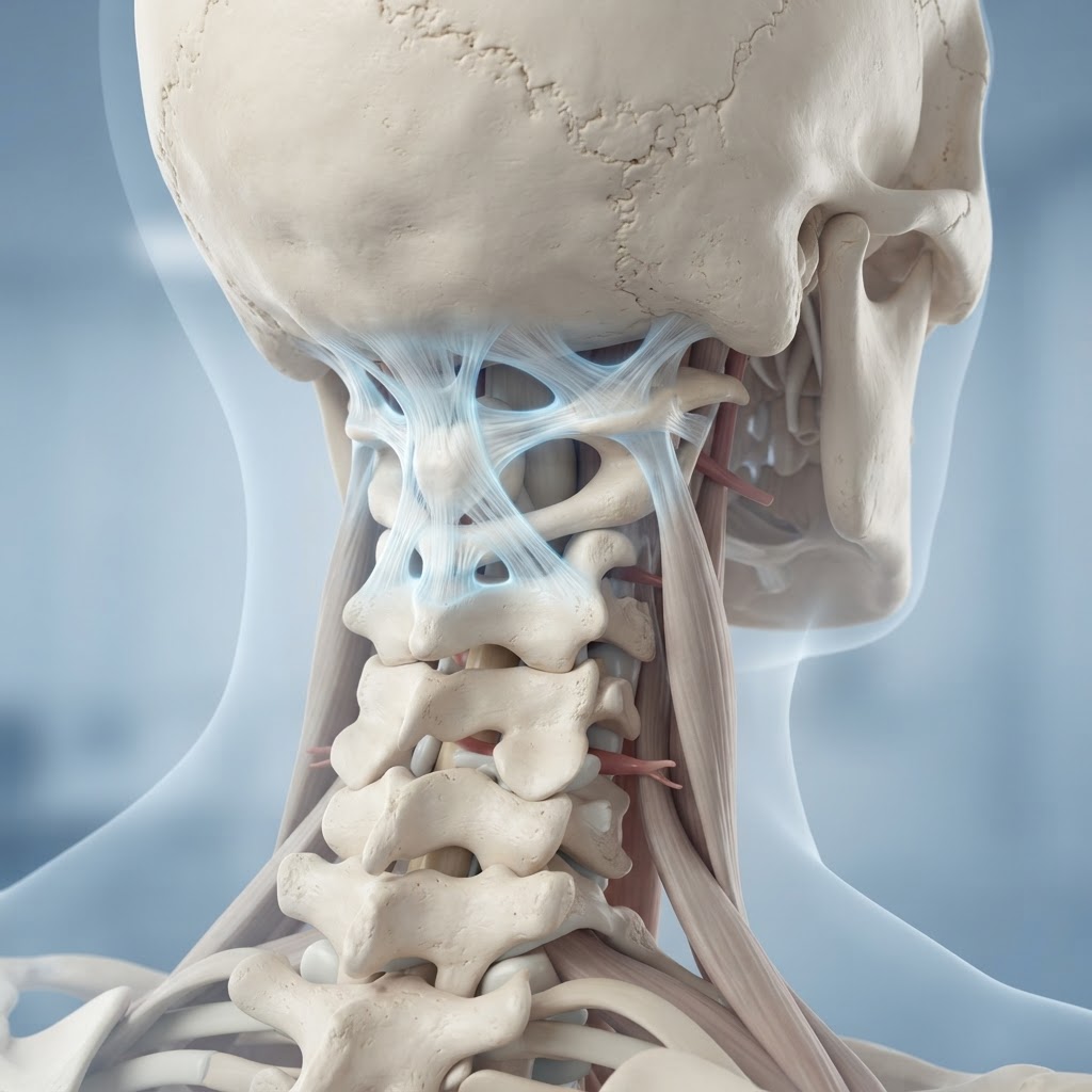
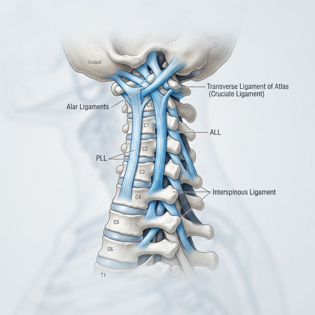
    Craniocervical instability is a mechanical condition involving the junction where the skull meets the cervical spine, specifically at the C0-C1 (atlanto-occipital) and C1-C2 (atlanto-axial) joints. In a healthy, well-functioning neck, a complex network of ligaments, including the alar and transverse ligaments, acts as a system of cables and anchors. These structures ensure that the heavy mass of the skull remains balanced securely on the delicate spinal column, protecting the brainstem and spinal cord from excessive movement during everyday activities like walking, turning the head, or even swallowing.

    When someone experiences CCI, these ligaments become overly loose—a state clinically known as ligament laxity. This loss of tension allows the skull to shift abnormally on the cervical vertebrae. Think of it as a loose hinge on a heavy door; because the hinge is not tight, the door sags and creates friction, misalignment, and stress on the surrounding hardware. In the human body, this “sagging” can place undue pressure on the medulla oblongata, the brainstem, and the delicate cranial nerves that exit the skull base. Because this area is the command center for involuntary bodily functions, the consequences of this instability are rarely localized just to the neck.

    Understanding the anatomy is vital to grasping why the condition feels so systemic. The upper cervical spine is unique in its mobility; it is designed to allow for rotation and nodding, but it lacks the bony locking mechanisms found in the lower lumbar spine. Consequently, it relies almost entirely on soft tissue for stability. When these soft tissues lose their inherent tension, the nervous system enters a state of constant high alert, attempting to stabilize the head using muscles that were never intended for such a heavy load. This leads to muscle spasms, myofascial trigger points, and a cycle of secondary pain that can make a localized problem feel like a full-body health crisis.

    Many experts emphasize that CCI is not merely a spinal issue but a structural foundation issue. When the skull is not properly supported, the entire vertebral column may attempt to compensate, leading to pelvic tilts, scoliosis, or secondary issues in the thoracic spine. Identifying CCI requires moving beyond standard diagnostic models that focus on bone density or disc herniation and instead looking at how the skull and spine behave during physical movement. Whether the cause is an inherent weakness in collagen or a traumatic event that overstretched the ligaments, the fundamental reality remains the same: the mechanical anchor of the nervous system is no longer functioning as intended.

    Common Symptoms of CCI: More Than Just Neck Pain

    The clinical presentation of craniocervical instability is notoriously broad, which is why patients often visit a dozen different specialists before finding a name for their experience. While chronic neck pain relief is the primary goal for many, the neurological symptoms of CCI are often what impact day-to-day functionality the most. Because the brainstem is situated directly at the craniocervical junction, any mechanical shift in the skull can compress or irritate these nerve tracts, leading to symptoms that mimic other conditions like dysautonomia, vestibular disorders, or even primary headaches.

    One of the most frequent CCI symptoms reported is postural orthostatic tachycardia syndrome (POTS) or other forms of autonomic dysfunction. Patients may notice that their heart rate spikes upon standing, or they may experience cold, clammy extremities and blood pressure instability. This occurs because the autonomic nervous system, which regulates heart rate and digestion, passes through the very area that is being compressed by the shifting skull base. When the “cables” are loose, the messaging system between the brain and the body experiences signal interference.

    Vision and balance are also frequently compromised. Many people with CCI report “visual snow,” double vision, or a feeling of being detached from their body, a phenomenon often described as brain fog. This happens because the brainstem acts as a relay station for visual information and proprioception—the body’s ability to sense its position in space. If the brainstem is irritated, the signals from the inner ear and the eyes are processed incorrectly, leading to a persistent sense of dizziness or an inability to focus on moving objects. These symptoms are often worse after periods of physical exertion or prolonged screen time, as the neck muscles fatigue and lose their protective support.

    Other common manifestations include:

    • Headache patterns: Specifically, occipital neuralgias or tension-type headaches that radiate from the base of the skull toward the eyes.
    • Dysphagia: Difficulty swallowing, or a feeling of a “lump in the throat” caused by proximity of the pharyngeal nerves to the unstable vertebrae.
    • Tinnitus: Persistent ringing or buzzing in the ears, often linked to the flow of cerebrospinal fluid or nerve irritation near the auditory pathways.
    • Sleep disturbance: Many patients find they cannot fall asleep unless their head is positioned in a very specific way, as the neck feels “too heavy” when lying down.

    It is important to note that these symptoms are often episodic. They may flare up after a long day or during periods of systemic inflammation and then subside with rest. This fluctuation is one of the most confusing aspects for patients, as it can make them feel like their symptoms are “all in their head.” However, the mechanical reality of the lax ligaments remains, regardless of whether the symptoms are present at any given moment.

    The Link Between Ehlers-Danlos Syndrome and CCI

    The correlation between EDS and CCI is one of the most discussed topics in modern physical medicine. Ehlers-Danlos Syndrome (EDS) is a group of genetic disorders that affect the body’s connective tissue. Connective tissue is essentially the “glue” that holds the body together, composed primarily of collagen. In individuals with EDS, the collagen is structurally flawed, making the ligaments, tendons, and skin much more stretchy and fragile than average. Because the ligaments in the upper neck are the primary stabilizers of the head, it is easy to see why those with connective tissue disorders are highly susceptible to developing craniocervical instability.

    When an individual has EDS and CCI, their body lacks the structural “toughness” to maintain the alignment of the C1 and C2 vertebrae. For the general population, a minor whiplash injury might lead to temporary stiffness; for an individual with EDS, that same injury can cause permanent ligament laxity because the tissue lacks the elasticity to “snap back” to its original length. This condition is often referred to as hypermobility, but when it affects the spine, it becomes a distinct clinical challenge that requires a more nuanced approach than standard physical therapy.

    The management of this specific intersection requires caution. Traditional aggressive manual adjustments (often called “cracking” the neck) can be potentially hazardous for someone with EDS and associated instability, as the ligaments are already compromised. Instead, experts generally suggest focusing on isometric stabilization and gentle, targeted strengthening of the deep neck flexors. Understanding that the instability is not a muscular weakness—but a tissue-integrity failure—is the first step toward creating a safer, more sustainable management plan.

    Approach Focus Best For
    Isometric Stabilization Building muscle tension without movement Acute phase, early strengthening
    Cervical Collars External structural support Severe symptom relief during flares
    Proprioceptive Training Retraining nerve-to-muscle pathways Chronic, long-term alignment support
    Manual Therapy (Gentle) Myofascial release and fluid flow Reducing compensatory muscle spasms

    Living with the combination of these conditions requires a paradigm shift. One must learn to listen to the body’s signals of instability—such as a sudden feeling of heaviness in the head or a brief loss of balance—and respond with rest rather than pushing through. By focusing on maintaining a neutral spine and utilizing supportive aids during high-activity periods, many individuals find that they can successfully manage the symptoms without needing more invasive interventions. The key is consistent, low-impact habits that prioritize the protection of the cervical junction above all else.

    Major Causes of Ligament Laxity in the Upper Neck

    Ligament laxity, the root cause of craniocervical instability, does not occur in a vacuum. While genetic predisposition like EDS plays a significant role, many cases are acquired through a combination of environmental factors, trauma, and chronic systemic issues. Understanding these neck instability causes is essential for developing a personalized strategy for natural relief. When we look at why ligaments lose their ability to support the head, we must look at both mechanical and biochemical contributors.

    Trauma remains a leading cause of acquired CCI. This is not restricted to high-impact injuries like car accidents or contact sports. While those events are obvious triggers for “whiplash-associated disorders,” repetitive micro-trauma can be just as damaging over time. For example, individuals who spend excessive hours in a forward-head posture—often called “text neck”—place constant, abnormal tension on the posterior ligaments of the cervical spine. Over years, this consistent strain can cause the ligaments to stretch and lose their resting tone, effectively creating a state of mechanical instability without a single, dramatic accident.

    Another major contributor is chronic systemic inflammation. Ligaments are not static; they are living tissues that require a stable internal environment to maintain their integrity. Conditions such as rheumatoid arthritis, ankylosing spondylitis, or even chronic undiagnosed systemic infections can degrade the quality of collagen within the ligaments. When the body is in a state of systemic inflammation, the enzymes responsible for tissue turnover can become overactive, leading to the breakdown of collagen fibers faster than the body can repair them. This process, often referred to as “ligamentous laxity due to inflammatory insult,” can leave the upper cervical spine vulnerable even in the absence of a direct injury.

    Furthermore, lifestyle factors such as nutritional deficiencies can impact how the body repairs its connective tissue. Vitamin C, for instance, is a critical cofactor for collagen synthesis. Similarly, magnesium and collagen-building amino acids are required to maintain the structural health of all soft tissues. A diet high in processed foods and deficient in these micronutrients may impair the body’s ability to recover from minor, daily strains. When you combine this lack of building blocks with the modern sedentary lifestyle, the cervical spine loses its natural resiliency. It is a slow, silent progression, but it is one that can be mitigated through proactive lifestyle adjustments and targeted nutritional support, which remain fundamental components of any natural CCI treatment plan.

    Diagnostic Challenges and When to See a Specialist

    One of the primary frustrations for those investigating craniocervical instability is the difficulty of achieving a clinical diagnosis. Standard imaging protocols, such as static X-rays, CT scans, and MRIs, are performed while the patient is lying completely still. While these tests are excellent for ruling out tumors, fractures, or disc herniations, they are often insufficient for identifying CCI. Because CCI is a dynamic condition—meaning it occurs when the head moves into certain positions—the instability is frequently invisible on a static scan.

    This diagnostic gap is why many patients feel gaslit by the medical system. A technician may perform a standard MRI and conclude that the cervical spine looks “normal,” while the patient is still suffering from severe neurological symptoms. To accurately assess the health of the craniocervical junction, specialists often need to employ dynamic imaging, such as upright or flexion-extension X-rays and MRIs. These allow the clinician to see exactly how the vertebrae shift when the head is tilted or rotated, which can reveal the telltale signs of instability that occur only during movement.

    When should you consider seeking a specialist? It is generally advised to pursue a consultation with a neurologist, neurosurgeon, or a physiatrist who specializes in hypermobility or cervical spine disorders if you experience the following:

    1. Symptoms that worsen significantly when you are upright but improve when you lie down.
    2. A history of unexplained autonomic symptoms, such as sudden heart rate changes or fainting.
    3. Persistent “clunking” or “grinding” sensations in the neck, especially during rotational movements.
    4. Neurological symptoms that do not respond to physical therapy or chiropractic adjustments and instead seem to flare up after such interventions.
    5. A known diagnosis of a connective tissue disorder like EDS or Marfan syndrome paired with chronic, refractory neck pain.

    Seeking out a provider who understands the nuance of CCI is crucial. You want to work with someone who asks about your specific triggers and considers your functional capacity rather than just looking at the shape of your bones on a film. If a specialist seems unfamiliar with the concept of dynamic imaging for cervical stability, it may be time to seek a second opinion. Because the craniocervical junction is so close to the brainstem, it is always best to prioritize practitioners who emphasize non-invasive, conservative management first, using surgical intervention only as a measure of last resort when stability cannot be achieved through other means. Empowerment begins with finding a medical partner who views your symptoms as a legitimate, measurable physiological challenge rather than a mystery to be ignored.

    Natural Strategies to Support Neck Stability

    When dealing with craniocervical instability, the primary goal is to minimize mechanical stress on the ligaments that connect the skull to the upper cervical spine. Natural management is rarely about “curing” the structural laxity, but rather about creating an environment where your remaining structural integrity is preserved and your neurological system is protected from irritation. A multi-modal approach is often the most successful strategy.

    The first natural strategy involves proprioceptive training. Individuals with CCI often experience a disconnect between their brain and the positioning of their head in space. This “proprioceptive deficit” can lead to erratic muscle movements or bracing patterns that further strain the cervical ligaments. Working with specialized physical therapists on gentle cervical proprioception—such as learning to hold the head in a neutral “stacked” position—can reduce the need for the muscles to over-contract in a futile attempt to compensate for ligamentous laxity.

    Thermal therapy, when applied correctly, can be a potent tool. While systemic inflammation is a hallmark of EDS and related collagen disorders, targeted thermal application can help manage the secondary muscular guarding that occurs when the neck becomes unstable. Many find that cold therapy (ice packs) is superior for acute flare-ups where inflammation is driving the pain, while moist heat is better for chronic, low-grade muscle tension. However, it is vital to avoid aggressive cervical traction or heavy heat packs that can cause the neck muscles to become too relaxed, potentially leading to further instability.

    Additionally, the use of targeted cervical orthotics or soft collars—under the supervision of a healthcare provider—can be a natural intervention to provide temporary external support. Rather than relying on them 24/7, which can lead to muscle atrophy, using them during high-exertion activities or long periods of sitting can allow the damaged ligaments to recover by preventing end-range shearing movements.

    The Role of Posture and Ergonomics in Managing CCI

    For someone with CCI, the “text neck” phenomenon is not just a nuisance; it is a mechanical threat. Every inch that the head migrates forward from its neutral alignment increases the load on the cervical spine by several pounds. When your ligaments are already lax, this increased weight creates a “hinge” effect at the craniocervical junction, leading to the hallmark CCI symptoms like dizziness, vision disturbances, and headaches.

    Optimizing your environment is essential. Your monitor should be positioned so that the top third of the screen is at eye level. This prevents the tendency to tilt the chin downward or extend the neck forward. If you use a laptop, a separate keyboard and a laptop stand are non-negotiable. Furthermore, your chair should provide robust lumbar support. When the lower back loses its natural curve (slumping), the head naturally follows, pushing the cervical spine into a vulnerable position.

    Sleeping posture is another critical factor often overlooked. Those with neck instability often find that sleeping with excessive pillows forces the neck into a flexed position all night long. A cervical pillow designed to support the neck’s natural lordosis, or even a rolled-up towel placed specifically under the neck, can keep the cervical spine neutral while sleeping on your back. Side sleeping requires a pillow thick enough to keep the neck aligned with the spine, preventing a lateral tilt that could irritate the C1-C2 joint.

    Safe Exercises for Strengthening the Neck Muscles

    Strengthening for CCI is a process of nuance, not intensity. Traditional “neck curls” or heavy resistance training can be counterproductive, potentially causing a traumatic subluxation if the ligaments are severely compromised. Instead, the focus must be on deep cervical flexors and scapular stabilization. The deep neck flexors—the muscles located in the front of the throat—are responsible for stabilizing the head. When these muscles are weak, the larger, more superficial neck muscles (the trapezius and sternocleidomastoid) take over, leading to chronic tightness and pain.

    The “chin tuck” is the gold standard exercise, but it must be performed correctly. While lying flat on your back, gently draw your chin inward as if making a double chin, while keeping the back of your head in contact with the surface. The goal is not to lift the head, but to shorten the muscles in the front of the neck. Hold for a few seconds and release. This isometric contraction strengthens the stabilizers without putting the cervical spine through an unsafe range of motion.

    Scapular retraction is equally important. Your shoulder blades act as the foundation for your neck. If your shoulders are rounded forward, your neck is structurally disadvantaged. Exercises that focus on pulling the shoulder blades together and downward—like rows with light resistance bands—can unload the cervical spine by improving overall thoracic posture.

    Exercise Type Primary Benefit Best For
    Isometric Chin Tucks Stabilizes deep neck flexors Beginners/Acute symptoms
    Scapular Retraction Unloads tension from the upper traps Improving thoracic alignment
    Eye-Movement Training Improves vestibular-cervical control Reducing dizziness/vertigo
    Seated Posture Correction Prevents end-range cervical shear Long-term daily stability

    Nutritional Support for Ligament and Collagen Health

    Ligament laxity is often exacerbated by systemic nutritional deficiencies that impair collagen synthesis. While diet cannot “tighten” a loose ligament, providing the body with the necessary building blocks is a fundamental step in supporting the connective tissue you have left. Collagen is the most abundant protein in the body, and it relies heavily on specific amino acids and vitamins for its structural integrity.

    Vitamin C is perhaps the most critical nutrient for collagen cross-linking. Without sufficient Vitamin C, the body cannot effectively bind collagen fibers together, which may contribute to the fragility seen in those with hypermobility spectrum disorders. Increasing intake of citrus fruits, bell peppers, and strawberries is a simple, natural way to support tissue health.

    Additionally, bone broth has become a staple for those with connective tissue issues because it contains naturally occurring collagen, proline, and glycine. These amino acids are the primary components of ligaments and tendons. Some experts also suggest that maintaining adequate hydration is vital; ligaments are largely composed of water and proteins, and dehydration can lead to increased stiffness and decreased elasticity, making the tissue more prone to micro-tears.

    Magnesium supplementation is also frequently discussed, as it plays a role in muscle relaxation and the prevention of the muscle spasms that often plague CCI patients. A magnesium-rich diet or transdermal magnesium (via Epsom salt baths) can help calm the nervous system and reduce the hyper-tonicity of the neck musculature, creating a more stable foundation for the cervical spine.

    Lifestyle Adjustments to Prevent Further Neck Strain

    Managing CCI requires a permanent shift in how you interact with your environment. High-impact activities are generally contraindicated for those with documented neck instability. Activities like contact sports, heavy lifting, or even activities that involve repetitive jarring, such as running on hard surfaces, can create significant shear forces on the cervical vertebrae.

    Learning to move your body as a single unit is a crucial skill. For example, if you need to turn to look at something behind you, don’t just rotate your neck. Turn your entire torso. This “en-bloc” turning minimizes the amount of rotation required at the atlantoaxial joint, protecting the vulnerable ligaments from twisting forces.

    Furthermore, managing the digital load is essential. Using voice-to-text software for long emails or documents can significantly reduce the amount of time you spend looking down at a screen. If you must use a phone, bring the device up to eye level rather than lowering your head to the device. These small, incremental changes to your daily habits can prevent the accumulation of micro-trauma that leads to chronic neck pain relief being so elusive.

    Frequently Asked Questions

    Can physical therapy cure craniocervical instability?

    Physical therapy is generally not considered a “cure” for structural ligamentous laxity, especially in cases related to conditions like EDS. However, it is an essential management tool. By strengthening the deep stabilizers of the neck, physical therapy can often reduce the severity of symptoms and prevent further degradation of the cervical alignment, allowing many individuals to improve their quality of life significantly.

    What are the first signs that I might have CCI?

    Early CCI symptoms often include a heavy feeling in the head, frequent “cracking” or popping sensations in the neck, persistent headaches that start at the base of the skull, and sudden episodes of dizziness or lightheadedness when changing positions. Many people also report a feeling that they need to physically support their head with their hand when sitting for long periods.

    Is surgery the only option for someone with CCI?

    Surgery, such as a cervical fusion, is typically considered a last resort for severe cases where neurological damage is imminent or quality of life is profoundly impacted despite comprehensive conservative management. Most experts recommend exhausting all natural, non-invasive, and regenerative medicine options before considering surgical intervention, as cervical fusion carries risks and long-term mechanical implications.

    How does Ehlers-Danlos Syndrome (EDS) affect my neck?

    EDS is a group of genetic disorders that affect the connective tissue. Because the ligaments in the body are largely composed of collagen, those with EDS have ligaments that are more prone to stretching or tearing. In the cervical spine, this systemic ligament laxity means the ligaments holding the skull to the spine are often too loose to keep the vertebrae in proper alignment, which is why CCI is common in the EDS community.

    Are there specific diagnostic tests for CCI?

    Diagnosing CCI can be difficult because traditional static imaging, like a standard MRI or X-ray while lying down, often fails to show the instability. Doctors typically look for dynamic imaging, such as an Upright MRI or Digital Motion X-ray (DMX), which captures the cervical spine while the head is in motion or under gravity. These tests are far better at identifying the degree of “slide” or instability between the vertebrae.

    Can I lead a normal life with craniocervical instability?

    Yes, many people with CCI lead fulfilling lives by making intentional lifestyle adjustments. While it may require giving up high-impact activities and prioritizing ergonomic health, learning how to stabilize the neck through exercise and supportive habits allows many individuals to manage their symptoms successfully and maintain a high level of daily function.

    Conclusion

    Craniocervical instability is a complex and often misunderstood condition, but it is one that can be managed with the right combination of patience, lifestyle discipline, and informed guidance. By understanding the mechanical causes of your neck instability—whether related to collagen disorders like EDS or simple wear and tear—you can move from a state of reactive pain management to a proactive strategy of stabilization and protection. Remember that your cervical spine is a delicate structure, and the small, consistent efforts you make today—through posture, targeted exercise, and nutritional support—are the best insurance against further decline. Take charge of your movement, listen to your body’s signals, and work closely with healthcare providers who understand the nuances of the craniocervical junction.

    If you suspect you are struggling with neck instability, start by tracking your symptoms and discussing them with a specialist who understands cervical biomechanics. You do not have to settle for chronic discomfort; informed action is your greatest tool for relief.

    By healthauthoritylife Editorial Team

    This article is for informational purposes only and is not a substitute for professional medical advice. Always consult your doctor before making changes to your diet, exercise routine, or treatment plan.
  • Cervical Instability: Symptoms, Causes, and Natural Support

    Cervical Instability: Symptoms, Causes, and Natural Support

    Key Takeaways

    • Cervical instability occurs when ligaments in the upper neck lose their integrity, failing to keep the vertebrae aligned during movement.
    • Common cervical instability symptoms often include chronic neck pain, dizziness, vertigo, and unusual neurological sensations.
    • Ligament laxity is frequently caused by trauma, repetitive strain, or underlying systemic connective tissue disorders.
    • The cervical spine serves as a primary hub for the nervous system, meaning instability can trigger wide-ranging physiological reactions beyond simple pain.
    • Improving cervical spine health requires a multidisciplinary approach focusing on stabilization, postural correction, and systemic support for connective tissue.

    The human neck is a marvel of biological engineering, balancing the weight of the skull atop a delicate stack of seven vertebrae while facilitating complex, multi-directional movement. However, when the soft tissues responsible for maintaining this architecture begin to falter, the results can be debilitating. Cervical instability is a condition that often remains under-diagnosed because it does not always show up clearly on standard imaging like X-rays or basic MRIs, which typically capture the spine in a static, resting position. For those suffering from the chronic, elusive symptoms associated with an unstable neck, finding answers is the first step toward reclaiming quality of life. In this comprehensive guide, the healthauthoritylife Editorial Team explores the intricacies of neck health, the impact of ligament laxity on your nervous system, and the pathways toward regaining stability and comfort.

    What Is Cervical Instability?

    At its core, cervical instability represents a structural compromise within the neck where the vertebrae are no longer supported by robust, healthy ligaments. Ligaments are the tough, fibrous bands of connective tissue that connect bone to bone; their primary role is to serve as the “check-reins” of your skeleton, preventing bones from sliding out of place or moving beyond their intended range of motion. When these ligaments become overstretched, torn, or chronically weakened—a state often referred to as ligament laxity—the cervical spine loses its structural integrity. This leads to excessive, aberrant motion between the vertebrae during normal head and neck movements.

    Unlike a catastrophic spinal injury, which is usually immediate and obvious, cervical instability is frequently a progressive, degenerative process. It is often described as the “invisible” injury because, while the patient feels the spine “shifting” or “clicking” with even subtle movements, the underlying structural instability may be missed during a standard, non-weight-bearing diagnostic scan. Many individuals live with the sensation that their head is “too heavy” for their neck, or that they have to constantly manipulate their neck to find a position of comfort.

    A subset of this condition is known as craniocervical instability (CCI). This occurs specifically at the junction where the skull meets the top of the spine, primarily involving the atlas (C1) and axis (C2) vertebrae. Because this area is the gateway for the brainstem and critical nerves, even microscopic amounts of excessive movement can have significant downstream effects on the entire body. The severity of cervical instability exists on a spectrum; in mild cases, it might manifest as intermittent soreness, while severe cases can lead to profound neurological dysregulation and chronic pain syndromes.

    To understand the mechanics of cervical instability, one must view the spine not just as a column of bones, but as a tension-loaded system. When the ligaments slacken, the surrounding muscles are forced to compensate by entering a state of chronic hypertonicity (tightness or spasm). This is the body’s attempt to create “muscular splinting” to protect the unstable segments. Unfortunately, this constant muscle guarding eventually leads to fatigue, trigger points, and a cycle of pain that further exacerbates the instability. Addressing this condition is not just about popping a joint back into place; it is about restoring the biological capacity of the tissues to hold the vertebrae securely, ensuring the neck can function safely through its full range of motion without compromising the nervous system.

    Common Symptoms of Cervical Instability

    The hallmark of cervical instability symptoms is their wide-reaching nature. Because the cervical spine is densely packed with nerve endings, vascular pathways, and structural supports, when it shifts, the entire body can be affected. Patients rarely present with neck pain alone; instead, they often report a constellation of seemingly unrelated issues that fluctuate in intensity throughout the day. The most frequent complaint is a chronic, deep ache in the neck or at the base of the skull, often described as a “grinding” or “catching” sensation when turning the head.

    Dizziness and vertigo are perhaps the most distressing symptoms. Because the upper cervical spine houses proprioceptive sensors—tiny receptors that tell your brain exactly where your head is in space—instability disrupts these signals. When the brain receives conflicting data about balance from the neck, it can trigger intense feelings of lightheadedness, disequilibrium, or a spinning sensation. Many individuals find that simple actions, such as looking up at a screen or turning to back up a car, immediately trigger a flare-up of these symptoms.

    Neurological symptoms are also common due to the proximity of the cervical vertebrae to the spinal cord and nerve roots. This can manifest as radiculopathy—pain, numbness, or tingling that radiates down the shoulders, arms, or even into the fingers. Some individuals experience “electric” sensations when flexing their neck forward, a phenomenon sometimes associated with nerve irritation. Furthermore, the vagus nerve, which runs through the cervical region, can be affected by structural shifts. This can lead to autonomic nervous system dysfunction, manifesting as heart palpitations, shortness of breath, digestive issues, or difficulty regulating body temperature.

    Headaches, specifically cervicogenic headaches, are another major marker. These are typically felt at the base of the skull or behind the eyes and occur when referred pain from the strained ligaments and tight neck muscles radiates into the head. Many patients note that their pain changes based on their posture. For instance, lying down might provide temporary relief as the gravity-induced stress is removed from the ligaments, while standing or sitting for prolonged periods exacerbates the discomfort. It is this posture-dependency that is a strong clinical indicator of instability.

    Finally, there is the psychological impact of chronic, unexplained physical symptoms. Living with persistent neck instability often leads to brain fog, fatigue, and difficulty concentrating. Because the body is constantly fighting to stabilize the neck, it remains in a state of high physiological arousal or “fight or flight,” which can lead to anxiety or burnout. Recognizing that these symptoms are linked by the common thread of neck dysfunction is the first step toward seeking targeted, effective relief.

    Root Causes: Why Ligaments Become Lax

    Understanding why ligaments lose their integrity is essential for effective long-term management. While “laxity” sounds like a simple wearing out of parts, the etiology is typically multifactorial, involving a mix of biomechanics, trauma, and systemic biology. At the top of the list is mechanical trauma. Whiplash injuries from motor vehicle accidents, contact sports, or even a hard fall are classic triggers for ligament damage. During a sudden impact, the head is whipped back and forth, forcing the ligaments beyond their elastic limit. If these ligaments fail to heal with their original tensile strength, the foundation of the neck remains permanently compromised.

    Beyond acute trauma, chronic repetitive strain is a silent culprit in modern life. The proliferation of digital technology has introduced “tech neck,” a condition where the head is held in a forward-leaning posture for hours every day. The weight of the human head is substantial; shifting it just a few inches forward significantly increases the gravitational load on the cervical vertebrae. Over months and years, this sustained tension causes the ligaments at the back of the neck to creep and stretch, eventually becoming elongated and unable to provide the necessary support to keep the cervical curve in its natural, protective alignment.

    Systemic factors also play a significant role. Ligaments are composed primarily of collagen—a complex protein that acts as the “glue” of the body. Genetics dictate the quality of the collagen a person produces. Some individuals have a naturally lower threshold for ligament laxity, which can sometimes be exacerbated by deficiencies in nutrients required for collagen synthesis, such as vitamin C, manganese, or amino acids like proline and glycine. When the body lacks the raw materials to repair connective tissue, even minor stressors can lead to permanent structural weakness.

    Inflammation is another significant contributor. Chronic, low-grade systemic inflammation can break down healthy tissue over time. Whether it stems from an autoimmune condition, gut dysbiosis, or high levels of oxidative stress, this internal environment makes it difficult for the body to maintain healthy connective tissues. When the surrounding tissues become inflamed, the ligaments may undergo a process known as collagen degradation, becoming weaker and more susceptible to injury.

    Finally, we must consider the role of pre-existing spinal conditions. Prior surgeries, such as fusions, can put undue stress on the segments directly above and below the surgical site, causing adjacent segment instability. Additionally, degeneration associated with aging can weaken the disc spaces, further shifting the burden of stabilization onto the ligaments. By identifying these root causes—whether they are mechanical, nutritional, or systemic—we can move away from merely masking pain and toward a more comprehensive, proactive approach to neck health.

    The Connection Between Connective Tissue and Neck Health

    To truly manage cervical spine health, one must view the neck not as a static stick, but as a dynamic, living system governed by the health of connective tissues. Connective tissue (fascia, ligaments, and tendons) is the body’s internal support network. In the cervical spine, this network is particularly dense. The fascia creates a web-like sheath around every muscle fiber, bone, and nerve, while ligaments provide the stability required to keep the spine safe during movement. When we speak about strengthening the neck, we are not just talking about superficial muscles; we are talking about the entire connective tissue complex.

    Connective tissue is “mechanosensitive,” meaning it changes its structure based on how we use it. If we move in healthy, varied ways, our tissues typically become more resilient and hydrated. If we move in limited, repetitive ways—or remain sedentary—the tissues can become “dehydrated” and brittle. This dehydration reduces the gliding capacity of the fascia, leading to stiffness and a reduced ability of the ligaments to “snap back” after being stretched. Maintaining the health of this tissue requires a combination of adequate hydration, nutrient-dense nutrition to support collagen cross-linking, and specific movement patterns that promote tissue remodeling.

    This biological perspective also sheds light on why some people recover from neck strain quickly while others develop chronic instability. It is a matter of tissue resilience. Someone with high-quality collagen and healthy circulation to their connective tissues will likely heal from minor strains before they turn into chronic laxity. Conversely, those with poor tissue turnover or systemic health challenges may find that their ligaments never fully tighten back up after an injury. Therefore, supporting connective tissue health is a holistic endeavor that involves managing systemic inflammation and providing the body with the metabolic resources it needs to maintain its structural integrity.

    Approach Primary Mechanism Best For
    Isometric Stabilization Engaging muscles without moving the joint Acute flare-ups and initial rehabilitation
    Postural Correction Restoring natural spinal curvature Long-term alignment and repetitive strain
    Connective Tissue Support Nutritional and lifestyle tissue building Systemic health and long-term resilience
    Manual Therapy Guided soft-tissue release Breaking up fascia restrictions

    How Cervical Instability Affects the Nervous System

    The nervous system is the body’s master controller, and the cervical spine serves as its primary highway. When cervical instability is present, the impact on this system is profound. The brainstem, which resides at the craniocervical junction, is responsible for vital functions such as breathing, heart rate, blood pressure, and digestion. Any structural misalignment or excessive movement in the atlas and axis vertebrae can introduce mechanical strain or “shear forces” on these delicate structures, potentially interfering with the transmission of neural impulses.

    One of the most concerning aspects of this is the effect on the autonomic nervous system. This system has two modes: sympathetic (“fight or flight”) and parasympathetic (“rest and digest”). When the neck is unstable, the body’s proprioceptive sensors send distorted signals to the brain. In many cases, the brain perceives this instability as a threat, potentially keeping the sympathetic nervous system in an elevated, reactive state. This explains why many patients with cervical instability report symptoms like heart rate variability, anxiety, and sleep disturbances—the body is essentially stuck in a state of high alert because it feels physically insecure.

    Additionally, cervical instability can impact the flow of cerebrospinal fluid (CSF). The circulation of this fluid is critical for clearing metabolic waste from the brain and cushioning the central nervous system. Experts generally believe that optimal fluid flow requires a patent, properly aligned spinal canal. If the cervical vertebrae are shifting or if the surrounding tissues are constantly inflamed, it can create a bottleneck effect, potentially hindering the efficient movement of CSF. This is a common hypothesis for why patients with this condition experience persistent brain fog and mental fatigue.

    There is also the matter of cranial nerve irritation. Several cranial nerves exit or travel near the upper cervical spine, including the trigeminal, facial, and vagus nerves. When the vertebrae are not holding their positions correctly, they can put pressure on these nerves, leading to symptoms as diverse as facial pain, tinnitus, visual disturbances, and swallowing difficulties. It is not always a matter of “compression” in the traditional sense; it is often a matter of irritation caused by the constant, micro-oscillatory motion of the unstable segments.

    For these reasons, addressing cervical instability is not merely about treating “neck pain.” It is about stabilizing the housing for the most critical components of the human body. By restoring structural integrity through a combination of strengthening, postural habits, and systemic support, we are effectively protecting the nervous system. This creates a feedback loop: as the neck becomes more stable, the nervous system receives fewer “danger” signals, the sympathetic overdrive begins to subside, and the body can finally shift into a restorative, parasympathetic state, which is necessary for genuine healing and long-term health.

    Diagnostic Challenges: What You Should Know

    One of the most persistent hurdles in addressing cervical instability is the complexity of achieving a definitive diagnosis. Many individuals suffering from chronic neck pain, headaches, and neurological symptoms find themselves in a diagnostic loop, often undergoing standard imaging like X-rays or basic MRIs that come back appearing “normal.” The primary reason for this is that traditional imaging is typically performed in a static, supine (lying down) position. In this state, the gravity-dependent nature of ligament laxity is often minimized, allowing the vertebrae to appear aligned even when they demonstrate significant instability under the weight of the head.

    To capture the true extent of craniocervical instability or segmental neck instability, practitioners sometimes rely on functional or dynamic imaging. This might include flexion-extension X-rays, which observe the spine as it moves, or upright, weight-bearing MRIs. These modalities aim to reveal the “hidden” shifts in the cervical vertebrae that only occur when the neck is tasked with supporting the head against gravity. Even then, these tests can be nuanced, requiring a specialist trained to look specifically for subtle millimeter shifts that indicate ligamentous laxity.

    Furthermore, because the symptoms of cervical instability—such as dizziness, tinnitus, visual disturbances, and brain fog—often mimic other neurological or vestibular conditions, patients are frequently misdiagnosed with migraines, tension-type headaches, or even anxiety disorders. The diagnostic process is rarely a simple “check-box” exercise. It often requires a clinical assessment that correlates specific physical movements and provocative testing with the patient’s lived experience of instability. Understanding that a “clear” scan does not necessarily equate to a stable cervical spine is a crucial first step for anyone navigating this journey.

    Natural Strategies to Support Cervical Stability

    Supporting cervical spine health requires a multi-faceted approach that addresses both the structural integrity of the connective tissues and the surrounding muscular support system. Because ligament laxity is often systemic or related to chronic inflammatory processes, natural strategies focus on creating an environment conducive to tissue repair and reducing the mechanical load on the cervical discs and ligaments.

    Anti-inflammatory nutrition serves as the foundation. Collagen-rich foods and nutrients that support connective tissue synthesis—such as Vitamin C, zinc, copper, and manganese—are often emphasized to encourage the body’s natural maintenance processes. Managing systemic inflammation is equally critical; high levels of chronic inflammation can degrade collagen over time, further loosening the ligaments that hold the cervical spine in place. Many individuals find that a diet focused on whole, nutrient-dense foods, while avoiding inflammatory triggers like processed sugars and excessive refined oils, helps to reduce the overall “pain load” on the neck.

    Supplements are also frequently discussed in the context of natural neck support. While not a cure, nutrients like magnesium for muscle relaxation, and specific herbs known for their potential to support joint and ligament health, are often integrated into wellness plans. Below is a comparison of various natural supportive interventions:

    Intervention Primary Benefit Best for
    Cervical Pillow (Contour) Maintains neutral spine alignment during sleep Reducing morning stiffness and nerve impingement
    Magnesium Glycinate Supports muscle relaxation and nervous system health Patients with chronic muscle guarding/spasms
    Collagen Peptides Provides building blocks for connective tissues Long-term structural maintenance
    Postural Correction Braces Provides tactile feedback to discourage slouching Temporary support during high-strain tasks
    Hydration & Electrolytes Supports disc hydration and nerve function Improving overall tissue resilience

    Targeted Exercises for Neck Strength and Posture

    Strengthening neck muscles is perhaps the most effective way to compensate for ligament laxity. When ligaments are loose, the muscles—specifically the deep cervical flexors—must work harder to act as “dynamic stabilizers” for the spine. However, the goal is not to “bulk up” the neck with aggressive resistance training, but rather to cultivate endurance and precise, neurological control of the small muscles that hold the vertebrae in place.

    The “Chin Tuck” is widely considered the gold standard for beginning cervical stabilization. By gently pulling the chin back (creating a “double chin” without tilting the head down), you engage the deep cervical flexors, which are often weak in individuals with poor posture. This exercise should be performed with a focus on form rather than force, holding for a few seconds and releasing. The key is to avoid engaging the superficial neck muscles (the sternocleidomastoids), which can lead to further tension if overused.

    Scapular retraction is another essential component. Because the cervical spine is physically connected to the thoracic spine, strengthening the muscles between the shoulder blades helps stabilize the foundation upon which the neck sits. When the shoulder blades are retracted and depressed, the neck is better positioned to maintain its natural cervical lordosis (the inward curve), reducing the strain on the upper cervical segments. Integrating gentle isometric holds and prioritizing stability over range of motion is the safest route to building the muscular corset needed for long-term relief.

    Lifestyle Adjustments to Prevent Further Strain

    Lifestyle modifications act as the “guardrails” for your recovery. Even the best exercise program can be undermined by daily habits that place the cervical spine in a compromised position for hours at a time. The most notorious culprit is “tech neck”—the habit of looking down at phones or tablets. This position significantly increases the effective weight of the head, placing massive leverage-based stress on the lower cervical vertebrae.

    Ergonomics in the workplace is equally critical. Screens should be at eye level, ensuring that the neck remains in a neutral, vertical alignment. If you work at a desk, consider using an external keyboard and mouse so your screen can be elevated to the correct height. Furthermore, frequent “movement breaks” are essential. Even a few minutes of standing up and gently resetting your posture can prevent the muscles from locking into a static, strained position.

    Sleep hygiene also plays a major role. Choosing a pillow that provides adequate support for the neck curve—and avoiding sleeping on the stomach, which forces the neck into a rotated position—can make a profound difference in recovery. For those with chronic instability, avoiding high-impact activities or heavy overhead lifting until the muscles are sufficiently conditioned is a necessary, albeit temporary, lifestyle adjustment to allow for healing.

    When to See a Specialist for Cervical Issues

    While natural support and strength training are powerful tools, there are “red flag” symptoms that demand immediate professional evaluation. Cervical instability, particularly when it progresses to craniocervical instability, can put pressure on the brainstem or spinal cord. Symptoms such as sudden, severe neurological deficits, loss of bladder or bowel control, significant stumbling or difficulty walking (gait disturbance), or electric-shock sensations running down the limbs are signs of an urgent medical situation.

    Furthermore, if you find that your neck pain is accompanied by unexplained weight loss, night sweats, or a history of trauma, these require investigation to rule out more serious underlying conditions. Even without “red flags,” if your symptoms are significantly impairing your ability to work, sleep, or participate in daily life despite consistent effort with natural protocols, it is time to consult with an orthopedic specialist, a physiatrist, or a neurologist specializing in the spine. These professionals can provide a formal diagnostic pathway, including imaging or non-surgical intervention options, to ensure that you are on the right path toward recovery.

    Frequently Asked Questions

    Can cervical instability be fully reversed naturally?

    While many individuals experience significant improvement in symptoms through strengthening and lifestyle changes, “reversing” ligament laxity depends on the severity of the damage. Ligaments do not have a robust blood supply like muscles, making them slow to heal. However, building strong muscular support can often functionally stabilize the spine, allowing for a return to full, pain-free activity.

    Is it safe to get a neck adjustment if I have suspected instability?

    Most experts advise caution. If you suspect ligament laxity or instability, high-velocity adjustments (the “cracking” sound often associated with chiropractic care) may be contraindicated, as they could potentially exacerbate the looseness of the joints. Low-force, non-manipulative care or specific therapeutic exercises are generally preferred until stability is established.

    Does a neck brace help with cervical instability?

    A neck brace can provide temporary relief by offloading the weight of the head and providing stability during flare-ups. However, prolonged use is often discouraged, as it can cause the stabilizing muscles of the neck to weaken further. Braces are typically viewed as a short-term aid rather than a long-term solution.

    How long does it take to see results from neck strengthening exercises?

    Recovery is a process of consistency. Most people begin to notice a decrease in the intensity of their pain after 4 to 8 weeks of daily, targeted stabilization exercises. It is essential to start slowly and prioritize form over intensity to avoid triggering a muscle spasm, which can set your progress back.

    What is the difference between neck pain and cervical instability?

    Neck pain is a general symptom that can arise from muscle strain, poor posture, or minor injury. Cervical instability, however, refers to a structural issue where the ligaments are unable to hold the vertebrae in their proper alignment during movement. This leads to chronic, recurring symptoms and a sensation that the neck is “weak” or “giving way.”

    Can stress make cervical instability symptoms worse?

    Yes, stress is a significant factor. When the body is under stress, it naturally triggers a “fight or flight” response, which often causes the muscles of the neck and shoulders to tighten and brace. For someone with underlying instability, this protective bracing can increase pressure on already sensitive joints, leading to increased pain and neurological symptoms.

    Conclusion

    Cervical instability is a complex and often misunderstood condition, but it is not something you have to navigate without a plan. By focusing on the structural foundation of your neck through targeted exercises, making proactive ergonomic adjustments, and supporting your body with the right nutritional tools, you can actively reduce the strain on your spine and reclaim your quality of life. The path to stability is built on consistency and patience—prioritizing the health of your cervical spine today will pay dividends in your mobility and comfort for years to come. If you feel your symptoms are plateauing or worsening, never hesitate to seek the guidance of a qualified healthcare professional who can provide the necessary imaging and expert support to guide your recovery.

    By healthauthoritylife Editorial Team

    This article is for informational purposes only and is not a substitute for professional medical advice. Always consult your doctor before making changes to your diet, exercise routine, or treatment plan.

  • NAFLD vs Bile Sludge: How Liver and Gallbladder Health Connect

    NAFLD vs Bile Sludge: How Liver and Gallbladder Health Connect

    Key Takeaways

    • The liver and gallbladder share a complex functional relationship where bile production and efficient secretion are critical for metabolic health.
    • Bile sludge acts as a precursor to gallstones and is often an overlooked indicator of impaired detoxification pathways.
    • There is a profound fatty liver gallbladder connection, as excess liver fat often disrupts the chemical composition and flow of bile.
    • Chronic inflammation serves as a bridge between NAFLD and bile sludge, creating a cycle of metabolic stress that affects the entire digestive tract.
    • Improving bile flow requires a multi-faceted approach involving dietary adjustments, hydration, and targeted lifestyle modifications to support liver function.

    The health of your digestive system is defined by a silent, constant collaboration between two vital organs: the liver and the gallbladder. While we often treat these systems as isolated parts of the body, they operate as a single, integrated axis. When this axis is compromised—most notably by the accumulation of fat in the liver and the subsequent thickening of bile—a cascade of health issues often follows. Navigating the relationship between NAFLD and bile sludge requires more than just a surface-level understanding of digestion; it demands an appreciation for how our internal chemistry dictates our energy levels, hormonal balance, and long-term metabolic health. In this article, the healthauthoritylife Editorial Team explores why the liver-gallbladder link is the linchpin of your well-being and how addressing stagnant bile might be the missing piece in your wellness journey.

    Understanding the Liver-Gallbladder Axis

    To understand the liver-gallbladder axis, it is helpful to view it as a high-capacity manufacturing and storage facility. The liver is the factory, constantly working to process nutrients, synthesize proteins, and detoxify substances. A primary byproduct of this metabolic activity is bile—a golden, watery fluid that serves two essential purposes: it aids in the emulsification and absorption of fats, and it acts as the primary vehicle for excreting waste products, including cholesterol and spent hormones, out of the body through the stool.

    The gallbladder, on the other hand, is the reservoir. Once the liver produces bile, it is concentrated and stored in the gallbladder until a meal—specifically one containing fat—triggers a release. When you eat, the gallbladder contracts, injecting a concentrated dose of bile into the small intestine. This intricate timing is where the “axis” functions at its best. If the liver is overburdened or the gallbladder is sluggish, this rhythm breaks down, leading to a bottleneck in your digestive process.

    Many people fail to realize that the gallbladder relies entirely on the liver’s input to maintain the correct chemical consistency of bile. If the liver is struggling with metabolic stressors—such as an excess intake of refined carbohydrates or high levels of oxidative stress—the bile it creates can become too thick or imbalanced. This is where the fatty liver gallbladder connection begins to show its cracks. When the bile is not properly formulated, the gallbladder struggles to perform its contraction-and-release cycle efficiently, leading to stasis. This is not just a localized digestive concern; because bile is responsible for binding to fat-soluble toxins, poor bile flow essentially keeps toxins circulating in your system longer than they should be, placing an additional tax on the liver. Furthermore, the bile flow improvement process is inherently tied to liver health. You cannot have a healthy gallbladder without a liver that is capable of producing thin, free-flowing bile. By supporting the liver’s ability to process fats and stabilize blood glucose, you are simultaneously alleviating the pressure on your gallbladder, creating a synergistic effect that promotes systemic detoxification and digestive comfort.

    What Is Bile Sludge and How Does It Form?

    Bile sludge, often referred to by medical professionals as biliary sludge, is a mixture of particulate solids that collect within the gallbladder. While healthy bile should remain a fluid, viscous liquid, sludge represents a transition state—a “mushy” collection of cholesterol crystals, calcium salts, and pigments. Over time, if this sludge is not cleared out regularly, it can aggregate into larger stones, potentially leading to obstruction or inflammation.

    The formation of sludge is rarely an acute event; it is almost always the result of chronic stagnation. When the gallbladder fails to empty completely during a meal, bile sits in the gallbladder for extended periods. During this time, water is reabsorbed through the gallbladder wall, causing the bile to become increasingly concentrated. As the concentration rises, the solubility of cholesterol decreases, causing it to precipitate out as microscopic crystals. These crystals then act as a base, attracting other debris and mucus to form the gritty, toothpaste-like consistency of sludge.

    Several factors accelerate this process. A diet low in dietary fiber is a primary culprit, as fiber helps bind to bile acids in the intestine and encourages their excretion, which signals the liver to produce fresh, thinner bile. Conversely, rapid weight loss, certain medications, and hormonal fluctuations can also influence how the gallbladder contracts. However, the most significant factor is often the chemical composition of the bile itself, which is dictated by the liver. When a person is experiencing NAFLD, the liver’s metabolic output is frequently altered. The lipid metabolism within a “fatty” liver can result in bile that is hyper-saturated with cholesterol, providing the perfect environment for sludge to develop. Understanding that bile sludge is not just an “annoying digestive issue” is the first step toward better health. It is a sign that your body’s natural elimination pathways are struggling to flush out the debris of daily metabolism. If left unaddressed, this sludge can contribute to a chronic state of inflammation that prevents the body from recovering from metabolic challenges, including the accumulation of liver fat itself.

    Approach Methodology Best For
    Nutritional Support Increasing soluble fiber and bitter herbs Daily bile thinning and maintenance
    Hydration Strategy Consistent intake of filtered water and electrolytes Improving total bile volume/viscosity
    Meal Timing Regulated, balanced meals to trigger contraction Preventing stagnation and buildup
    Targeted Lipids Incorporating healthy fats like phospholipids Short-term bile flow improvement

    The Connection Between NAFLD and Stagnant Bile

    The link between NAFLD and bile sludge is a classic “chicken or the egg” scenario in metabolic medicine. We know that the liver is responsible for clearing lipids and maintaining metabolic homeostasis. When that system is overloaded—as is the case with NAFLD—the liver’s ability to manufacture healthy, free-flowing bile is frequently diminished. This, in turn, affects the gallbladder, which becomes a repository for the thick, poor-quality bile that the struggling liver produces. This is the fatty liver gallbladder connection in practice.

    When the liver is burdened by excess fat, it undergoes cellular stress that impacts its enzymatic functions. Bile acids, which are synthesized from cholesterol, are essential for digestion. In a fatty liver, the balance of these acids is often disrupted. The resulting bile is less effective at emulsifying fats, which means that the dietary fats you consume are harder to digest. This leads to a vicious cycle: poor digestion means the body struggles to get the necessary nutrients to repair the liver, while the gallbladder continues to store sluggish bile that contributes to systemic toxicity. This relationship highlights why addressing NAFLD requires more than just targeting liver fat; you must also address the efficiency of the entire biliary tree.

    Furthermore, the inflammation generated by NAFLD acts as a irritant to the biliary system. The liver doesn’t just output bile; it outputs inflammatory markers that circulate through the biliary duct system. This chronic low-grade inflammation can cause the gallbladder to become hypersensitive or, conversely, less responsive to the signals that tell it to contract. When the gallbladder does not empty effectively, the bile sits and thickens, leading to sludge. This stagnation isn’t just a side effect; it’s a contributor. Stagnant bile increases the concentration of substances that can be toxic to the liver cells, effectively encouraging more fat accumulation. If you are working toward improving liver health, ignoring the gallbladder is a common oversight that often leads to a plateau in results. By focusing on bile flow improvement, you assist the liver in its detoxification duties, allowing it to offload its burden more effectively. This creates the physiological breathing room the liver needs to mobilize stored fat and begin the process of structural repair. Ultimately, the health of your liver is inextricably tied to the flow of bile; when one fails, the other almost certainly suffers.

    Common Symptoms of Poor Bile Flow

    Recognizing the signs of stagnant bile is an essential skill for anyone managing their metabolic health. Because bile is central to digestion, the symptoms of poor flow often manifest in the gastrointestinal tract, but they can also ripple out to affect your skin, energy, and hormonal balance. Many individuals dismiss these symptoms as “typical” digestive discomfort, but when they occur frequently, they are often a signal that your liver-gallbladder axis is in need of support.

    One of the most classic signs of poor bile flow is post-prandial discomfort, particularly after eating fatty meals. If you find yourself feeling unusually bloated, nauseated, or sluggish after a meal that includes even healthy fats, it is a strong indicator that your gallbladder is not receiving or secreting the necessary bile to break down those fats effectively. This discomfort can sometimes present as a dull, localized ache under the right rib cage, where the gallbladder resides. While this is not always a sign of gallstones, it is a hallmark of “fullness” or pressure in the gallbladder associated with sludge.

    Beyond digestive distress, stagnant bile symptoms frequently include changes in stool quality. Because bile pigments are responsible for the brown color of stool, poor flow can lead to pale, light-colored, or floating stools, which often indicate that fat is not being properly digested and is instead being excreted. On a systemic level, poor bile flow can affect your skin and energy. Since bile is the primary route for the body to excrete excess cholesterol and fat-soluble toxins, a backup in this system can lead to an increased toxic load, which often manifests as chronic fatigue, skin breakouts, or even a feeling of being “toxic” or “heavy.”

    Furthermore, those with chronic poor bile flow often report difficulty with fat-soluble vitamin absorption. Vitamins A, D, E, and K require bile for assimilation. Even if you are taking high-quality supplements, a lack of adequate bile flow can render them ineffective, leading to signs of deficiency such as dry skin, brittle nails, or immune system fatigue. Many people overlook these symptoms, assuming they are just signs of getting older or being stressed, but they are often highly manageable through dietary changes and lifestyle adjustments. By tracking these symptoms alongside your diet, you can begin to identify the specific triggers that contribute to your stagnation, allowing for a more tailored approach to liver detox tips and biliary support. Understanding these markers is a powerful tool for taking control of your internal health.

    How Chronic Inflammation Impacts Bile Production

    Chronic inflammation is the invisible orchestrator of metabolic dysfunction. When we discuss the liver and gallbladder, it is essential to view inflammation not just as a symptom of disease, but as a driving force that actively changes the chemistry of your body. In the context of NAFLD, the liver is almost always in a state of chronic, low-grade inflammation. This inflammatory environment fundamentally alters the production and composition of bile at the cellular level.

    Under normal conditions, the liver maintains a precise balance of bile acids, phospholipids, and cholesterol. This ratio ensures that the bile remains liquid and flows easily through the biliary ducts. However, inflammatory cytokines—signaling molecules released by the immune system in response to liver fat and other stressors—disrupt these pathways. They can downregulate the transporters responsible for pumping bile into the ducts, causing a backup of bile into the liver cells, a condition known as cholestasis. When bile flow is impeded at the source, the liver effectively “stews” in its own waste products, which further fuels the inflammatory response.

    This inflammatory cascade also affects the gallbladder’s sensitivity. Inflammation alters the nerve signals that regulate gallbladder contraction, often leading to a situation where the gallbladder becomes less responsive to the hormonal signals (such as cholecystokinin) that occur after eating. If the gallbladder doesn’t contract when needed, the bile sits stagnant, becoming more concentrated and susceptible to sludge formation. The connection here is cyclical: inflammation leads to poor bile flow, and poor bile flow leads to the accumulation of toxic compounds that cause even more inflammation. This is why many effective liver detox tips focus on reducing systemic inflammation rather than just “flushing” the organs.

    Mitigating this inflammation is the key to breaking the cycle. This involves addressing the root causes of the stress on the liver, which usually includes managing blood sugar, reducing intake of pro-inflammatory oils, and increasing the consumption of antioxidant-rich foods. Furthermore, certain phytonutrients have been shown to help calm the biliary tree and support healthy bile production. By lowering the total inflammatory load on the liver, you allow the hepatocytes (liver cells) to return to their normal functional rhythm. When the liver is no longer fighting a constant battle against inflammation, it can dedicate more energy to producing high-quality, free-flowing bile. This restoration of function is essential for the reversal of NAFLD symptoms and the long-term health of the gallbladder. As the bile quality improves, the gallbladder is better able to do its job, and the digestive system begins to operate with the efficiency it was designed to have, ultimately supporting better metabolic health and improved overall vitality.

    Testing for Bile Issues and Fatty Liver

    Because the symptoms of NAFLD and bile sludge often overlap—such as upper-right quadrant discomfort, digestive distress, and fatigue—clinical diagnostic testing is essential to differentiate between the two or identify a comorbid condition. When you visit a healthcare professional, they typically initiate testing with a combination of blood panels and non-invasive imaging to assess the structural and functional health of the hepatobiliary system.

    Blood work is often the first line of defense. Liver function tests (LFTs) measure enzymes such as ALT (alanine aminotransferase) and AST (aspartate aminotransferase), which often elevate when the liver cells are inflamed due to fatty infiltration. A lipid panel and a fasting glucose test are also standard, as NAFLD is frequently tied to metabolic syndrome. To specifically look at bile health, clinicians may check serum bilirubin and alkaline phosphatase levels; elevated alkaline phosphatase can sometimes indicate biliary obstruction or the presence of sludge.

    Imaging serves as the “gold standard” for visualizing the liver and gallbladder directly. An abdominal ultrasound is the most common diagnostic tool for identifying both conditions. It can reveal “bright liver” echoes, which indicate the presence of fat, while simultaneously spotting “sludge balls” or gallbladder microlithiasis. If the ultrasound results are inconclusive, an HIDA scan (cholescintigraphy) may be utilized to measure the ejection fraction of the gallbladder, helping to determine if the organ is contracting effectively to move bile into the small intestine.

    Dietary Strategies to Improve Bile Thinning

    Dietary modification is arguably the most powerful tool for reversing the fatty liver gallbladder connection. Stagnant bile is often the result of poor fat metabolism and a lack of specific nutrients that stimulate the liver to produce thin, free-flowing bile. The goal is to move from a pro-inflammatory diet to one that acts as a natural “choleretic,” or bile-flow stimulator.

    Cruciferous vegetables are perhaps the most important addition to your plate. Broccoli, cauliflower, Brussels sprouts, kale, and arugula contain compounds that support phase II liver detoxification. By increasing your intake of these bitter greens, you provide the liver with the sulfur-containing precursors necessary to produce high-quality bile. Furthermore, bitter herbs—such as dandelion greens, radicchio, and endive—trigger the gustatory nerves in the mouth, which signal the gallbladder to prepare for contraction, essentially “priming” your digestive tract before a meal.

    Hydration cannot be overstated. Bile is approximately 97% water. When a person is chronically dehydrated, the liver struggles to synthesize bile with the correct viscosity, leading to sludge. Increasing your water intake, particularly by adding lemon or apple cider vinegar, can aid in the thinning process. The acidity of these additives helps mimic the stomach acid required to stimulate the CCK (cholecystokinin) hormone, which is responsible for gallbladder contraction.

    Healthy fats are often feared by those with gallbladder issues, but they are necessary to prevent stagnation. If the gallbladder does not receive a fat trigger, it does not empty. However, the type of fat matters. Incorporating medium-chain triglycerides (MCTs) from coconut oil or omega-3 fatty acids from wild-caught salmon can provide the liver with the fuel it needs without putting the same strain on the biliary tract as trans-fats or heavy processed oils.

    Dietary Component Action Mechanism Best For
    Bitter Greens Stimulates gustatory nerve reflex for bile release Immediate digestive priming
    MCT Oil Bypasses biliary system, reduces liver load Energy support without gallbladder stress
    Cruciferous Veggies Provides sulfur for bile production Long-term liver detox support
    Hydration + Lemon Optimizes bile viscosity Daily maintenance of bile flow

    Lifestyle Habits to Support Optimal Bile Flow

    Lifestyle habits dictate the rhythmic nature of your liver and gallbladder health. The gallbladder operates on a circadian-like rhythm, emptying most effectively during scheduled meal times. Skipping meals or intermittent fasting for too long without proper preparation can actually cause bile to sit in the gallbladder for extended periods, thickening into sludge. To maintain flow, aim for consistent, moderate-sized meals that include a small amount of healthy fat to keep the “biliary pump” active.

    Stress management is another critical, yet often overlooked, component. The gallbladder is governed by the parasympathetic nervous system (the “rest and digest” state). When you are under chronic stress, your body enters a sympathetic (“fight or flight”) state, which shuts down digestive secretions. Eating while stressed or on the move effectively guarantees poor bile release. Practicing deep, diaphragmatic breathing before meals can switch your system into a parasympathetic state, ensuring the sphincter of Oddi—the valve that releases bile—relaxes to allow flow.

    Movement, specifically core-activating exercise, acts as an internal massage for the liver and gallbladder. Walking, yoga, and gentle twists help compress and release the abdominal organs, physically encouraging the movement of lymph and bile. Even ten minutes of low-impact movement after a meal can aid in stabilizing blood sugar and promoting the mechanical release of bile from the gallbladder.

    When to Consult a Professional About Liver Health

    While lifestyle changes are effective for mild NAFLD and minor bile sludge, there are “red flag” symptoms that mandate an immediate consultation with a gastroenterologist or hepatologist. If you experience persistent abdominal pain, particularly in the upper right quadrant that radiates to the shoulder, this could indicate a blocked bile duct or acute gallbladder inflammation.

    Seek medical attention if you notice changes in your stool color (pale or clay-colored stools) or if your skin or the whites of your eyes develop a yellow tint (jaundice). These are signs that bile is not reaching the intestines and is instead backing up into the bloodstream, which is a medical emergency. Additionally, if you have been managing your diet for several months with no improvement in liver enzyme panels or if you experience unexplained, rapid weight loss, professional diagnostic testing is necessary to rule out advanced scarring (fibrosis) of the liver.

    Natural Supplements That Support Liver and Gallbladder Synergy

    Supplementation should be viewed as an adjunct to diet rather than a replacement. When used strategically, specific compounds can assist in thinning bile and protecting liver cells from the damage associated with fat accumulation.

    TUDCA (Tauroursodeoxycholic acid): A water-soluble bile salt that is widely regarded for its ability to thin bile and protect liver cells from programmed cell death. It is often recommended by those looking to clear biliary congestion.

    Milk Thistle (Silymarin): This herb is a classic liver protectant. It supports the production of glutathione, the body’s master antioxidant, which helps the liver process toxins that would otherwise contribute to fatty liver disease.

    Beet Root Extract: Beets contain betaine, a compound that supports liver function and helps the liver process fats more efficiently. It is also thought to help improve the fluidity of bile.

    Ox Bile: For those who have sluggish digestion or have had their gallbladder removed, ox bile supplementation can provide the necessary bile acids to ensure the breakdown and absorption of fats, preventing the bloating and stagnation often associated with poor fat metabolism.

    Frequently Asked Questions

    Can bile sludge be reversed naturally?

    In many cases, yes. By focusing on a diet that stimulates regular gallbladder contractions and thinning bile, many individuals report a reduction in sludge. This involves eating regular, healthy meals, staying hydrated, and potentially utilizing targeted supplements under guidance.

    Is fatty liver the same as having a bad gallbladder?

    No, they are different conditions, but they are closely linked. Fatty liver involves the accumulation of fat in liver cells, while bile sludge involves the thickening of digestive fluid in the gallbladder. Because both rely on the same pathway for metabolism, one often puts stress on the other.

    Does coffee help with bile flow?

    Some evidence suggests that moderate coffee consumption can be beneficial for liver health and may stimulate gallbladder contractions. However, it should not be considered a substitute for a balanced diet and proper hydration.

    What does it feel like to have stagnant bile?

    Common symptoms include a feeling of heaviness or pressure under the right rib cage, bloating after meals (especially fatty ones), nausea, light-colored stools, and a general feeling of sluggishness or lethargy, particularly in the afternoon.

    Can I have fatty liver if I don’t drink alcohol?

    Yes, that is the definition of Non-alcoholic fatty liver disease (NAFLD). It is typically caused by metabolic factors like insulin resistance, high sugar intake, and a sedentary lifestyle, rather than alcohol consumption.

    How long does it take to clear gallbladder sludge?

    The timeline varies depending on the severity of the sludge and how strictly you adhere to dietary and lifestyle changes. Some people notice improvement in digestive symptoms within a few weeks, while significant changes in liver or gallbladder imaging may take several months of consistent effort.

    Conclusion

    The connection between your liver and gallbladder is the cornerstone of your metabolic health. NAFLD and bile sludge are not merely isolated issues; they are signals from your body that your digestive and detoxification pathways require support. By prioritizing nutrient-dense, bitter-rich foods, maintaining consistent eating patterns, and managing your stress, you can significantly influence your internal environment to promote thin, free-flowing bile and a healthy, fat-free liver. Remember, the journey to wellness is built on daily, sustainable habits. Start by adding one bitter vegetable to your dinner tonight or committing to a post-meal walk. If you are concerned about persistent symptoms, consult with a healthcare professional to establish a personalized plan that addresses your unique needs.

    By healthauthoritylife Editorial Team

    This article is for informational purposes only and is not a substitute for professional medical advice. Always consult your doctor before making changes to your diet, exercise routine, or treatment plan.

  • Chronic Costochondritis: Symptoms, Causes, and Natural Relief

    Chronic Costochondritis: Symptoms, Causes, and Natural Relief

    Key Takeaways

    • Chronic costochondritis is a condition characterized by persistent, non-cardiac chest wall pain arising from inflammation at the junction of the ribs and breastbone.
    • Common costochondritis symptoms often include localized tenderness and sharp, stabbing sensations that worsen with deep breaths, coughing, or specific physical movements.
    • While the exact origin is frequently idiopathic, triggers often involve repetitive strain, poor posture, respiratory infections, and high levels of emotional stress.
    • Rib pain management requires a multi-faceted approach, balancing physical therapy, ergonomic adjustments, and lifestyle modifications to reduce inflammation of the ribs.
    • Natural relief for costochondritis typically centers on gentle mobility exercises, thermal therapy, and systemic anti-inflammatory support to address the root cause of discomfort.

    For those living with persistent chest pain, the diagnostic journey is often long and fraught with anxiety. When cardiac issues have been ruled out, many individuals find themselves grappling with a diagnosis that is frequently misunderstood: chronic costochondritis. This condition, defined by inflammation of the ribs where they meet the sternum, can turn even the simplest tasks—like taking a deep breath, reaching for a coffee mug, or rolling over in bed—into painful obstacles. Unlike acute episodes that may resolve with rest, the chronic iteration of this condition can persist for months or even years, profoundly impacting an individual’s quality of life. Understanding the nuances of this condition is the first step toward reclaiming comfort and mobility. By exploring the underlying drivers, from structural imbalances to systemic inflammation, we can move beyond mere symptom management and toward comprehensive, sustainable healing strategies.

    What Is Chronic Costochondritis?

    At its core, chronic costochondritis is a musculoskeletal condition manifesting as persistent tenderness and inflammation of the ribs, specifically at the costochondral junctions—the areas where the bony portion of the ribs connects to the cartilage that attaches to the breastbone (sternum). While the medical community often categorizes costochondritis as a self-limiting condition that resolves within a few weeks, some individuals experience a far more stubborn version. In these cases, the inflammation fails to subside, leading to long-term discomfort that characterizes the chronic form of the condition.

    The primary nature of this ailment is mechanical rather than internal or organ-based. The joints affected are synovial, meaning they are designed for movement. However, when these joints become irritated or inflamed, the subtle expansion and contraction of the rib cage that occurs with every breath becomes a source of recurring pain. Chronic costochondritis can affect one or multiple levels of the ribs, with the second through fifth ribs being the most common sites of inflammation.

    Unlike cardiac-related chest pain, which is typically described as a crushing, pressure-like sensation radiating to the arm or jaw, the pain associated with costochondritis is often pinpointed and reproducible. A hallmark of the condition is that it can usually be recreated or intensified by pressing directly on the affected cartilage. This characteristic is crucial for distinguishing it from other thoracic pain syndromes. Chronic sufferers often report that the pain is not merely a temporary annoyance but a fluctuating experience that can diminish their ability to engage in daily activities, exercise, or even get a restful night of sleep.

    It is important to understand that chronic costochondritis is often an “invisible” condition. Because standard medical imaging like X-rays, MRIs, or CT scans often appear normal—as these tests are generally designed to look for structural bone damage or tumors rather than subtle cartilage inflammation—patients may feel dismissed. However, the absence of findings on a scan does not negate the presence of the pain. The inflammation is likely microscopic at the cellular level or related to the tension within the connective tissues surrounding the joints. For many, the condition becomes chronic because the initial insult—whether it be a bout of coughing, a heavy lifting injury, or prolonged poor posture—is never fully addressed, allowing the joint to remain stuck in a cycle of irritation. Recognizing the condition as a chronic, valid, and treatable musculoskeletal challenge is essential for building a long-term plan for rib pain management.

    Common Symptoms of Costochondritis

    Navigating the symptom profile of costochondritis can be confusing, primarily because the discomfort often mimics other more serious medical issues. Recognizing the specific patterns of your pain is vital for effective communication with healthcare providers and for tailoring your approach to natural relief for costochondritis. The symptoms are generally localized, meaning they stay within a specific area of the chest, yet the intensity can range from a dull, persistent ache to a sharp, stabbing sensation that stops you in your tracks.

    The most frequently reported symptom is tenderness along the sternum, specifically near the ribs. If you press your fingers into the side of your breastbone and feel an immediate, sharp reproduction of your chest pain, you are likely dealing with costochondritis. This “reproducibility” is a key diagnostic feature. Along with this tenderness, patients often experience pain that fluctuates based on physical movement. Because the ribs are part of the kinetic chain of the torso, movements that involve twisting the trunk, reaching across the body, or even lifting light objects can cause a flare-up. Deep breathing is a major trigger; as the rib cage expands, the inflamed cartilage at the joints is stretched, which can cause a sudden, sharp spike in pain.

    Many individuals also report a sense of “tightness” or a “heaviness” in the chest. This is often mistaken for heart-related anxiety, but in the context of costochondritis, it is usually related to the muscles surrounding the rib cage (such as the pectoralis muscles) becoming tight in a protective response to the underlying joint pain. Over time, this muscle guarding can lead to secondary pain in the back, particularly between the shoulder blades, as the body attempts to compensate for the restricted chest movement.

    Sleep disruption is a common, though often overlooked, symptom. Because positions that compress the chest or involve rolling onto the side can put direct pressure on the inflamed joints, many sufferers find it impossible to find a comfortable sleeping position. This lack of restorative sleep can, in turn, lower the pain threshold, making the daily experience of the condition feel significantly worse. Furthermore, the emotional toll of dealing with persistent chest wall pain should not be underestimated. The anxiety of not knowing the cause—or fearing the worst—can manifest as a tightening of the chest wall muscles, further exacerbating the symptoms of the condition. While the pain is mechanical, the holistic experience is deeply tied to both physical movement and systemic physiological stress.

    Approach Mechanism Best for
    Thermal Therapy Increases local circulation and relaxes tense muscle tissue around the rib cage. Reducing acute muscle guarding and stiffness.
    Gentle Mobility Prevents joint “locking” and maintains flexibility of thoracic ligaments. Preventing long-term immobility and chronic stiffness.
    Ergonomic Adjustment Reduces mechanical stress on the sternum caused by posture-related compression. Addressing desk-job induced triggers and chronic flare-ups.

    What Causes Persistent Chest Wall Inflammation?

    When searching for the root cause of chronic costochondritis, one must look at the mechanical environment of the torso. While acute costochondritis often follows a viral illness—where the inflammation of the respiratory tract causes intense, frequent coughing that strains the rib joints—chronic cases typically arise from a more complex interplay of factors. Often, the initial injury never fully heals because of recurring mechanical strain, creating a chronic inflammatory feedback loop.

    One primary contributor is repetitive strain. Individuals whose occupations or hobbies involve repetitive upper-body motion, heavy lifting, or bracing the chest are at a higher risk. For example, athletes involved in rowing, swimming, or weightlifting often experience these issues due to the significant mechanical stress placed on the thoracic cage. Even beyond sports, the “tech neck” era has introduced new triggers. Prolonged hunching over smartphones and computers causes the pectoral muscles to become chronically tight and shortened. These muscles pull the shoulders forward, which in turn puts constant, abnormal tension on the front of the rib cage and the sternum. Over months or years, this constant pulling stress on the costochondral joints can prevent them from ever achieving a state of rest, keeping them in a perpetual state of low-grade inflammation.

    Systemic factors also play a significant role. Some individuals have a predisposition toward systemic inflammation, which can make it harder for the body to resolve minor tissue injuries. Conditions that affect the connective tissues can leave the cartilage at the rib joints more susceptible to irritation. Furthermore, an underlying lack of mobility in the thoracic spine can shift the burden of movement onto the more delicate rib joints. If the mid-back (thoracic spine) becomes stiff, the rib cage must work harder to compensate during movement and breathing. This shift in kinetic responsibility often exceeds what the small costochondral joints are designed to handle, leading to chronic breakdown and discomfort.

    Another often overlooked cause involves nutritional and lifestyle deficiencies. Chronic inflammation is often fueled by an imbalanced diet high in processed foods, which can increase the body’s inflammatory markers. When the body is already dealing with systemic inflammation, the capacity for healing local musculoskeletal injuries is significantly reduced. Addressing these internal factors, alongside the external mechanical ones, is crucial for anyone seeking lasting natural relief for costochondritis. The goal is to identify which of these levers—postural, mechanical, or systemic—are most relevant to your individual experience so that a targeted rib pain management strategy can be implemented.

    The Link Between Stress and Costochondritis Flares

    The connection between emotional stress and chronic costochondritis is profound, yet it is rarely the first thing people consider when they feel a flare-up. To understand this, one must look at how the human body processes stress through the “fight or flight” mechanism. When an individual experiences high levels of stress, the nervous system triggers a series of physiological responses designed to prepare the body for action. One of the most immediate changes is a shallow, rapid breathing pattern and a bracing of the core muscles.

    This stress-induced breathing pattern is detrimental to the health of the rib cage. Rapid, shallow breathing uses the accessory muscles of the neck and upper chest rather than the diaphragm. This forces the rib cage to expand and contract in a suboptimal way, putting uneven pressure on the costochondral joints. When this occurs over extended periods—as is common during high-stress work weeks or difficult life events—it puts consistent, repetitive stress on the very cartilage that is already inflamed. Furthermore, the body naturally adopts a “protective” posture under stress, characterized by rounded shoulders and a collapsed chest. This postural collapse effectively compresses the space where the ribs meet the sternum, adding mechanical friction to already inflamed tissue.

    Beyond the mechanical aspect, stress is a major driver of systemic inflammation. When the body is in a state of chronic sympathetic nervous system activation, cortisol levels remain elevated. While cortisol is an anti-inflammatory hormone in short bursts, chronic exposure to high levels can lead to a dysregulated inflammatory response. This makes it difficult for the body to repair the micro-tears and inflammatory lesions that constitute the “chronic” part of chronic costochondritis. Essentially, the body’s internal environment becomes less conducive to healing.

    Many patients note that their rib pain management efforts are significantly less effective during periods of high stress. They may be performing their exercises and using ergonomic tools, but the underlying physiological state of their body is working against them. This is why addressing the nervous system is a vital component of natural relief for costochondritis. Techniques such as diaphragmatic breathing, progressive muscle relaxation, and mindfulness are not just “relaxing” activities; they are direct medical interventions that encourage the body to shift out of the defensive, contracted state and into a parasympathetic “rest and digest” mode. By calming the nervous system, you reduce the physical bracing and the systemic inflammation that keep the ribs in a state of perpetual irritation.

    How Posture Affects Rib Cage Pain

    In our modern environment, posture is perhaps the most significant modifiable factor in the management of chronic costochondritis. The human body is designed for a neutral, upright alignment, yet the vast majority of our daily tasks—driving, typing, scrolling, and cooking—involve a forward-leaning posture. This is often referred to as “upper crossed syndrome.” In this postural pattern, the muscles of the chest (pectorals) become tight and short, while the muscles of the upper back become weak and overstretched. Because the ribs are physically attached to the front (sternum) and the back (spine), this imbalance creates a constant, tug-of-war effect on the rib cage.

    When the shoulders are chronically rounded forward, the entire rib cage is compressed. This compression decreases the space for the ribs to move properly, and it creates a permanent tension on the costochondral joints at the front of the body. Think of it like a hinge that is being held at a slight, unnatural angle; if you move that hinge thousands of times a day while it is forced out of alignment, it will inevitably become inflamed and painful. For those with chronic costochondritis, this postural stress acts as a perpetual injury, preventing the body’s natural inflammatory cascade from ever settling down.

    Improving postural awareness is not about forcing yourself into a rigid “military” stance, which can actually cause more muscle fatigue and pain. Instead, it is about finding a functional, relaxed neutral. This begins with the thoracic spine. If the upper back is stiff and hunched, it is physically impossible to keep the chest open. Exercises that focus on thoracic extension—moving the spine backward—are essential for relieving the pressure on the sternum. When the spine is properly aligned, the rib cage is lifted and free, allowing for more expansive, diaphragmatic breathing that does not stress the front joints.

    Ergonomics in the workplace or home office also play a critical role. Many people unknowingly exacerbate their chest wall pain by having a workstation that encourages a forward slump. Simple adjustments, such as raising a computer monitor to eye level so the neck does not drop forward, or using a lumbar support chair to maintain the natural curve of the lower back, can have a cumulative effect on reducing chest pain. Furthermore, taking “micro-breaks” every thirty minutes to open the chest, roll the shoulders back, and perform a few deep, intentional breaths can break the cycle of postural strain. By minimizing the mechanical loading on the ribs throughout the day, you provide the inflamed cartilage with the window of opportunity it needs to begin the healing process. Posture is not just a cosmetic issue; it is a fundamental pillar of natural rib pain management that must be addressed to achieve long-term relief from chronic costochondritis symptoms.

    Natural Anti-Inflammatory Approaches for Relief

    Managing chronic costochondritis requires a multifaceted approach, primarily focused on cooling the systemic and localized fire of inflammation. When the costosternal joints are perpetually irritated, the body’s natural defense mechanism is to flood the area with inflammatory markers, which ironically exacerbates the sensation of rib pain. Addressing this through natural, anti-inflammatory protocols can significantly dampen the pain signaling in the chest wall.

    Dietary modification is often the first line of defense. Foods high in omega-3 fatty acids, such as wild-caught salmon, chia seeds, walnuts, and flaxseeds, are essential for regulating the body’s inflammatory response. Conversely, reducing intake of refined sugars, processed carbohydrates, and trans fats—which are known to trigger systemic inflammation—can alleviate the burden on the rib cage tissues. Many individuals find that incorporating turmeric (specifically with black pepper to enhance absorption) and ginger into their daily routine provides a mild but consistent analgesic effect, helping to reduce the intensity of chronic costochondritis flares.

    Topical applications also play a critical role in rib pain management. While oral medications carry side effects, topical anti-inflammatory gels or essential oils diluted in carrier oils can be applied directly to the site of the inflammation. For instance, magnesium oil or Epsom salt baths are frequently recommended to encourage muscle relaxation and decrease the muscular guarding that often accompanies chest wall pain. Magnesium is a vital mineral that assists in nerve function and muscle relaxation, providing a soothing effect on the intercostal muscles that surround the inflamed cartilage.

    Furthermore, cold and heat therapy remain cornerstone natural interventions. During an acute flare, cold packs can help constrict blood vessels and reduce the inflammatory swelling around the sternum. Once the acute intensity subsides, transitioning to moist heat—such as a warm compress or a heated rice sock—can increase blood flow to the area, promoting tissue healing and loosening the stiffened thoracic joints.

    Method Mechanism of Action Best For
    Omega-3 Supplementation Systemic anti-inflammatory response Long-term inflammation reduction
    Magnesium Topical Spray Muscle fiber relaxation Relieving secondary muscle tightness
    Turmeric/Curcumin Natural COX-2 inhibition Managing daily baseline pain
    Moist Heat Therapy Increased localized circulation Improving joint mobility post-flare

    Gentle Stretches to Improve Chest Mobility

    One of the primary costochondritis triggers is thoracic immobility. When we hunch over desks, look down at phones, or maintain poor posture, the rib cage remains in a compressed state. This chronic compression places undue stress on the costosternal joints, keeping them in a state of agitation. Improving chest mobility is essential to “opening up” the rib cage and distributing the mechanical load more evenly across the entire thoracic structure.

    The doorway stretch is perhaps the most effective exercise for those suffering from chronic costochondritis symptoms. By placing your forearms against the frame of a doorway at a 90-degree angle and gently leaning forward until you feel a light stretch in the pectoral region, you can counteract the forward-rounded posture that often worsens rib pain. It is imperative, however, to perform this movement with caution; never push through sharp pain, as this can lead to further irritation of the cartilage.

    Thoracic extensions over a foam roller represent another excellent modality. By placing a foam roller horizontally across the mid-back and gently arching backward—ensuring the head is supported—you can encourage the ribs to expand and the intercostal muscles to lengthen. This movement helps reset the rib joints. It is vital to perform these stretches slowly and deliberately, focusing on deep, diaphragmatic breathing. The rhythm of your breath acts as an internal pump for the rib cage; as you inhale, the ribs naturally expand, and as you exhale, they contract. Practicing this breath-work while in a stretched position can facilitate deeper release in the chest wall.

    Always remember that consistency, rather than intensity, is the goal. Stretching for five minutes twice a day is far more beneficial for the delicate cartilage of the ribs than a single, aggressive session that might trigger a painful backlash. If a particular stretch causes a “stabbing” sensation, discontinue it immediately and consult with a physical therapist to ensure the movement is being executed with correct alignment.

    Lifestyle Adjustments to Prevent Future Pain

    Preventing the recurrence of chest wall pain involves auditing your daily environment and habits. Chronic costochondritis is often a lifestyle-linked condition, meaning that how you sit, sleep, and carry your belongings significantly impacts the health of your costosternal joints. One of the most overlooked factors is sleep position. Sleeping on your stomach or curled in a tight “fetal” position can place pressure on the chest and keep the ribs in a compressed state for several hours, leading to stiffness and pain upon waking.

    Switching to back sleeping with a pillow under the knees—or side sleeping with a pillow between the arms to prevent the shoulders from rolling forward—can make a substantial difference in managing rib pain. Throughout the day, evaluate your workspace ergonomics. If you work at a computer, ensure your monitor is at eye level so that your neck and shoulders remain neutral. Rounded shoulders are a frequent contributor to costochondritis, as they shorten the muscles of the anterior chest, pulling on the already sensitive sternal attachments.

    Weight management and load bearing are also factors to consider. Carrying heavy bags, such as a laptop bag or backpack slung over one shoulder, creates an asymmetrical load on the rib cage. The constant tugging of straps against the thoracic muscles can act as a catalyst for inflammation. Opt for a double-strap backpack, ideally with a chest strap, to distribute weight evenly across the torso. If you must carry a bag, consider swapping shoulders frequently or utilizing luggage with wheels to remove the strain from your upper body entirely.

    Finally, stress management is a non-negotiable component of healing. Chronic stress often leads to subconscious shallow breathing and sustained muscle tension in the chest and shoulders. Techniques such as progressive muscle relaxation, meditation, or mindfulness can help you become aware of when you are holding tension in your chest. By consciously dropping your shoulders and taking deep, full breaths, you can prevent the development of the “guarding” reflex that often leads to prolonged, chronic rib pain.

    When to See a Doctor for Chest Pain

    While chronic costochondritis is a recognized and manageable condition, it is critical to distinguish it from other potentially serious medical events. Chest pain is a symptom that should never be ignored or automatically dismissed as “just costochondritis,” especially if the nature of the pain changes or if new symptoms emerge.

    Seek emergency medical attention immediately if your chest pain is accompanied by pressure, crushing sensations, or a feeling of heaviness that radiates to the jaw, neck, back, or down the arms. Furthermore, if you experience shortness of breath, dizziness, cold sweats, or a rapid, irregular heartbeat, these can be signs of cardiac involvement and require urgent evaluation. Do not assume your existing diagnosis of costochondritis covers new, atypical symptoms.

    Additionally, you should schedule a non-emergency appointment with a healthcare provider if you notice any of the following: a high fever associated with the chest pain, unexplained swelling or redness over the sternum, or pain that is getting progressively worse despite your best efforts at natural relief. You should also seek professional guidance if the pain is disrupting your ability to eat, sleep, or maintain basic daily functions for more than a few weeks. A medical professional can help perform an updated assessment to rule out underlying autoimmune conditions or other inflammatory pathologies that might be mimicking the symptoms of costochondritis.

    Frequently Asked Questions

    Is chronic costochondritis considered a permanent condition?

    While the term “chronic” implies a long-lasting condition, it does not necessarily mean the pain is permanent. For many people, symptoms fluctuate over time. With appropriate lifestyle changes, physical therapy, and anti-inflammatory strategies, many individuals successfully manage the condition to the point where it no longer interferes with their daily life or enters a state of long-term remission.

    Can stress make costochondritis feel worse?

    Yes, stress is a significant factor in exacerbating chest wall pain. When the body is under stress, it produces cortisol and increases muscle tension, particularly in the shoulders and chest. Furthermore, stress often leads to shallow, rapid breathing, which prevents the ribs from moving through their full range of motion, potentially causing increased stiffness and pain at the costosternal joints.

    What is the difference between costochondritis and Tietze syndrome?

    The primary difference is the presence of swelling. Costochondritis typically involves pain in the chest wall without visible swelling or inflammation that can be seen on the surface of the skin. Tietze syndrome, a rarer condition, is characterized by visible, palpable swelling at the costosternal joints. Both share similar pain sensations, but the diagnostic criteria differ due to the presence of visible inflammation in Tietze syndrome.

    Can certain sleeping positions trigger a flare-up?

    Absolutely. Sleeping on your stomach or in a curled, hunched position can compress the rib cage for extended periods, preventing proper blood flow and creating mechanical stress on the sternum. Experts typically recommend back sleeping with adequate neck and back support or side sleeping with a pillow between the arms to prevent the shoulders from collapsing forward onto the chest.

    Are there specific exercises I should avoid?

    During a flare, you should avoid heavy lifting, high-impact activities like running or jumping, and any exercises that involve heavy pectoral engagement, such as bench presses or push-ups. These movements put direct strain on the rib joints. Even during periods of lower pain, it is best to focus on mobility and gentle stretching rather than high-intensity chest resistance training until you are fully pain-free.

    How long does it usually take for inflammation to subside?

    The duration of inflammation varies significantly from person to person. Some individuals may notice improvements within a few weeks of implementing anti-inflammatory lifestyle changes, while others with chronic cases may require several months of consistent physical therapy and dietary adjustments. Patience is required, as cartilage is a slow-healing tissue with limited blood supply compared to muscles.

    Conclusion

    Living with chronic costochondritis is undoubtedly a challenge, but it is not a condition that has to define your daily existence. By understanding that your chest wall pain is a signal from your body to prioritize mechanical alignment, reduce systemic inflammation, and manage stress, you can regain control. Whether it is through the gentle application of moist heat, the careful practice of thoracic mobility stretches, or simply adjusting your sleeping posture, small changes often yield the most significant long-term results.

    The journey to relief is rarely linear, but with diligence and a proactive approach, you can diminish the intensity and frequency of your symptoms. If you are struggling with persistent pain, do not hesitate to reach out to a qualified healthcare professional to create a comprehensive plan tailored to your unique needs. Start by implementing one or two of the strategies mentioned in this guide today—your rib cage will thank you for the extra attention and care.

    By healthauthoritylife Editorial Team

    This article is for informational purposes only and is not a substitute for professional medical advice. Always consult your doctor before making changes to your diet, exercise routine, or treatment plan.
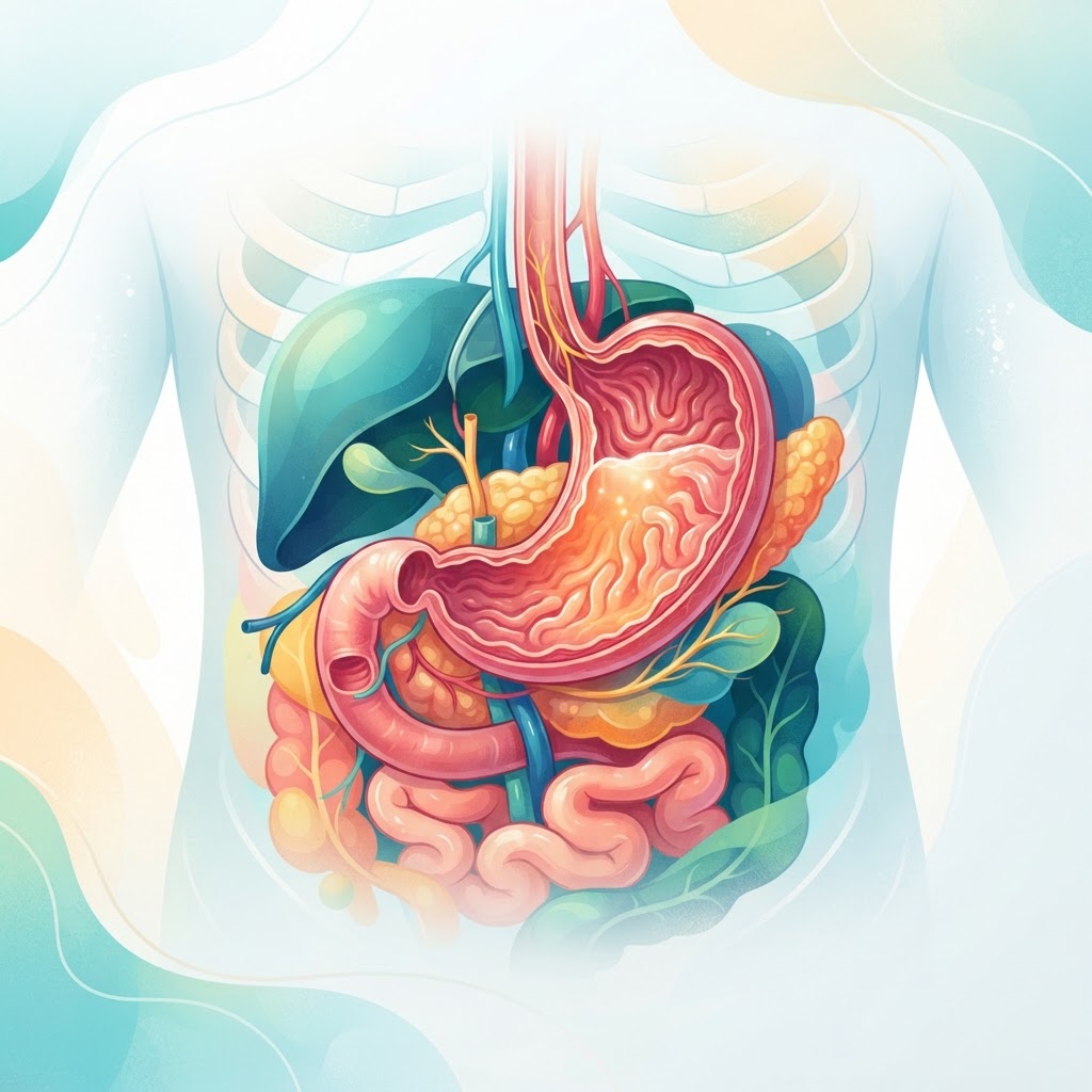
  • Chronic Urticaria: Causes, Triggers, and Natural Relief Tips

    Chronic Urticaria: Causes, Triggers, and Natural Relief Tips

    Key Takeaways

    • Chronic urticaria is defined by hives lasting six weeks or longer, often without a singular, obvious cause.
    • Mast cell activation plays a central role in releasing inflammatory mediators that trigger itchy skin patches.
    • Leaky gut and systemic inflammation are increasingly recognized as primary drivers of persistent skin reactivity.
    • Managing triggers—ranging from diet to stress—is often more effective than relying solely on symptomatic medication.
    • A low-histamine lifestyle and gut-healing protocols are powerful tools for managing flare-ups naturally.

    Living with skin that seems to betray you can be an exhausting, isolating experience. For those navigating the persistent itch and swelling of chronic hives, the journey often feels like a never-ending cycle of trial and error. Whether you are dealing with spontaneous flares or the specific patterns of dermatographic urticaria, the frustration is real. When your skin inflammation persists for weeks or months, it is time to look deeper than surface-level solutions. At the healthauthoritylife Editorial Team, we believe that understanding the physiological roots of these outbreaks is the first step toward reclaiming your comfort. This guide explores the complex mechanisms behind chronic urticaria, uncovering how your internal environment—from your gut microbiome to your immune system—directly influences your skin health.

    What Is Chronic Urticaria and Why Does It Happen?

    Chronic urticaria, often referred to simply as chronic hives, is a medical condition characterized by the recurrent appearance of itchy, raised welts (wheals) or deeper skin swelling (angioedema) that persists for six weeks or longer. Unlike acute hives, which typically appear suddenly as a reaction to a specific allergen like a bee sting or a certain medication, chronic hives often lack a clear, singular cause. This unpredictability makes the condition particularly challenging to manage, as patients often find themselves searching for a “trigger” that may not be an external substance at all, but rather an internal state of immune imbalance.

    To understand why this happens, we must look at the skin as an active immune organ rather than a passive barrier. In individuals with chronic hives, the immune system—specifically the mast cells—is in a state of hyper-vigilance. These cells, which reside in the tissues throughout the body, are responsible for releasing histamine and other chemical mediators in response to perceived threats. In chronic urticaria, these mast cells are essentially “trigger-happy.” They release their inflammatory payload without an actual foreign invader, leading to the vasodilation and fluid leakage that creates the characteristic red, raised, and itchy hives. While some cases are linked to underlying autoimmune conditions, where the body creates antibodies that target its own mast cells, many others remain “idiopathic,” meaning the exact mechanism remains unclear. However, modern functional health perspectives suggest that these flare-ups are rarely random; they are usually the final signal of systemic inflammation attempting to express itself through the largest organ in the body: the skin.

    Another common presentation is dermatographic urticaria, where the skin becomes so sensitive that the mere act of stroking, rubbing, or applying pressure to the skin results in raised, red lines. This is essentially a localized version of mast cell hypersensitivity. The physical stimulus triggers a disproportionate release of inflammatory chemicals right at the site of contact. For those struggling with this, the skin barrier appears fragile and prone to reacting to even the mildest friction, such as tight clothing or even a bedsheet. Understanding that chronic urticaria is an expression of systemic signaling rather than a localized skin “rash” is the pivot point for finding effective relief. By shifting the focus from suppressing the symptoms on the skin to calming the immune system internally, many individuals find that they can significantly reduce the frequency and severity of their outbreaks.

    Common Internal Triggers for Persistent Hives

    When searching for the root cause of chronic hives, it is tempting to scan the room for pollen or check the ingredient label of your latest meal. While external contact allergens are common culprits for acute cases, chronic urticaria is frequently driven by internal stressors that keep the immune system on high alert. If the immune system is already overburdened by internal factors, it becomes much more reactive to minor environmental triggers that would normally be ignored. One of the most prevalent yet overlooked triggers is sub-clinical infection. Persistent, low-grade infections—such as those involving common bacteria in the sinuses, the dental roots, or the digestive tract—can provide a constant stimulus to the immune system. Because the body is constantly “on the hunt” for these pathogens, the mast cells become primed for action, leading to a state of chronic inflammation that manifests on the skin.

    Another major driver is hormonal fluctuation. We often hear of “stress hives,” but the mechanism goes deeper than just feeling overwhelmed. Cortisol, the primary stress hormone, is meant to be anti-inflammatory; however, when the body experiences chronic stress, the cells can become desensitized to cortisol, leading to an unregulated inflammatory response. Furthermore, thyroid dysfunction is a well-documented internal trigger. There is a documented association between autoimmune thyroid conditions and chronic urticaria, suggesting that when the endocrine system is struggling, the skin is one of the first places to reflect that imbalance. Addressing these internal factors requires a comprehensive look at your health history.

    The following table illustrates the various triggers and the primary approach to managing them, helping you categorize where your own sensitivities might lie:

    Trigger Category Example Best For
    Dietary Histamines Aged cheeses, fermented foods Immediate reduction of load
    Chronic Inflammation Undiagnosed dental or sinus issues Long-term systemic resolution
    Physical Pressure Dermatographic urticaria, tight belts Protective barrier maintenance
    Emotional Stress Work pressure, life transitions Nervous system regulation

    Beyond infections and hormones, the accumulation of environmental toxins—such as heavy metals or mold exposure in the home—can act as a persistent irritant. Your liver, which is responsible for processing these toxins, can become overwhelmed. When the liver is congested, the body may attempt to shunt toxins out through the skin. If your skin is already prone to inflammation, this additional load can trigger a hive outbreak. Identifying these internal triggers is not about finding one single “villain,” but rather about peeling back the layers of your unique health puzzle to identify which systems require the most support.

    The Connection Between Mast Cell Activation and Hives

    To truly grasp why hives persist, one must understand the role of mast cells. These are not merely passive cells waiting for an allergen; they are sophisticated sentinels of the innate immune system. Mast cells are found in high concentrations in areas that interface with the outside world—the skin, the respiratory tract, and the gut lining. Under normal conditions, they provide a protective function, releasing histamine and other mediators to help fight off invaders. However, in cases of chronic urticaria, these cells experience what is known as “mast cell activation.” This is a state where the cells become unstable and degranulate (release their internal contents) far too easily, or in response to triggers that should be benign.

    This activation is not just about histamine. While histamine is the most famous culprit—responsible for the itch, the heat, and the swelling—mast cells contain a vast pharmacy of other inflammatory substances, including leukotrienes, prostaglandins, and various cytokines. When a mast cell is triggered, it releases a “cocktail” of these chemicals. This is why standard antihistamines, which only block the histamine receptor, are often only partially effective for many people. If your mast cells are releasing leukotrienes as well, a simple H1-blocker will leave the underlying inflammation largely unaddressed. This is a critical realization for anyone seeking natural relief for hives, as it suggests the solution must involve stabilizing the mast cell membrane itself rather than just mopping up the histamine after it has already been released.

    What causes this instability? Research suggests that the “threshold” for mast cell degranulation can be lowered by a variety of factors. Nutrient deficiencies, particularly in minerals like magnesium or vitamins like B6 and vitamin C, can leave these cells fragile. Conversely, high levels of systemic oxidative stress can oxidize the cellular membranes of mast cells, making them more prone to bursting. Think of your mast cells like a balloon filled with water. If the balloon (the cell membrane) is strong and thick, it takes a significant amount of force (an allergen) to make it pop. But if the balloon is thin and brittle due to systemic inflammation or nutrient depletion, even a gentle poke—like a change in ambient temperature or a stressful thought—can cause it to burst and release its contents. Natural support strategies for chronic hives, therefore, focus on “strengthening the balloon.” This involves optimizing nutritional status, reducing the burden of oxidative stress, and supporting the body’s natural anti-inflammatory pathways to keep the mast cells quiet and stable.

    How Leaky Gut Contributes to Skin Inflammation

    The relationship between your digestive tract and your skin is profound, often referred to by experts as the “gut-skin axis.” If you are dealing with chronic hives, your gut should be one of the first areas to investigate. A healthy gut lining acts as a highly selective barrier, allowing nutrients to pass into the bloodstream while keeping undigested food particles, toxins, and bacteria safely inside the digestive tract. When this barrier is compromised—a condition commonly called “leaky gut” or increased intestinal permeability—large particles can slip through into the bloodstream. This is a red flag for the immune system, which interprets these particles as foreign invaders and mounts an inflammatory defense.

    Because the gut is the largest immune organ in the body, when it is under siege, the entire immune system becomes activated. This systemic inflammation rarely stays contained in the gut. The immune cells, primed and angry from dealing with digestive debris, circulate throughout the body. When they encounter the skin, they can initiate or exacerbate inflammatory skin conditions. Furthermore, many of the mast cells mentioned previously are located right in the lining of the gut. When the gut barrier is damaged, these mast cells are constantly being provoked by gut-derived irritants, keeping them in a perpetual state of readiness to release histamine. This creates a vicious cycle: the gut is inflamed, the mast cells are agitated, and the skin reacts with hives.

    Addressing the gut is not just about changing what you eat; it is about restoring the integrity of the mucosal lining. Chronic inflammation, overuse of certain medications, and a diet high in processed foods can all erode this protective layer. Conversely, identifying and removing food sensitivities is a vital step. When you have a leaky gut, you may develop temporary sensitivities to foods that you previously tolerated well. By rotating these foods out and incorporating gut-healing nutrients—such as collagen, L-glutamine, and bone broth—you can provide the raw materials necessary for the gut to repair itself. As the gut lining heals, the “leaking” stops, the systemic immune burden drops, and for many, the skin begins to quiet down. This is the cornerstone of sustainable, long-term relief for chronic urticaria.

    Dietary Strategies to Reduce Histamine and Skin Flare-ups

    Dietary modification is often the most accessible tool for managing chronic hives. However, it is important to distinguish between an “allergy diet” and a “low-histamine diet.” While you may have specific food allergies, a low-histamine approach is different; it focuses on reducing the total load of histamine in your system, giving your body a chance to clear its backlog. Histamine is found naturally in many healthy foods, particularly those that have been aged, fermented, or stored for long periods. If your mast cells are already unstable, these foods act like adding fuel to a fire. The goal of a low-histamine strategy is not to eliminate histamine forever, but to lower the systemic levels until your skin clears, then gradually reintroduce foods to find your personal threshold.

    High-histamine foods to consider temporarily limiting include aged cheeses, cured meats like salami and pepperoni, fermented vegetables (like sauerkraut and kimchi), kombucha, certain vinegars, and alcoholic beverages, especially red wine and beer. It is also helpful to be mindful of “histamine liberators”—foods that do not necessarily contain high levels of histamine but trigger your own mast cells to release more. Common culprits include citrus fruits, tomatoes, egg whites, and some artificial food dyes. While these are nutritious foods for the general population, they can be significant hurdles for those currently experiencing active hives. Keeping a simple food and symptom journal can be an eye-opening exercise; you might discover that a seemingly “healthy” bowl of fermented vegetables is actually the trigger for your afternoon flare-up.

    Beyond avoiding triggers, focus on the “freshness factor.” Histamine levels increase in foods as they age or sit in the refrigerator. Opting for freshly prepared meals rather than leftovers can significantly lower your histamine intake. Furthermore, focus on increasing your intake of natural mast cell stabilizers and anti-inflammatory foods. Quercetin, a bioflavonoid found in red onions, apples, and kale, is well-regarded for its ability to stabilize mast cells. Including foods rich in vitamin C and omega-3 fatty acids can also help to soothe skin inflammation from the inside out. Remember that this dietary phase is meant to be a therapeutic tool, not a life sentence. As you resolve the underlying issues—healing the gut and balancing the immune system—many people find that their tolerance for these foods naturally returns, allowing them to enjoy a broader, more varied diet once again.

    The Role of Nutrient Deficiencies in Skin Health

    The skin acts as a protective barrier and an internal mirror, often reflecting the nutritional status of the body. When dealing with chronic urticaria, the skin is in a state of heightened surveillance and inflammation. If the body lacks the raw materials necessary for cellular repair and immune regulation, the inflammatory response to triggers can become exaggerated and persistent. Nutrient deficiencies may not be the sole cause of chronic hives, but they frequently act as exacerbating factors that keep the immune system in a state of hyper-responsiveness.

    Vitamin D deficiency is among the most commonly discussed nutritional factors in dermatology. Vitamin D is not merely a vitamin; it functions as a potent hormone that modulates immune function. Many individuals with skin inflammation show lower levels of serum vitamin D, and ensuring optimal levels may help stabilize mast cells. When mast cells are unstable, they release histamine more readily, leading to the itchy, raised welts characteristic of urticaria.

    Similarly, the B-vitamin complex, particularly B12 and folate, plays a critical role in DNA synthesis and the repair of skin cells. A deficiency in these vitamins can hinder the skin’s ability to recover from the damage caused by frequent breakouts. Additionally, trace minerals like zinc and selenium are essential for antioxidant defense. Zinc, in particular, is vital for maintaining the structural integrity of the skin and supporting the body’s natural anti-inflammatory pathways. Without adequate zinc, the skin may be more susceptible to the irritating effects of environmental triggers, including friction-based conditions like dermatographic urticaria.

    Omega-3 fatty acids, often classified as essential nutrients, are fundamental in managing the inflammatory cascade. A high ratio of Omega-6 to Omega-3 fatty acids in the modern diet is thought to promote systemic inflammation. Increasing the intake of high-quality Omega-3s—found in cold-water fish, flaxseeds, and walnuts—can help dampen the chemical signaling that tells mast cells to degranulate. By focusing on nutrient density, individuals can provide their immune system with the resources it needs to switch from a reactive state to a resilient one.

    Lifestyle Habits to Lower Systemic Inflammation

    Managing chronic hives treatment requires a holistic approach that goes beyond topical creams. Lifestyle habits form the foundation of systemic health and can dictate the body’s inflammatory baseline. Reducing systemic inflammation often involves identifying and removing “hidden” sources of irritation in daily life.

    One of the most effective ways to lower inflammation is through circadian rhythm regulation. Sleep is the time during which the body performs intensive repair on the skin barrier. Chronic sleep deprivation elevates cortisol levels, which can further disrupt immune function and worsen mast cell activation. Maintaining a consistent sleep-wake cycle helps regulate the hormonal environment, potentially reducing the frequency of hive outbreaks.

    Hydration also plays a critical role. When the body is dehydrated, the skin’s protective moisture barrier weakens, making it more permeable to environmental allergens. Drinking filtered water throughout the day helps the lymphatic system clear metabolic waste and inflammatory mediators. Furthermore, physical movement, though potentially challenging for those with dermatographic urticaria, is essential. Low-impact activities like walking or restorative yoga can improve circulation and reduce the stress-induced inflammation that often triggers hives, provided the exercise environment is temperature-controlled and free of abrasive clothing.

    Dietary choices remain the most significant lifestyle lever. An anti-inflammatory diet focused on whole, unprocessed foods—such as colorful vegetables, berries, and lean proteins—limits the intake of common histaminergic triggers. While high-histamine foods (like aged cheeses, fermented vegetables, and processed meats) affect individuals differently, a temporary reduction in these foods can help determine if diet is a primary driver of symptoms. Keeping a detailed food and symptom diary can help uncover these hidden triggers, turning dietary management from a guessing game into a structured wellness strategy.

    Supplement Category Mechanism of Action Best for
    Quercetin Natural mast cell stabilizer Reducing acute histamine release
    Vitamin D3 Immune system modulation Long-term skin barrier resilience
    Omega-3 Fatty Acids Reduces pro-inflammatory cytokines Lowering overall body inflammation
    Probiotics Supports gut-immune axis Improving chronic digestive-related hives
    Magnesium Glycinate Nervous system relaxation Reducing stress-triggered outbreaks

    Natural Supplements That Support Immune Balance

    When considering natural relief for hives, supplements are often viewed as a way to provide targeted support to the immune system. The goal here is not to suppress the immune system, but to bring it back into balance. Quercetin, a flavonoid found in onions, apples, and berries, is widely recognized for its ability to act as a natural mast cell stabilizer. By inhibiting the release of histamine from mast cells, quercetin may help reduce the severity of the inflammatory response during a breakout.

    Probiotics also play a surprisingly large role in skin health. There is a strong connection between the gut microbiome and immune system reactivity. Research suggests that a diverse gut microbiome helps train immune cells to distinguish between harmless stimuli and actual threats. In some cases, addressing gut dysbiosis can significantly reduce the frequency of chronic hives. Choosing a probiotic strain that is histamine-neutral—meaning it does not produce histamine during fermentation—is an important consideration for those sensitive to high-histamine environments.

    Vitamin C is another essential supplement for those dealing with chronic skin inflammation. Beyond its role in collagen synthesis, Vitamin C acts as a natural antihistamine in high doses. It helps speed up the breakdown of histamine in the blood, potentially shortening the duration of hives. However, it is vital to source these supplements from high-quality, reputable manufacturers, as purity and bioavailability can vary significantly. Always start with lower doses to monitor for any adverse reactions, as even natural substances can trigger sensitive immune systems.

    Stress Management Techniques to Calm Nervous System Reactivity

    The link between stress and chronic urticaria is well-documented in the wellness community. Chronic stress keeps the body in a state of “fight or flight,” a sympathetic nervous system response that releases adrenaline and cortisol. In a person prone to hives, this state of heightened alert can make the body hypersensitive, lowering the threshold required to trigger a hive breakout. Managing this reactivity is a crucial, if often overlooked, component of chronic hives treatment.

    Breathwork is perhaps the most accessible tool for calming the nervous system. Techniques like “box breathing” or “4-7-8 breathing” stimulate the vagus nerve, which activates the parasympathetic nervous system—the body’s “rest and digest” mode. By consciously slowing the breath, you signal to your brain that the environment is safe, which can directly inhibit the release of stress-related chemicals that aggravate mast cells.

    Meditation and mindfulness practices are also highly effective. These practices do not just reduce stress in the moment; over time, they restructure how the brain responds to stressors. For those suffering from the physical discomfort of hives, body-scanning techniques can help detach the mind from the sensations of itching and burning, preventing the “itch-scratch-itch” cycle from escalating. Additionally, setting boundaries in daily life—such as limiting exposure to high-stress work environments or digital stimuli—can prevent the cumulative load of stress that often leads to a physical outbreak.

    When to Seek Professional Help for Chronic Skin Issues

    While many individuals successfully manage their chronic hives through lifestyle adjustments, there are clear indicators that suggest professional intervention is necessary. If hives persist for more than six weeks, they are clinically classified as chronic. At this stage, it is important to work with an allergist or dermatologist to rule out underlying systemic conditions, such as autoimmune thyroid disorders, chronic infections, or other inflammatory pathologies.

    Seek medical attention immediately if you experience “angioedema,” which involves deep swelling under the skin—often in the lips, eyelids, or throat. If you feel any difficulty breathing, throat tightness, or a sensation of your tongue swelling, this is a medical emergency that requires urgent care. Furthermore, if hives are accompanied by fever, joint pain, or significant fatigue, these may be signs of a more complex systemic issue that cannot be resolved with lifestyle changes alone. A professional can conduct blood work, skin prick tests, or patch testing to determine the precise nature of the irritation and provide a tailored plan that integrates medical treatments with your natural wellness goals.

    Frequently Asked Questions

    What is the difference between acute and chronic urticaria?

    Acute urticaria typically resolves within six weeks and is often triggered by a specific allergy, viral infection, or medication reaction. Chronic urticaria lasts for six weeks or longer and is frequently idiopathic, meaning the specific cause is often difficult to identify and may involve complex immune system dysregulation.

    Can stress really cause hives to appear on the skin?

    Yes, emotional and physical stress triggers the release of neurotransmitters and hormones that can prompt mast cells to degranulate. This “stress-induced urticaria” occurs when the nervous system becomes overactive, leading to skin inflammation even in the absence of a traditional environmental allergen.

    Is dermatographic urticaria a lifelong condition?

    Dermatographic urticaria, where the skin raises into welts after light pressure or scratching, is often persistent but manageable. While it can last for years, many people find that the sensitivity of their skin decreases over time as they reduce systemic inflammation and address underlying triggers.

    What are the most common foods that trigger hives in sensitive individuals?

    Common triggers include high-histamine foods like aged cheeses, processed meats, fermented foods (such as sauerkraut), shellfish, and certain artificial food dyes or preservatives. However, “trigger foods” are highly individual; a food that causes hives for one person may be perfectly safe for another.

    Can natural antihistamines replace prescription medication?

    Natural antihistamines, such as quercetin, Vitamin C, and certain herbal preparations, can support the body’s inflammatory response, but they should not be used to replace essential medication without consulting a doctor. They are best viewed as complementary tools to help manage triggers alongside a professional treatment plan.

    Are there specific laundry products that aggravate chronic hives?

    Yes, many conventional laundry detergents contain fragrances, enzymes, and optical brighteners that are common skin irritants. Switching to fragrance-free, hypoallergenic detergents and avoiding fabric softeners can significantly reduce physical irritation for those with hyper-sensitive skin.

    Conclusion

    Navigating the complexities of chronic urticaria requires patience, observation, and a commitment to understanding your body’s unique language. By addressing nutrient deficiencies, prioritizing systemic calm, and making intentional lifestyle choices, you can create an internal environment that is less reactive and more resilient. While the road to relief may feel daunting, small, consistent changes—from identifying your personal triggers to calming your nervous system—can yield significant improvements in your quality of life. Remember, your skin is your body’s biggest organ; treating it with care, both inside and out, is the ultimate form of wellness.

    If you have found these strategies helpful, consider sharing this guide with others who may be seeking natural ways to manage their skin health. Start your journey today by keeping a simple symptom diary to better understand your specific triggers.

    By healthauthoritylife Editorial Team

    This article is for informational purposes only and is not a substitute for professional medical advice. Always consult your doctor before making changes to your diet, exercise routine, or treatment plan.

  • Low Stomach Acid: Symptoms, Causes, and Natural Support

    Low Stomach Acid: Symptoms, Causes, and Natural Support

    Key Takeaways

    • Hypochlorhydria, or low stomach acid, is often overlooked as a root cause for chronic digestive distress and systemic nutrient deficiencies.
    • Common signs like bloating, acid reflux, and undigested food in stool can be misleading, as they frequently mimic high-acid symptoms.
    • The condition is often triggered by factors such as chronic stress, H. pylori infection, aging, and the frequent use of acid-suppressing medications.
    • Stomach acid is the primary gateway for nutrient absorption, meaning its deficiency can lead to anemia, bone density issues, and fatigue.
    • Restoring gastric health involves a multi-faceted approach, focusing on lifestyle modifications and gentle, natural support to improve stomach acid naturally.

    When you sit down to enjoy a meal, you likely focus on the flavor, the social connection, or perhaps the nutritional value of your food. However, your body is simultaneously initiating one of the most complex chemical reactions in the human system: the production of hydrochloric acid (HCl). This powerful substance is the unsung hero of your digestive tract, acting as a gatekeeper for nutrients and a security system against pathogens. When this mechanism falters, the resulting condition—hypochlorhydria—can create a domino effect of health challenges that extend far beyond your stomach. Many individuals suffer for years, misinterpreting the subtle, and sometimes not-so-subtle, signs of low stomach acid symptoms as unrelated issues like general fatigue, skin conditions, or mysterious bloating. In this comprehensive guide, the healthauthoritylife Editorial Team explores the intricacies of gastric acidity, shedding light on the underlying drivers and practical steps for finding sustainable low hydrochloric acid relief.

    What Is Hypochlorhydria and Why It Matters for Digestion

    Hypochlorhydria is the medical term used to describe a state where the stomach produces an insufficient amount of hydrochloric acid. To understand why this matters, one must first appreciate the stomach’s intended environment. The human stomach is designed to be highly acidic, typically maintaining a pH level between 1.5 and 3.5. This extreme acidity is not a flaw of design but a critical feature necessary for the first stage of protein breakdown. When we ingest food, the stomach must denature proteins—essentially unraveling their complex shapes—so that enzymes like pepsin can begin breaking them down into absorbable amino acids.

    When acid levels are low, this biological assembly line stalls. Digestion becomes an inefficient, laborious process rather than a seamless flow. Many people mistakenly believe that their digestive issues stem from having “too much” acid, leading them to reach for antacids or proton pump inhibitors (PPIs). While these medications can provide temporary comfort for burning sensations, they may inadvertently exacerbate the root problem if the actual cause is hypochlorhydria. Understanding the nuance between true acid excess and the symptoms of low acid is the first step toward reclaiming digestive health.

    Beyond protein digestion, your stomach acid acts as a crucial barrier to the outside world. The environment created by healthy stomach acid is meant to neutralize bacteria, fungi, and parasites that inevitably enter our systems through the food we eat. If your internal pH is too high—meaning it is not acidic enough—these opportunistic pathogens can survive their transit through the stomach and colonize the small intestine. This transition, often referred to as dysbiosis, is a primary driver of many modern gastrointestinal woes. By maintaining the integrity of this “acid bath,” you are not just helping your digestion; you are supporting your entire immune system. Recognizing that low stomach acid is a systemic health concern rather than just a “tummy ache” is essential for long-term vitality. It dictates how well you process energy, how effectively you protect yourself from infection, and how efficiently you utilize the vitamins and minerals you consume every single day.

    Common Symptoms of Low Stomach Acid You Should Not Ignore

    The clinical presentation of hypochlorhydria is notoriously deceptive. Because the body tries to compensate for poor digestion in various ways, the signs of low stomach acid often mirror those of high acidity, leading to widespread misdiagnosis and ineffective self-treatment. One of the most common, yet frequently misunderstood, symptoms is a heavy, sluggish feeling immediately following a meal. If you feel like your food is “sitting like a brick” in your stomach for hours, it is a hallmark indicator that your stomach is failing to break down the bolus of food efficiently. This stagnation leads to fermentation, which produces gas, bloating, and excessive burping.

    Another paradox involves acid reflux or heartburn. While these are typically associated with an excess of acid, they are frequently caused by low stomach acid. In a healthy system, high acidity signals the lower esophageal sphincter (LES) to close tightly after food passes into the stomach. When acid levels are insufficient, this signal may fail to trigger correctly, allowing stomach contents to splash upward into the esophagus. Furthermore, the slow transit time caused by low acid allows food to linger and ferment, creating pressure that pushes the LES open. Understanding that heartburn can be a cry for more acid rather than less is a paradigm shift that can change the trajectory of one’s health.

    Physical manifestations of this deficiency also appear outside the digestive tract. Because digestion is a energy-intensive process, a failing system often results in systemic fatigue. Individuals may notice their hair, skin, and nails becoming brittle or thinning, as the body struggles to break down the proteins and minerals (like zinc and iron) necessary for structural maintenance. You might also notice undigested food particles in your stool; this is a clear sign that the chemical “maceration” required for proper nutrient extraction simply did not happen. Some people also experience a diminished appetite, particularly in the morning, as the stomach remains full from the previous day’s inadequately processed meals. Keeping a food-and-symptom diary can be a highly effective way to identify these patterns, helping you and your healthcare provider distinguish between transient indigestion and the persistent nature of hypochlorhydria.

    Approach Mechanism Best For
    Mindful Eating Calming the nervous system to stimulate gastric juices Stress-induced low acid
    Bitter Herbs Triggering vagus nerve receptors to signal acid production Functional, mild deficiencies
    Betaine HCl Directly supplementing stomach acid during meals Clinically confirmed low acid
    Apple Cider Vinegar Providing supplemental acidity to aid initial breakdown Occasional indigestion relief

    Underlying Causes and Risk Factors for Low Hydrochloric Acid

    Understanding the root hypochlorhydria causes is the key to preventing the condition from recurring. The production of stomach acid is a delicate physiological act, requiring a calm nervous system and a balanced endocrine profile. One of the most prominent contributors in modern society is chronic stress. When we are in a state of “fight or flight,” our sympathetic nervous system takes over, prioritizing blood flow to our muscles and heart while simultaneously downregulating digestive processes. Eating on the go, staring at screens, or rushing through meals keeps the body in this state, effectively suppressing the release of gastrin, the hormone that tells the stomach to produce acid.

    Aging is another significant, albeit natural, factor. As we grow older, the lining of the stomach can undergo changes that lead to reduced gastric secretion. Experts generally observe that the capacity to produce HCl often tapers off after the age of 50. This, combined with a potential decrease in the absorption efficiency of the stomach lining, creates a perfect storm for nutrient deficiencies in older populations. Furthermore, certain infections play a major role. The bacterium Helicobacter pylori is notorious for its ability to colonize the stomach and create an environment that actually lowers acid production to suit its own survival. While H. pylori is a common occurrence, when it overgrows, it can damage the specialized cells responsible for acid secretion.

    Medication use also frequently precipitates low stomach acid. Acid-suppressing drugs, such as H2 blockers and proton pump inhibitors, are among the most commonly prescribed pharmaceuticals. While they are intended to mitigate symptoms, they effectively “turn off” the stomach’s acid production. Long-term use of these drugs often makes it difficult for the stomach to resume its natural rhythm once the medication is stopped, leading to a cycle of dependence. Additionally, certain nutrient deficiencies—specifically zinc, B-vitamins, and iodine—can prevent the stomach from having the raw materials necessary to synthesize hydrochloric acid. It is a biological irony that a deficiency in nutrients can lead to a condition that prevents the absorption of those same nutrients. Addressing these underlying factors, whether they are environmental, dietary, or medication-related, is essential for anyone seeking long-term relief.

    The Crucial Role of Stomach Acid in Nutrient Absorption

    If the stomach is the kitchen of the body, then hydrochloric acid is the primary stove. Without it, the food you ingest remains raw and largely inaccessible to your bloodstream. The most critical role of stomach acid is the breakdown of dietary proteins into amino acids. Proteins are complex, folded structures that require a highly acidic environment to denature. Without this acidity, the body cannot liberate the amino acids needed to build and repair muscles, synthesize hormones, and create neurotransmitters. Over time, this leads to a state of systemic depletion, where you are eating enough calories but starving for high-quality building blocks.

    Beyond protein, stomach acid is the key to unlocking minerals. Many vital minerals, including iron, calcium, magnesium, and zinc, exist in food in forms that require low pH to become soluble and absorbable in the small intestine. For example, the conversion of ferric iron (non-heme) into ferrous iron (the form the body can actually use) is highly dependent on sufficient HCl. This is why many people with hypochlorhydria experience persistent anemia despite taking iron supplements; the iron is present in their diet, but they lack the internal chemistry to access it. The same logic applies to calcium, which is essential for bone density; without enough acidity to ionize calcium, bone health can suffer, leading to increased risk of fractures and osteopenia as the years progress.

    Vitamins are similarly affected. Vitamin B12, for instance, requires a specific, multi-step process involving stomach acid and intrinsic factor. Hydrochloric acid separates B12 from the dietary proteins it is bound to, allowing it to eventually be absorbed in the ileum. When acid levels are chronically low, B12 deficiency becomes common, leading to symptoms like neurological issues, memory lapses, and profound lethargy. Essentially, your stomach acid determines the “bioavailability” of your diet. You could eat a perfectly healthy, organic, whole-food diet, but if your gastric function is impaired, you are essentially experiencing a high-cost version of malnutrition. Recognizing this link is a powerful motivator for focusing on digestive health as the foundation of your overall nutritional status.

    How Low Stomach Acid Affects Your Gut Microbiome

    The gut microbiome is a complex, delicate ecosystem that relies on a specific sequence of environments to thrive, and the stomach acts as the ultimate filter for the entire digestive tract. In a healthy individual, the highly acidic environment of the stomach serves as a sterilizing agent. It effectively reduces the microbial load of the food we consume, ensuring that we aren’t introducing harmful bacteria or fungi into the delicate environment of the small intestine. When stomach acid levels are low, this filter is compromised, and the small intestine—which should have a relatively low concentration of bacteria—becomes vulnerable to colonization by opportunistic microbes.

    This condition is often referred to as Small Intestinal Bacterial Overgrowth (SIBO). When bacteria move into the small intestine where they don’t belong, they start to feast on the nutrients meant for you. They ferment carbohydrates and fibers that were intended for digestion, producing byproducts like hydrogen, methane, or hydrogen sulfide gases. This leads to the classic “low acid” symptoms of bloating, gas, and abdominal distension, often occurring shortly after eating. Because these microbes are effectively stealing your nutrients, they further contribute to the systemic fatigue and malabsorption associated with hypochlorhydria.

    Furthermore, the shift in the microbiome can alter the way you feel on a daily basis. Since a significant portion of the body’s neurotransmitters, such as serotonin, are produced in the gut, a dysbiotic environment can have ripple effects on mood, sleep, and cognitive function. If the microbial population is out of balance, the body may experience increased inflammation, which can affect the integrity of the gut lining, often referred to as “leaky gut.” This further triggers immune system responses, leading to systemic inflammation. By focusing on how to improve stomach acid naturally, you are doing more than just helping your digestion; you are effectively policing the borders of your gut microbiome, ensuring that the right microbes stay in the right places, and helping to foster a healthier, more diverse, and resilient intestinal environment. This holistic view of the digestive system highlights why addressing low acid is a foundational step in any gut-healing journey.

    Testing Methods to Identify Low Stomach Acid Levels

    Determining whether your digestive struggles stem from hypochlorhydria requires a systematic approach. Because low stomach acid symptoms often overlap with those of high stomach acid (GERD or heartburn), guessing is rarely the right strategy. Several methods exist to help you gauge your current gastric environment, ranging from home-based observations to clinical diagnostic procedures.

    The most accessible starting point is the Baking Soda Challenge. While not a clinical diagnosis, many functional practitioners use it as an initial screening tool. By mixing a quarter-teaspoon of baking soda in four ounces of cold water on an empty stomach and timing how long it takes to burp, you can gain a subjective look at your acid production. A healthy stomach typically reacts with the bicarbonate to produce gas within two to three minutes. If you do not burp within five minutes, it may suggest that your stomach acid levels are insufficient to trigger the necessary chemical reaction.

    For those seeking more objective evidence, a Heidelberg Gastric Acid Test is considered the gold standard. This involves swallowing a small, tethered capsule that transmits pH readings from inside your stomach. It provides a real-time assessment of how your body responds to the ingestion of a test meal. Because it is highly precise, it effectively rules out other conditions that mimic low hydrochloric acid relief symptoms, such as hiatal hernias or gastric ulcers.

    Another diagnostic route involves assessing protein digestion markers through comprehensive stool analysis. When stomach acid is low, the gastric-phase breakdown of complex proteins is incomplete. Undigested muscle fibers or connective tissue visible in a stool test can serve as a strong indicator that your gastric secretions are not operating at the necessary acidity level to initiate proper nutrient extraction.

    Natural Dietary Changes to Support Healthy Gastric Secretion

    Your diet acts as the foundation for your digestive health. If you are struggling with low stomach acid symptoms, the way you eat is just as important as what you eat. The goal is to create an environment that encourages the stomach to produce sufficient hydrochloric acid while minimizing factors that suppress it.

    Start by incorporating “bitter” foods into your meals. Bitter compounds—found in arugula, dandelion greens, radicchio, watercress, and kale—naturally stimulate the vagus nerve. When these compounds hit the tongue, they trigger a reflex that signals the stomach to prepare for incoming food by increasing acid secretion. Consuming a small bitter salad or a splash of lemon juice in warm water before your main meal can act as a natural digestive “primer.”

    It is also essential to optimize your mineral intake, specifically zinc. Zinc is a critical component in the production of hydrochloric acid; without it, the gastric lining cannot manufacture the necessary secretions. Focus on zinc-rich foods such as pumpkin seeds, oysters, grass-fed beef, and lentils. Additionally, ensure you are not over-hydrating during meals. Excessive water consumption while eating can dilute existing stomach acid, making it harder for your body to break down dense proteins. Aim to drink liquids between meals rather than during them.

    Consider the timing and consistency of your meals. Intermittent fasting or erratic eating schedules can disrupt the body’s circadian rhythm of acid production. Eating at predictable times helps the body “anticipate” food, allowing it to begin the cephalic phase of digestion—the process where the mere thought, smell, or sight of food triggers the initial release of enzymes and acid before you have taken your first bite.

    Supplements and Enzymes That Aid Digestive Acid Production

    When dietary adjustments are not enough to provide low hydrochloric acid relief, targeted supplementation can fill the gap. These interventions are designed to mimic the body’s natural processes while supporting the stomach’s ability to recover its own function.

    Supplement Function Best For
    Betaine HCl with Pepsin Directly acidifies the stomach and aids protein breakdown. Immediate relief for heavy, bloated, or “stuck” feeling after meals.
    Gentian Root (Bitter Herbs) Stimulates natural gastric juice secretion. Long-term support and appetite stimulation.
    Digestive Enzymes (Broad Spectrum) Breaks down carbohydrates, fats, and proteins. General digestive efficiency and nutrient absorption.
    Zinc Carnosine Supports the mucosal lining of the stomach. Repairing gastric damage and inflammation.

    Betaine Hydrochloride (HCl) is perhaps the most widely recognized supplement for hypochlorhydria management. Taken during a meal containing protein, it provides the acidic environment necessary for pepsin—the enzyme that breaks down protein—to activate. It is crucial to start with a low dose and monitor for any burning sensations, as this indicates you may have taken more than your stomach currently requires. Always pair this with a high-quality digestive enzyme blend to ensure comprehensive breakdown of all macronutrients.

    If you prefer a herbal approach, gentian root or ginger tinctures are excellent alternatives. These are traditionally known as “stomachics”—herbs that strengthen the stomach’s functional capacity. Unlike HCl, which provides the acid itself, bitters encourage your body to produce its own. For those experiencing the signs of low stomach acid along with mild stomach lining irritation, Zinc Carnosine is an exceptional addition, as it helps fortify the gastric wall, allowing it to function more effectively over time.

    Lifestyle Habits to Improve Your Overall Digestive Function

    Digestive health is inextricably linked to the nervous system. The “rest and digest” state, managed by the parasympathetic nervous system, is a physiological requirement for adequate acid production. When you eat in a state of stress, your body activates the “fight or flight” (sympathetic) response, which physically shunts blood flow away from the stomach and suppresses the release of gastric juices.

    One of the most effective lifestyle changes is the practice of mindful eating. Slowing down, chewing your food thoroughly, and removing digital distractions while eating allows your body to remain in a parasympathetic state. Chewing is not just about breaking food into smaller pieces; it is a critical signaling mechanism. The more you chew, the more time your body has to recognize the chemical composition of the food and prepare the appropriate amount of acid and enzymes.

    Physical activity, specifically light walking after a meal, can also aid in digestion. However, avoid intense exercise immediately after eating, as this forces the body to divert energy to the muscles rather than the gut. Additionally, prioritize your sleep hygiene. Poor sleep is a massive stressor on the endocrine system, and research indicates that chronic sleep deprivation can negatively impact the hormone levels that regulate digestive secretion. By regulating your circadian rhythm, you help stabilize the hormones that keep your digestive system operating on a predictable, healthy schedule.

    When to Consult a Professional About Chronic Digestive Issues

    While natural interventions are powerful, there are situations where you must seek the expertise of a gastroenterologist or a functional medicine practitioner. If you have been attempting to improve your digestion for several weeks without success, or if your symptoms are progressive, professional oversight is mandatory.

    Seek medical attention immediately if you experience “red flag” symptoms. These include unexplained weight loss, difficulty swallowing (dysphagia), persistent vomiting, or the presence of blood in your stool (which may appear black and tarry). These are not signs of simple low stomach acid; they require diagnostic imaging and blood work to rule out more serious pathology such as H. pylori infections, gastric ulcers, or structural issues within the GI tract.

    Furthermore, if you are currently taking prescription medication—specifically Proton Pump Inhibitors (PPIs) or H2 blockers—do not discontinue or modify your regimen without your doctor’s explicit guidance. These medications are often prescribed for long-term conditions, and stopping them suddenly can lead to a “rebound effect,” where the stomach overproduces acid to compensate, causing severe discomfort. A healthcare professional can help you develop a safe, tapered plan to transition off these medications if appropriate.

    Frequently Asked Questions

    Can low stomach acid cause acid reflux?

    Paradoxically, yes. Low stomach acid can lead to poor protein digestion, which causes food to sit in the stomach longer than it should. This fermentation produces gas and pressure that pushes stomach contents upward into the esophagus. Because the stomach is not acidic enough to trigger the closure of the lower esophageal sphincter, this backflow occurs easily, resulting in symptoms often mistaken for high acid.

    Is it safe to take Betaine HCl if I have an ulcer?

    No, it is generally not recommended to take Betaine HCl if you have an active ulcer, gastritis, or if you are taking anti-inflammatory medications like ibuprofen or aspirin. Acid supplements can irritate open sores in the stomach lining. Always verify the health of your gastric lining with a professional before introducing acidic supplements to your protocol.

    How long does it take to fix low stomach acid naturally?

    The timeline varies significantly depending on the individual, the underlying cause, and adherence to lifestyle changes. Some people notice improvements in bloating and energy levels within a few days of dietary modifications and targeted support. However, healing the gastric mucosa and restoring consistent, natural acid secretion often takes several months of consistent habit changes.

    What are the most common signs of low stomach acid?

    Common signs include chronic bloating shortly after eating, feeling like food is “stuck” in the middle of your chest, undigested food particles in the stool, brittle fingernails, iron deficiency anemia, and hair loss. Many people also report a persistent feeling of being “heavy” or overly full even after eating small, light meals.

    Should I avoid coffee if I have low stomach acid?

    Coffee is highly acidic, but it is also a stimulant. While it may temporarily increase gastric sensations, it can also disrupt the parasympathetic nervous system if consumed in excess or on an empty stomach. If you struggle with hypochlorhydria, try drinking your coffee after a protein-rich meal rather than before, or switch to lower-acid options like cold brew to see if your digestive comfort improves.

    Can H. pylori cause low stomach acid?

    Yes, an H. pylori bacterial infection is a common cause of hypochlorhydria. The bacteria thrive in the stomach and can damage the cells responsible for producing hydrochloric acid. If you suspect an infection, a urea breath test or a stool antigen test conducted by your doctor is necessary, as this typically requires targeted treatment rather than just digestive support supplements.

    Conclusion

    Managing low stomach acid is a journey of understanding how your body interacts with the fuel you provide it. By recognizing the subtle signs of hypochlorhydria—such as persistent bloating, nutrient malabsorption, and the sensation of delayed gastric emptying—you can take proactive steps to restore your digestive rhythm. Whether through the mindful integration of bitter foods, the strategic use of digestive enzymes, or lifestyle habits that favor your parasympathetic nervous system, healing is entirely possible. Your digestive tract is remarkably resilient, and with the right approach, you can eliminate the discomfort of poor digestion and optimize your body’s ability to nourish itself. If you are struggling with persistent issues, do not hesitate to reach out to a professional to ensure your approach is tailored to your specific clinical needs. Start small, be consistent, and listen to your body’s signals as you transition toward better health.

    By healthauthoritylife Editorial Team

    This article is for informational purposes only and is not a substitute for professional medical advice. Always consult your doctor before making changes to your diet, exercise routine, or treatment plan.

  • Chronic Peripheral Edema: Causes, Symptoms, and Natural Relief

    Chronic Peripheral Edema: Causes, Symptoms, and Natural Relief

    Key Takeaways

    • Chronic peripheral edema is the persistent accumulation of fluid in the tissues of the lower limbs, often signaling underlying systemic health issues.
    • Identifying specific leg swelling causes is essential for effective management, ranging from simple lifestyle factors to complex cardiovascular or venous conditions.
    • Lymphatic system efficiency plays a critical role in fluid regulation; supporting this system can be key to long-term relief.
    • Natural remedies for edema, such as dietary adjustments and movement, can complement medical treatment but should not replace professional diagnostics.
    • Persistent swelling that does not resolve with elevation or lifestyle changes requires a comprehensive medical evaluation to rule out serious concerns like heart or kidney disease.

    Waking up to heavy, tight legs or noticing that your socks leave deep, painful indentations around your ankles at the end of the day can be both physically uncomfortable and emotionally taxing. While many people dismiss these signs as simple fatigue or the inevitable result of a long day spent on their feet, persistent swelling in the lower extremities—medically known as chronic peripheral edema—is often the body’s way of signaling that something in its internal fluid balance is askew. Whether you are dealing with subtle puffiness or significant, ongoing discomfort, understanding the mechanics of why fluid pools in your extremities is the first step toward reclaiming your comfort and mobility. This guide explores the multifaceted nature of chronic peripheral edema, moving beyond simple symptoms to look at the structural and systemic factors at play, offering evidence-based insights on how to manage and potentially alleviate this common, yet often misunderstood, health concern.

    What Is Chronic Peripheral Edema?

    At its core, chronic peripheral edema refers to the abnormal accumulation of fluid in the interstitial spaces—the tiny gaps between your body’s cells—specifically within the lower limbs. While healthy bodies are adept at maintaining a delicate equilibrium of fluid distribution, edema occurs when the systems responsible for fluid return, pressure regulation, or capillary filtration become overwhelmed or impaired. Unlike acute swelling, which might occur following an injury or a temporary bout of illness, chronic peripheral edema is characterized by its persistent nature. It is not a disease in itself but rather a clinical sign that the body is struggling to manage its fluid homeostasis.

    To understand why this happens, it is helpful to visualize the circulatory system as a complex network of highways. Arteries pump blood away from the heart, delivering oxygen and nutrients, while veins are tasked with returning that fluid—along with waste products—back to the heart and kidneys to be processed. Under normal conditions, small amounts of fluid leak from the capillaries into the tissues. This fluid is then typically reabsorbed by the veins or collected by the lymphatic system. Chronic peripheral edema develops when this “drainage” system fails to keep pace with the fluid leakage. This imbalance can occur due to increased pressure within the veins, a failure of the one-way valves that prevent blood from backflowing, or an obstruction within the lymphatic channels. When fluid stagnates, it creates the characteristic “pitting” seen in many cases, where pressing a finger into the skin leaves a temporary depression. Over time, this fluid buildup can lead to skin changes, such as thickening, discoloration, or a loss of suppleness, making it vital to address the root cause early on.

    The prevalence of this condition varies significantly across different demographics, but it is frequently seen in individuals who spend long hours standing or sitting, as gravity naturally encourages fluid to pool in the lower limbs. However, age, genetics, and comorbid health conditions often exacerbate these tendencies. The term “chronic” is key here; it implies that the swelling has been present for weeks or even months, requiring more than just a fleeting change in habits to resolve. By viewing edema as a symptom rather than a standalone condition, we can better appreciate the importance of investigating why the fluid is not circulating as it should. This investigation involves looking at the integrity of the vascular walls, the health of the heart, the function of the kidneys, and the efficiency of the lymphatic drainage system, all of which act as the body’s internal maintenance crew. Understanding this mechanism is the bedrock for finding effective fluid retention relief, ensuring that your interventions target the specific physiological bottleneck causing the distress.

    Common Symptoms of Lower Limb Swelling

    Recognizing the nuances of lower limb swelling is essential for differentiating between transient, gravity-induced puffiness and more significant chronic edema. While the primary indicator is visible distension of the skin, the physical manifestation of chronic peripheral edema often includes a cluster of secondary symptoms that can significantly impact daily quality of life. The most classic sign is pitting edema. If you press firmly on the swollen area—such as the shin or ankle—and notice that the indentation remains for several seconds after you release pressure, this is a hallmark clinical sign that fluid has pooled in the tissues. This is different from non-pitting edema, which is often associated with more complex lymphatic blockages or skin changes that make the tissue feel firm or woody to the touch.

    Beyond the simple change in appearance, individuals often report a sensation of “heaviness” or fatigue in the legs, particularly in the late afternoon or evening. This heaviness can make walking, climbing stairs, or even simple tasks like putting on footwear feel difficult. The skin itself may become taut, shiny, or stretched, which can cause significant discomfort or a feeling of tight pressure. In some cases, the area may feel warm or look slightly reddened, though it is important to distinguish this from the localized heat associated with infections like cellulitis, which often appears with pain, fever, or spreading redness. Chronic swelling can also lead to changes in the skin texture; it may become dry, flaky, or develop a brownish, rust-colored pigment, often referred to as hemosiderin staining, which occurs when blood cells leak into the tissues and break down.

    It is also common to experience a reduced range of motion in the ankles and feet. Because the skin around the joint is stiff with fluid, simple movements like flexing the foot upward (dorsiflexion) can feel restricted. Many people also report a dull, aching sensation that intensifies the longer they remain in one position. These symptoms are rarely uniform; they may shift from one leg to the other or vary in intensity based on the time of day, diet, or level of physical activity. Tracking these variations is an incredibly useful tool for anyone attempting to reduce water retention naturally, as it allows for the identification of triggers. For instance, noticing that symptoms are worse after consuming high-sodium meals or during particularly humid weather can provide actionable data for your health journey. Keeping a simple log of these symptoms—noting when they start, where they are localized, and what activities seem to aggravate or alleviate them—is one of the most proactive steps you can take before speaking with a professional, as it provides a clear picture of the chronicity and behavior of the swelling.

    Approach Mechanism Best For
    Compression Garments External pressure gradient Daily management of venous-related swelling
    Elevation Gravity-assisted drainage Quick, immediate relief after prolonged standing
    Lymphatic Massage Manual stimulation of lymph flow Reducing tissue congestion and fluid stasis
    Sodium Reduction Osmotic balance optimization Preventing systemic water retention long-term

    Underlying Causes: Why Your Body Retains Fluid

    When searching for the root causes of leg swelling, it is helpful to categorize them into mechanical, systemic, and lifestyle-driven factors. Often, chronic peripheral edema is not caused by a single issue but by a combination of these elements working in tandem. The most common structural culprit is chronic venous insufficiency (CVI). In a healthy leg, the veins have tiny, one-way valves that help push blood back up toward the heart against gravity. When these valves weaken or become damaged—often due to age, prolonged sitting, or hereditary factors—the blood pools in the lower veins. This increased venous pressure forces fluid out of the vessels and into the surrounding tissues, leading to the familiar swelling we recognize as edema.

    Systemic factors, however, can also create a landscape where edema thrives. The heart, for instance, acts as the primary pump for the entire circulatory system. If the heart is not pumping as efficiently as it should—a common issue in congestive heart failure—blood flow slows down, leading to a “backup” in the veins, which manifests as swelling in the legs and feet. Similarly, the kidneys play a critical role in regulating fluid levels by balancing sodium and water. If kidney function is compromised, the body may struggle to excrete excess fluid and sodium, causing both to accumulate in the body’s tissues. Thyroid issues can also be a hidden culprit, as an underactive thyroid can slow down the body’s metabolic processes and lead to generalized or localized water retention.

    Lifestyle-driven leg swelling causes often revolve around our daily habits, specifically those that challenge the body’s natural fluid circulation. A diet excessively high in sodium is perhaps the most notorious; because salt attracts water, an abundance of it in the bloodstream forces the body to retain extra fluid to keep the concentration of sodium in check. Similarly, excessive alcohol consumption can contribute to dehydration and subsequent fluid retention, as the body struggles to maintain balance. Physical inactivity is another major contributor; the muscles in our calves act as a “second heart,” helping to pump blood back up the legs whenever we walk or move. When we sit for hours at a time, we effectively deactivate this calf-muscle pump, allowing fluid to pool in the lower extremities without resistance. Furthermore, certain medications, particularly those used to manage blood pressure, inflammation, or hormonal balance, have well-documented side effects that include edema. If you suspect your medication may be the cause, it is vital to consult with your provider before making any changes. By understanding these diverse causes, we move away from blaming our bodies and toward identifying the specific mechanisms that we can influence through lifestyle adjustments, medical consultation, or targeted supportive therapies.

    The Link Between Lymphatic Health and Edema

    The lymphatic system is often the unsung hero of fluid regulation. While the circulatory system gets most of the attention for moving blood, the lymphatic system acts as the body’s essential drainage and filtration network. It is a one-way street of vessels that picks up excess fluid, proteins, bacteria, and waste from the tissues and returns them to the bloodstream. When this system functions optimally, you rarely notice it, but when it becomes overwhelmed or sluggish, it is a primary driver of persistent swelling. Understanding the role of the lymphatic system is crucial because it requires a different approach to relief than pure venous or cardiovascular-related swelling.

    The lymphatic system does not have a central pump like the heart. Instead, it relies on movement, muscle contractions, and deep breathing to circulate its fluid, known as lymph. When we are sedentary, this fluid can stagnate, leading to a condition that feels heavy and often resistant to standard measures like mere elevation. Chronic peripheral edema frequently involves a degree of lymphatic compromise, particularly if the swelling is long-term. This is why techniques like manual lymphatic drainage for legs are so highly recommended by specialists. These specific, light, rhythmic strokes are designed to stimulate the superficial lymphatic vessels, encouraging them to pick up the excess fluid that has collected in the skin and subcutaneous layers and guide it toward the lymph nodes for processing.

    Supporting your lymphatic health goes beyond massage, however. Because the lymphatic system is so heavily reliant on your body’s internal movement, regular low-impact exercise—such as walking, swimming, or rebounding (using a small trampoline)—acts as a natural catalyst for lymph flow. Furthermore, hydration is paradoxical but essential; while it may seem counterintuitive to drink more water when you are “retaining” fluid, your body needs adequate water to keep the lymphatic fluid at the right consistency. If you are dehydrated, the lymph becomes thicker and harder for your body to move through the narrow vessels. Additionally, the lymphatic system is a key part of your immune system. Chronic inflammation, often caused by poor diet or lack of sleep, can “clog” these pathways, making them less efficient at clearing out the interstitial debris that contributes to edema. By prioritizing anti-inflammatory habits and consistent, gentle movement, you create a more hospitable environment for the lymphatic system to do its job, which is a cornerstone for anyone looking to reduce water retention naturally. Integrating lymphatic support into your daily routine is not just about reducing swelling; it is about keeping your body’s detoxification pathways clear and fully functional.

    When to See a Doctor for Persistent Swelling

    While many cases of lower limb swelling can be managed with lifestyle adjustments, there are certain “red flag” symptoms that mandate a professional medical evaluation. It is important to approach your health with a balance of caution and empowerment. If your swelling is new, sudden, or occurs in only one leg without a clear cause, it should be investigated promptly. Sudden, asymmetrical swelling can sometimes be a sign of a deep vein thrombosis (DVT), a blood clot that requires immediate medical attention to prevent more serious complications. Similarly, if your swelling is accompanied by chest pain, shortness of breath, dizziness, or a rapid, irregular heartbeat, do not wait; seek emergency care, as these can be signs of heart-related distress that requires clinical stabilization.

    Even if the swelling is not sudden, persistent edema that fails to improve with home care warrants a diagnostic workup. A health professional will typically start by taking a detailed history to rule out medication-induced swelling, as many common prescriptions—including some calcium channel blockers or non-steroidal anti-inflammatory drugs (NSAIDs)—can cause the body to hoard fluid. They may perform physical exams to assess the “pitting” of the tissue, check for signs of venous insufficiency, or look for systemic indicators of organ strain. Blood tests, such as checking your kidney and liver function or reviewing your electrolyte balance, can help clarify whether the edema is a local tissue issue or a manifestation of a systemic, internal condition.

    Do not feel that you are being “alarmist” by seeking answers. In fact, early diagnosis is often the most effective way to prevent the skin changes and tissue hardening that can make chronic peripheral edema more difficult to treat in the long run. If you have been living with swelling for months and have exhausted basic remedies, a doctor can help you develop a personalized plan that might include prescription diuretics, diagnostic imaging like a venous ultrasound, or a referral to a lymphedema specialist. Your goal is to move beyond temporary relief and into a state of sustainable health. When you speak to a professional, bring a list of your symptoms, a record of when the swelling is at its worst, and a full list of any supplements or medications you are currently taking. This level of preparation allows for a more efficient consultation and ensures that your care team has all the necessary information to distinguish between manageable fluid retention and underlying health concerns that require targeted intervention.

    Natural Anti-Inflammatory Foods for Fluid Reduction

    When addressing chronic peripheral edema, your dietary choices serve as the foundation for reducing systemic inflammation and encouraging healthy fluid movement. Inflammation acts as a barrier to efficient lymphatic drainage; when tissues are inflamed, they tend to hold onto excess interstitial fluid, exacerbating swollen ankles and heavy legs. By focusing on whole, anti-inflammatory foods, you can support your body’s natural ability to regulate internal fluid pressure.

    Potassium-rich foods are essential in this process. Sodium and potassium work in a delicate balance within your cells to manage water levels. If your diet is high in processed sodium and low in potassium, your body may struggle to flush out excess fluids. Incorporating potassium-dense foods like dark leafy greens, avocados, bananas, and sweet potatoes helps counteract the water-retention effects of salt. Magnesium also plays a pivotal role; it acts as a mild vasodilator and supports healthy blood vessel function, ensuring that circulation remains smooth and unobstructed.

    Omega-3 fatty acids are another cornerstone of an edema-reducing diet. Found abundantly in walnuts, chia seeds, flaxseeds, and fatty fish, these essential fats are known to modulate the inflammatory response in the body. By reducing the “stickiness” of the blood and improving vascular health, omega-3s can significantly reduce the pressure exerted on vessel walls, which often leads to fluid leakage into the surrounding tissues.

    Finally, we must consider the role of natural diuretics. While pharmaceutical diuretics must be managed by a physician, certain plant-based foods can gently assist the kidneys in filtering waste and fluid. Asparagus, celery, cucumber, and parsley contain natural compounds that increase urine output slightly without depleting essential electrolytes as aggressively as synthetic medications. Including these in your daily meals can provide subtle, consistent fluid retention relief.

    Daily Habits to Improve Circulation and Reduce Edema

    Chronic peripheral edema often thrives in an environment of stagnation. Whether your daily routine involves long periods of sitting at a desk or standing behind a counter, gravity continuously pulls fluid toward the lower extremities. To combat this, you must integrate daily habits that prioritize blood flow and lymphatic return.

    The most effective habit for managing leg swelling causes is the practice of elevation. By elevating your legs above the level of your heart for fifteen to twenty minutes daily, you allow gravity to assist the venous system in returning pooled blood to the core. This is not merely about comfort; it is a mechanical necessity for someone struggling with chronic issues. If you spend your day at a desk, consider using a footrest or a small stool to keep your feet elevated slightly, and take frequent “circulation breaks” where you stand and engage your calf muscles.

    Compression therapy is another vital habit. Medical-grade compression stockings work by applying graduated pressure—tightest at the ankle and gradually decreasing as they move up the calf. This external pressure prevents fluid from accumulating in the tissues of the lower leg. It is important to note that these should be fitted properly; wearing stockings that are too tight at the top can actually create a tourniquet effect, worsening the very issue you are trying to solve.

    Temperature control also impacts vessel dilation. While a hot bath can be relaxing, extreme heat can cause blood vessels to widen, potentially leading to increased fluid leakage in those with compromised venous valves. Ending your shower with a cool rinse on your legs—from your feet upward—can help constrict vessels and tone the skin, promoting a “pumping” effect that encourages fluid movement back toward the torso.

    Effective Movement and Stretching for Lymphatic Flow

    Unlike the circulatory system, which has the heart as a central pump, the lymphatic system relies entirely on muscle contraction and deep breathing to move fluid through the body. When you lead a sedentary life, your lymphatic flow slows down, leading to congestion in the extremities. Implementing targeted movement is essential for natural edema management.

    The “calf pump” is perhaps the most critical exercise for those suffering from chronic peripheral edema. The calf muscles are often referred to as the “second heart.” Every time you flex your foot or walk, these muscles contract, squeezing the deep veins and forcing blood and lymph upward. Walking is an excellent low-impact activity, but you can enhance its effectiveness by focusing on a full heel-to-toe gait, which maximizes calf engagement.

    Lymphatic drainage for legs can also be stimulated through dry brushing or gentle, rhythmic movement. Using a soft-bristled brush on dry skin before a shower, brushing in long, sweeping motions toward the heart, helps stimulate the lymphatic vessels located just beneath the skin. This simple daily ritual can assist in mobilizing stagnant fluid.

    Consider the following comparison of physical activities that specifically benefit the lymphatic and venous systems:

    Activity Impact on Lymphatic Flow Best For
    Rebounding (Mini-trampoline) High Gentle joint impact and whole-body lymph stimulation
    Swimming Moderate-High Hydrostatic pressure naturally compresses the legs
    Yoga (Inversions) Moderate Using gravity to aid venous return
    Brisk Walking Moderate Daily maintenance of calf muscle pump
    Leg Elevation Low (Passive) Immediate relief after long periods of activity

    In addition to active exercise, deep diaphragmatic breathing is an overlooked tool. When you breathe deeply, the movement of the diaphragm creates a pressure change in the thoracic cavity that “sucks” lymph fluid into the main lymphatic ducts. Incorporating five minutes of belly breathing each morning can significantly improve your internal fluid dynamics.

    Herbal Supports for Fluid Balance

    Nature provides a variety of botanicals that have been used for centuries to support vascular integrity and fluid balance. When considering herbal support, the goal is not to force the body into a state of dehydration, but to tone the vascular walls and improve the efficiency of the kidneys.

    Horse chestnut (Aesculus hippocastanum) is widely recognized in European traditional practices for its potential to support venous health. It contains a compound called aescin, which is believed to help strengthen capillary walls and reduce their permeability. By making these vessels less prone to leaking, horse chestnut may help address the root cause of certain types of leg swelling. It is often found in both oral supplement form and topical creams.

    Dandelion leaf is another traditional ally. Unlike many synthetic diuretics that strip the body of potassium, dandelion leaves are naturally high in this essential mineral. This creates a balanced effect, where the herb helps the body eliminate excess water while simultaneously replenishing the electrolyte that prevents further imbalance.

    Butcher’s broom (Ruscus aculeatus) is frequently used for its potential to improve circulation. It contains ruscogenins, which may help constrict blood vessels and prevent the pooling of blood in the lower limbs. While research is ongoing regarding the specific dosages and long-term effects, many individuals report that incorporating these herbs into their regimen, under the guidance of a professional, provides noticeable support for fluid retention relief.

    Managing Salt Intake and Hydration for Edema Relief

    One of the most persistent myths regarding fluid retention is that the answer is to stop drinking water. In reality, dehydration can cause the body to enter a “survival” state, where it holds onto every drop of moisture it can find, leading to increased fluid retention. Proper, consistent hydration is the most effective way to signal to your body that it is safe to release stored fluids.

    Managing salt intake, however, is a different matter. Modern diets are often inundated with sodium, which acts like a sponge in the extracellular space. When sodium levels in the blood are too high, the body pulls water from the cells to dilute the salt, increasing the volume of fluid outside of your cells—exactly where you see the swelling in your ankles and feet. Reducing salt intake does not mean your food must be bland; it means shifting away from processed, pre-packaged, and restaurant-prepared foods, which are the primary culprits for excess dietary sodium.

    Try the following strategies to balance your hydration and sodium intake:

    1. Track your hidden salt: Look at labels for bread, soups, and salad dressings. You may find that your “healthy” lunch is contributing significantly to your edema.
    2. Prioritize mineral water: Opt for high-quality spring water that contains naturally occurring minerals, which are more easily utilized by the body than distilled water.
    3. Infuse your water: If you find plain water difficult to drink, infuse it with lemon, cucumber, or mint. These additions provide a mild, natural diuretic boost and make staying hydrated more enjoyable.
    4. Use salt alternatives: Utilize fresh herbs, spices, citrus zest, and vinegars to flavor your cooking. These ingredients provide vibrant flavor without the fluid-retaining impact of sodium.

    Frequently Asked Questions

    Is chronic peripheral edema a sign of heart failure?

    While chronic peripheral edema can be a symptom of heart failure, it is not the only cause. Swelling can also result from venous insufficiency, kidney issues, medication side effects, or lymphedema. Because heart-related swelling often involves other symptoms like shortness of breath or fatigue, it is essential to have any persistent swelling evaluated by a physician to rule out serious underlying conditions.

    Can drinking too much water make my swelling worse?

    Generally, drinking enough water is necessary to help your kidneys flush out excess sodium. However, in cases of severe kidney or heart disease, a doctor may actually recommend fluid restriction. For the average person, reducing water intake rarely helps with edema and often makes the body hoard fluids, so it is important to drink an adequate amount based on your health profile.

    How can I tell if my swelling is related to my veins or my lymph system?

    Venous edema typically occurs at the end of the day, affects both legs, and is often accompanied by skin discoloration or a feeling of heaviness. Lymphedema, which involves the lymphatic system, may feel firmer to the touch, often affects the top of the foot or toes, and may not pit (dent) as easily as venous swelling. Both conditions require a professional diagnosis to determine the best treatment path.

    Are compression stockings safe for everyone?

    Compression stockings are generally safe, but they should be avoided or used with extreme caution by individuals with severe peripheral artery disease or certain skin infections. The pressure could potentially restrict blood flow if the underlying arteries are damaged. Always consult with a healthcare professional to ensure compression is appropriate for your specific vascular health.

    How long does it take to see results from natural remedies?

    Natural remedies such as dietary adjustments, herbal support, and daily movement typically require consistency over several weeks. Unlike medication that might provide immediate symptomatic relief, natural approaches work by improving the body’s underlying efficiency. Most people begin to notice a difference in skin texture and reduced morning swelling within two to four weeks of steady implementation.

    Is it normal for my swelling to be worse in the summer?

    Yes, heat often exacerbates edema. Warm temperatures cause blood vessels to dilate (widen) to help the body cool down. In someone with compromised circulation, this dilation allows more fluid to seep into the surrounding tissues. It is very common to experience more pronounced swelling during hot weather, making it especially important to stay hydrated and keep the legs cool during the summer months.

    Conclusion

    Managing chronic peripheral edema is a journey toward better understanding the intricate systems of your body. By acknowledging that your legs are not just “swollen,” but rather signaling a need for better fluid dynamics, you can take proactive steps to restore balance. Through the combination of an anti-inflammatory diet, consistent movement, mindful hydration, and the gentle support of natural botanicals, you can significantly improve your comfort and vascular health.

    Do not wait for your symptoms to escalate. Start today by incorporating one simple habit—such as elevating your legs for fifteen minutes or swapping one processed meal for a whole-food, potassium-rich alternative. Small, consistent changes create the momentum needed for long-term health. If your swelling persists, continues to worsen, or is accompanied by pain, redness, or shortness of breath, consult with a healthcare provider immediately to ensure you have a comprehensive plan tailored to your needs.

    By healthauthoritylife Editorial Team

    This article is for informational purposes only and is not a substitute for professional medical advice. Always consult your doctor before making changes to your diet, exercise routine, or treatment plan.
  • Peripheral Arterial Disease: Symptoms, Causes, and Natural Relief

    Peripheral Arterial Disease: Symptoms, Causes, and Natural Relief

    Key Takeaways

    • Peripheral Arterial Disease is a narrowing of the arteries that reduces blood flow to the limbs, often signaling systemic vascular health issues.
    • The most common indicator is claudication, or muscle pain during activity that subsides with rest.
    • Primary risk factors include smoking, diabetes, hypertension, and sedentary lifestyle choices.
    • Chronic inflammation plays a critical role in the progression of plaque buildup, known as atherosclerosis.
    • Managing PAD requires a multi-faceted approach combining medical guidance with proactive lifestyle modifications to improve blood flow naturally.

    For many, the first sign of a vascular concern is not a dramatic event, but a subtle, recurring ache in the legs during a routine walk. This sensation, often dismissed as a mere side effect of aging or fatigue, is frequently an early warning sign of Peripheral Arterial Disease (PAD). As a condition that affects millions globally, PAD is far more than a localized circulation issue; it serves as a vital indicator of your overall cardiovascular health. Understanding the mechanisms behind this disease—and learning how to support your body through natural relief strategies—is the first step toward restoring mobility and protecting your long-term vitality. In this comprehensive guide, the healthauthoritylife Editorial Team explores the intricacies of PAD, providing the insights you need to take control of your vascular journey.

    What is Peripheral Arterial Disease?

    Peripheral Arterial Disease, or PAD, is a chronic circulatory condition in which narrowed blood vessels reduce blood flow to the limbs. It is a subset of atherosclerosis, a systemic process where fatty deposits, known as plaque, accumulate along the inner walls of the arteries. While we often associate heart health with the coronary arteries, PAD focuses on the peripheral system—the highways of blood vessels that supply the arms, but most commonly the legs and feet, with oxygenated blood.

    When these peripheral arteries become constricted or blocked, the muscles and tissues in the extremities do not receive the oxygen-rich blood necessary to function efficiently, especially during physical movement. Think of it like a kink in a garden hose; when the flow is restricted, the output at the end of the line suffers. In the human body, this restriction creates a “demand-supply mismatch.” Your muscles demand more fuel (oxygen) when you walk, climb stairs, or exercise, but the narrowed arteries cannot supply it quickly enough, leading to discomfort.

    It is important to understand that PAD is rarely an isolated event. It is frequently considered a “canary in the coal mine” for broader cardiovascular disease. Individuals diagnosed with PAD are statistically more likely to have underlying issues in the arteries supplying the heart and the brain. Therefore, addressing PAD is not just about relieving leg pain; it is a vital component of protecting yourself against systemic threats like heart attack and stroke. The condition often progresses slowly, starting with minor structural changes in the arterial lining that, over time, limit the vessel’s elasticity and width.

    From a biological standpoint, the inner lining of the artery, called the endothelium, becomes damaged. This damage triggers an immune response where cholesterol and other substances begin to cluster at the site of the injury. Over many years, these clusters harden into plaque, stiffening the artery walls—a process often referred to as “hardening of the arteries.” As the plaque grows, the channel through which blood flows, known as the lumen, narrows significantly. When the narrowing reaches a critical threshold, even minor exertion triggers the classic symptoms of restricted circulation. By recognizing what PAD is early on, individuals can pivot from a path of progression to a path of maintenance and, in many cases, significant improvement through lifestyle adjustments and targeted support.

    Common Symptoms and Warning Signs of PAD

    The hallmark symptom of PAD is claudication. This term refers to muscle pain, cramping, or a feeling of heaviness that occurs in the legs during physical exertion and typically resolves after a few minutes of rest. Many people describe it as an ache in the calf, thigh, or buttock that forces them to stop mid-stride. It is a predictable pattern: you walk a certain distance, the pain starts, you pause, and the pain dissipates as the oxygen demand of the muscles drops back to baseline levels.

    However, PAD symptoms are not universal. While some experience intense claudication, others may report “atypical” symptoms. These can include persistent leg fatigue, a sense of weakness that makes walking difficult, or a feeling that the legs are “giving out.” Furthermore, because the restriction of blood flow can affect the extremities in various ways, symptoms often present in the feet and toes rather than the legs. Patients may notice coldness in one lower leg or foot compared to the other, or a persistent tingling, pins-and-needles sensation, often described as paresthesia.

    As the condition progresses, skin changes become more apparent. The skin on the lower legs may become thin, shiny, and brittle. You might notice a loss of hair on the feet or lower legs, which is a common sign that the local tissue is not receiving sufficient nutrient-rich blood to sustain hair follicles. In more advanced stages of PAD, you might experience sores or ulcers on the toes or heels that refuse to heal, even with standard wound care. Because circulation is sluggish, the body’s natural ability to repair itself is severely compromised, making these non-healing wounds a serious warning sign that requires professional attention.

    It is also essential to watch for changes in skin color. If your legs turn pale when elevated but deep red or purplish when hanging down—a phenomenon known as dependent rubor—it is a strong indication of compromised vascular health. Some individuals also experience “rest pain,” where the pain persists even when lying down at night. This is a sign that the arterial narrowing is so significant that the legs cannot get enough blood even when at rest, which is often considered a clinical red flag. Recognizing these early and middle-stage warning signs allows for earlier intervention, which is significantly more effective than attempting to manage severe, advanced-stage ischemia. Keeping a log of when pain occurs, where it is located, and what movements trigger it can be an invaluable tool when discussing your health with a provider.

    Approach Primary Focus Best For
    Supervised Exercise Therapy Controlled walking to promote collateral blood flow Improving walking distance and claudication symptoms
    Anti-Inflammatory Nutrition Reducing systemic markers of vascular stress Long-term arterial wall health and plaque stabilization
    Foot and Skin Care Preventing micro-trauma and secondary infections Protecting tissue integrity in patients with poor circulation

    Primary Causes and Risk Factors

    Peripheral Arterial Disease rarely appears out of nowhere. It is almost always the culmination of long-term risk factors that damage the integrity of the vascular system. At the core of PAD causes is atherosclerosis, the gradual accumulation of plaque. However, understanding what accelerates this accumulation is the key to prevention and management. Lifestyle factors, environmental exposures, and underlying health conditions all act as catalysts for the narrowing of the arteries.

    Smoking remains the single most significant preventable risk factor for PAD. The chemicals in tobacco smoke are incredibly destructive to the endothelium, the delicate inner lining of the blood vessels. When inhaled, these toxins cause the arteries to tighten and spasm, immediately reducing blood flow. Simultaneously, smoking increases the “stickiness” of the blood and promotes the buildup of plaque. Research consistently shows that those who smoke are at a exponentially higher risk of developing PAD and experiencing a more rapid progression of the disease compared to non-smokers. Quitting is often the most impactful step one can take to stabilize vascular health.

    Diabetes is another powerful driver of vascular issues. Elevated blood sugar levels act like an abrasive substance within the bloodstream, damaging the arterial walls and encouraging the formation of plaque. Furthermore, individuals with diabetes often experience “diabetic neuropathy,” which can mask the pain of PAD. Because the nerves in the feet may be damaged, a person might have severe circulation issues but not feel the warning pain, leading to delayed diagnosis and a higher risk of serious complications like foot ulcers. Managing blood glucose levels is not just about avoiding sugar spikes; it is a critical strategy for protecting the physical architecture of your arteries.

    High blood pressure and high cholesterol also play pivotal roles. Hypertension places constant, excessive mechanical stress on the arterial walls, creating micro-tears that the body attempts to repair with plaque. High cholesterol, specifically high levels of LDL (low-density lipoprotein), provides the raw material for those plaques to grow. While these are common, they are often silent, meaning they can progress for years without causing noticeable symptoms. Finally, age and sedentary behavior are factors to consider. As we age, our arteries naturally lose some of their elasticity, becoming stiffer and more susceptible to blockage. A sedentary lifestyle exacerbates this by failing to challenge the circulatory system, which can result in the vessels becoming less efficient at dilating when necessary. By identifying which of these risk factors apply to you, you can begin to create a targeted strategy—such as dietary adjustments, activity planning, and routine monitoring—to mitigate your personal risk profile.

    How PAD Affects Your Cardiovascular System

    To understand the impact of PAD on the cardiovascular system, one must view the body as a vast, interconnected network of pressurized pipes. When a segment of this network—the peripheral arteries—becomes constricted, it forces the entire system to work harder to compensate. The cardiovascular system is designed to be highly responsive, but PAD forces it into a state of constant, inefficient adaptation. This ripple effect reaches the heart, the lungs, and the smaller micro-vessels located deep within your tissues.

    The primary concern is the increased workload placed on the heart. If your peripheral arteries are narrowed, the heart must pump with greater force to push blood through the resistance created by the plaque. Over time, this can contribute to structural changes in the heart itself, such as the thickening of the heart muscle. This increased demand is particularly dangerous for individuals who may already have undiagnosed coronary artery disease. If the heart is struggling to pump against high resistance while the coronary arteries are also struggling to feed the heart muscle with oxygen, the risk of a major cardiac event significantly escalates.

    Furthermore, PAD affects the “micro-circulation”—the smallest capillaries that deliver oxygen and nutrients directly to the skin, muscles, and nerves. When the larger “trunk” arteries are blocked, the micro-vessels downstream often widen in a desperate attempt to maintain flow. However, this compensatory dilation can only go so far. Eventually, the capacity for autoregulation is lost. This is why individuals with PAD are prone to feeling cold, experiencing slow wound healing, and feeling a persistent sense of lethargy in their limbs. The cardiovascular system is essentially trying to perform a high-performance task with a restricted fuel line.

    There is also a profound systemic effect related to how the body processes oxygen. Because blood delivery is unreliable, the tissues in the lower legs and feet may become accustomed to an environment low in oxygen, leading to metabolic changes. This can result in localized muscle atrophy—the wasting away of muscle tissue—because the body cannot maintain the muscle fibers without a steady supply of nutrients. This creates a vicious cycle: the less you move because of pain, the weaker your muscles become, and the less efficient your circulation becomes, as the “muscle pump” (the contraction of calf muscles that helps return blood to the heart) is not being utilized. Understanding this impact is crucial because it reinforces why physical movement, even when limited, is a pillar of vascular health. It’s not just about comfort; it is about keeping the entire circulatory circuit engaged and functional.

    The Role of Inflammation in Vascular Health

    For many years, the medical community viewed atherosclerosis and PAD primarily as “cholesterol problems”—an issue of too much fat building up in the pipes. While lipids are certainly involved, modern science has shifted its focus to a far more dynamic factor: chronic, low-grade inflammation. Inflammation is the body’s natural response to injury, intended to protect and repair, but in the context of vascular health, it can become a destructive force that keeps the cycle of PAD in motion.

    Think of inflammation as a “smoldering fire” within the vessel walls. When the endothelium is irritated by high blood pressure, tobacco toxins, or excess blood sugar, it sends out chemical distress signals. These signals recruit immune cells, such as macrophages, to the area. These cells ingest the cholesterol that has become trapped in the vessel wall, but they can become overwhelmed. As these immune cells die, they release further inflammatory chemicals that attract even more immune cells, creating a self-sustaining cycle of inflammation and plaque growth.

    This is why simply lowering cholesterol levels is sometimes not enough to resolve vascular issues. If the underlying fire of inflammation is still burning, the arterial walls remain susceptible to further damage and plaque destabilization. A plaque that is “inflamed” is much more likely to become unstable or rupture, potentially causing a blockage that leads to an acute medical crisis. Therefore, a modern approach to improving blood flow naturally must prioritize cooling down this systemic inflammation.

    Dietary choices, stress management, and physical activity are the primary levers we have to modulate this inflammatory response. A diet high in processed foods, trans fats, and added sugars acts as “fuel” for inflammation, keeping the vascular system in a state of high alert. Conversely, an anti-inflammatory diet—rich in omega-3 fatty acids, vibrant vegetables, antioxidants, and fiber—can help downregulate the body’s inflammatory signaling. Similarly, chronic stress releases cortisol and other hormones that have been shown to exacerbate inflammation in the vascular lining. By addressing the root causes of systemic inflammation, you are doing more than just clearing the arteries; you are creating an internal environment where the vessels can actually heal and maintain their elasticity. Recognizing inflammation as a primary antagonist in the PAD story is a profound shift that empowers you to look beyond pills and procedures, focusing instead on the daily habits that foster a calm, healthy, and resilient circulatory system.

    Natural Lifestyle Strategies to Improve Blood Flow

    When managing chronic peripheral arterial disease (PAD), the primary objective is to restore, maintain, and protect blood flow to the lower extremities. While medical interventions are sometimes necessary, lifestyle modifications serve as the bedrock of long-term vascular health. The most effective strategy for individuals experiencing claudication is structured exercise therapy. Unlike casual walking, structured exercise involves guided sessions designed to push the muscles to the point of discomfort, which encourages the body to develop collateral circulation—a process where small, secondary blood vessels grow to bypass blocked arteries.

    To implement this effectively, aim for 30 to 45 minutes of moderate-intensity activity, such as brisk walking, at least three to four times per week. The goal is to reach a level of pain that forces you to pause, rest until the pain subsides, and then resume activity. This “walk-rest-walk” cycle is the gold standard for improving walking distance and endurance in PAD patients. Consistency is vital; it often takes several weeks of regular activity before significant improvements in claudication symptoms are noticed.

    Beyond exercise, smoking cessation is arguably the single most important lifestyle change. Tobacco use significantly damages the lining of the arteries (the endothelium), promotes plaque buildup, and causes blood vessels to constrict, which severely worsens poor circulation. Even exposure to secondhand smoke can have detrimental effects on vascular walls. Utilizing resources such as nicotine replacement therapy, support groups, or counseling can drastically improve outcomes for those struggling to quit.

    Temperature management also plays a subtle but meaningful role. Cold temperatures cause vasoconstriction, which can exacerbate the numbness and pain associated with PAD. Keeping the legs and feet warm with layers and avoiding direct exposure to extreme cold can help prevent sudden spikes in discomfort. Additionally, skin care is paramount; because PAD often leads to reduced sensation, individuals are at a higher risk of developing sores or ulcers that go unnoticed. Inspecting your feet daily for cuts, blisters, or changes in skin color is a non-negotiable habit for anyone with compromised circulation.

    Best Dietary Changes to Support Arterial Health

    Dietary patterns influence the progression of arterial plaque. The most recommended approach for those seeking to improve blood flow naturally is the Mediterranean diet. This eating pattern emphasizes nutrient-dense, whole foods that support cardiovascular function while reducing systemic inflammation. By swapping processed fats for unsaturated fatty acids, you can stabilize blood pressure and improve cholesterol markers, both of which are critical factors in the management of PAD.

    Incorporate a variety of colorful vegetables and fruits into every meal. The antioxidants found in berries, leafy greens, and cruciferous vegetables help protect the delicate inner lining of the arteries from oxidative stress. Specifically, look for foods rich in nitrates, such as beets and arugula. Once consumed, the body converts dietary nitrates into nitric oxide, a molecule that helps signal the blood vessels to relax and widen, effectively promoting better blood flow.

    Fiber intake is another essential component of arterial health. Soluble fiber, found in oats, beans, lentils, and flaxseeds, acts like a sponge, binding to cholesterol in the digestive tract and helping to remove it from the body before it can be deposited in the arterial walls. Maintaining healthy cholesterol levels is fundamental to preventing the progression of the blockages that cause PAD symptoms.

    Finally, be mindful of sodium intake. Excessive salt leads to fluid retention and increased blood pressure, which puts additional strain on the vascular system. Opt for herbs, spices, garlic, and citrus juices to flavor foods instead of salt. Garlic, in particular, has been long recognized in traditional wellness for its potential to support circulation and heart health. By focusing on whole, unprocessed foods, you create an environment that encourages vascular longevity.

    Nutrient Category Examples Best For
    Nitrate-rich foods Beets, spinach, kale Natural vasodilation
    Omega-3 Fatty Acids Salmon, walnuts, chia seeds Reducing arterial inflammation
    Soluble Fiber Oats, legumes, apples Lowering LDL cholesterol
    Antioxidants Blueberries, dark chocolate, pecans Protecting vascular endothelium

    Essential Supplements for Circulation and PAD

    While food should always be the foundation of a healthy lifestyle, certain supplements have been identified for their potential to support vascular health and improve blood flow. However, it is important to note that supplements are not a cure for PAD and should never replace prescribed medications or essential lifestyle changes.

    L-arginine and L-citrulline are often discussed for their role in the production of nitric oxide. As previously mentioned, nitric oxide is essential for vasodilation, which allows for better blood flow through narrowed arteries. Some individuals find that these supplements help manage the intensity of claudication during light exercise. Always start with a lower dose to assess tolerance, as these can affect blood pressure.

    Omega-3 fatty acids, specifically EPA and DHA, are widely used for their anti-inflammatory properties. By helping to keep the blood slightly thinner and reducing the risk of clot formation, omega-3s support better vascular circulation. Similarly, Vitamin E is often cited for its role in protecting the blood vessels from oxidative damage, though it should be used cautiously, especially if you are already taking blood-thinning medication, as it may increase bleeding risk.

    Magnesium is another mineral that supports healthy circulation by promoting the relaxation of smooth muscles within the blood vessel walls. Many people do not get enough magnesium through diet alone, and supplementation may help reduce cramps and discomfort in the legs. Coenzyme Q10 (CoQ10) is also frequently used to support energy production in cells and has been studied for its potential to improve the function of the endothelium, the internal lining of the heart and blood vessels.

    Always speak with a healthcare provider before adding these to your regimen. Many supplements interact negatively with common medications used for PAD, such as blood thinners (anticoagulants) or blood pressure medication, which can lead to dangerous complications.

    When to Seek Medical Attention for Leg Pain

    While managing PAD through lifestyle is encouraged, recognizing the warning signs of a medical emergency is vital for limb preservation. If you experience sudden, severe leg pain that does not improve with rest, this is a red flag. Unlike the typical claudication pain—which occurs during movement and subsides when you stop—critical limb ischemia is a serious condition where blood flow is so restricted that pain persists even while sitting or sleeping.

    Look out for “the four P’s” of acute limb ischemia: Pain, Pallor (pale skin), Pulselessness (difficulty feeling a pulse in the foot), and Paresthesia (a tingling or “pins and needles” sensation). If your foot or leg feels cold to the touch or if the skin turns a bluish or mottled color, you should seek emergency medical care immediately.

    Additionally, pay attention to any non-healing wounds. A minor scrape, cut, or blister on the foot or toe that refuses to heal after several weeks is a sign that your circulation is insufficient to support the body’s repair process. Such ulcers are highly susceptible to infection, which can spread rapidly in the presence of poor blood flow. If you notice swelling, redness, or a foul odor coming from a leg wound, consult a physician promptly to prevent complications such as gangrene.

    Finally, do not dismiss “rest pain.” If you are being woken up at night by pain in your feet or lower legs, it suggests that your PAD is advancing. While mild claudication can often be managed conservatively, the onset of rest pain usually indicates that arterial blockages have reached a stage where surgical or interventional vascular procedures may be required to restore flow and protect the tissue.

    Frequently Asked Questions

    Is PAD considered a life-threatening condition?

    While PAD itself is a chronic, manageable condition, it is a significant marker of systemic atherosclerosis. Individuals with PAD are at a statistically higher risk for heart attack and stroke. If left untreated, severe PAD can lead to non-healing ulcers and, in extreme cases, amputation. However, with consistent lifestyle management and medical care, the risks can be significantly lowered.

    Can walking really help if it causes me pain?

    Yes, but it must be done correctly. The pain is a signal that your muscles are not receiving enough oxygenated blood. By walking until the pain starts, then resting until it goes away, you encourage the body to form “collateral circulation”—new, smaller pathways that bypass the primary blockages. This is a standard, medically backed strategy for increasing walking distance.

    What is the difference between PAD and varicose veins?

    PAD involves the arteries, which carry oxygen-rich blood away from the heart, while varicose veins involve the veins, which carry oxygen-poor blood back to the heart. PAD typically results in pain during activity and pale or cool skin, whereas varicose veins often present as bulging, twisted vessels and may cause aching or a heavy feeling in the legs.

    Does a family history of heart disease mean I will get PAD?

    A family history of cardiovascular disease increases your risk, but it does not guarantee that you will develop PAD. Genetics play a role, but environmental factors such as diet, exercise, and smoking habits are significant contributors. Understanding your family history should serve as a prompt to adopt preventative measures earlier in life.

    Why do my feet feel cold even in a warm room?

    Cold feet are a common symptom of poor circulation. When arteries are narrowed by plaque, the volume of warm, oxygenated blood reaching the extremities is reduced. This lack of blood flow prevents the feet from maintaining a normal temperature. If this is accompanied by numbness or pain, it is advisable to speak with a healthcare professional.

    Is surgery always the final outcome for PAD patients?

    No, surgery is not inevitable. Many individuals manage PAD successfully for decades through aggressive lifestyle changes, such as smoking cessation, structured exercise, and dietary modifications, along with medications to control cholesterol and blood pressure. Surgery or stenting is typically reserved for cases where symptoms severely impact quality of life or where there is a risk of tissue loss.

    Conclusion

    Peripheral Arterial Disease is a serious condition, but it is not a diagnosis that limits your future to a sedentary life. By understanding the causes—ranging from plaque buildup in the arteries to systemic lifestyle factors—you gain the power to take action. Improving your circulation is a journey that starts with the small, daily choices you make: committing to a structured walking plan, nourishing your body with nitrate-rich and heart-healthy foods, and prioritizing the health of your vascular system through the removal of harmful habits like smoking.

    Your blood vessels are the highways of your body, and keeping them clear and flexible is essential for vitality. While professional medical guidance is necessary to monitor the progression of PAD, your commitment to natural lifestyle strategies remains the most potent tool in your kit for long-term health. Start today by taking a short, intentional walk, reviewing your pantry for heart-supporting additions, and scheduling a check-up with your healthcare provider to discuss a personalized plan. Better circulation leads to better living; take the first step toward reclaiming your mobility and overall wellness now.

    By healthauthoritylife Editorial Team

    This article is for informational purposes only and is not a substitute for professional medical advice. Always consult your doctor before making changes to your diet, exercise routine, or treatment plan.

  • Peripheral Neuropathy: Symptoms, Causes, and Natural Relief

    Peripheral Neuropathy: Symptoms, Causes, and Natural Relief

    Key Takeaways

    • Peripheral neuropathy occurs when the peripheral nerves are damaged, disrupting communication between the brain and the body.
    • Early identification of peripheral neuropathy symptoms, such as numbness or tingling, is critical for preventing long-term nerve degeneration.
    • High blood sugar levels are a leading factor in nerve damage, making blood glucose management a cornerstone of neuropathy treatment.
    • Lifestyle modifications, including anti-inflammatory nutrition and regular movement, provide effective natural nerve pain relief.
    • Protecting the peripheral nervous system requires a holistic approach that targets both systemic causes and localized nerve sensitivity.

    The human nervous system is an intricate network of communication, acting as the body’s internal highway system. When this infrastructure begins to degrade, the consequences are often felt as agonizing sensations, debilitating weakness, or profound numbness. Peripheral neuropathy is a condition that affects millions, yet it remains one of the most misunderstood chronic ailments in modern health. Whether you are searching for answers regarding burning feet causes or attempting to navigate the complex world of neuropathy treatment, understanding the foundational mechanics of nerve damage is your first step toward regaining quality of life. This guide, compiled by the healthauthoritylife Editorial Team, explores the multifaceted nature of nerve pain and provides actionable strategies for managing your long-term health.

    Understanding Peripheral Neuropathy: What Is Happening to Your Nerves?

    To understand peripheral neuropathy, one must first visualize the structure of the nervous system. The peripheral nervous system acts as a relay, carrying information from the brain and spinal cord to every other part of the body, including the skin, muscles, and organs. When these nerves are functioning correctly, they facilitate movement, sensation, and automatic bodily functions. However, when these nerves are damaged, the signals sent to the brain become distorted, weakened, or completely blocked, leading to the complex array of sensations we categorize as nerve pain.

    Neuropathy is essentially a breakdown in signaling. Imagine a copper wire that has become frayed; the electricity still flows, but it sparks, flickers, or fails to reach the destination. In the human body, this translates to the classic symptoms associated with nerve health issues. Damage can occur in different ways: axonal degeneration, where the nerve fiber itself breaks down; or demyelination, where the protective sheath—the myelin—surrounding the nerve is stripped away. The causes of nerve pain are diverse, ranging from systemic metabolic conditions and toxic exposures to physical trauma and genetic predispositions. Because these nerves span the entire body, the manifestations of damage are equally varied.

    Typically, nerve damage starts at the most distant points from the body’s center, often appearing in the toes or feet. This is referred to as “stocking-glove” distribution. As the damage progresses, it may move up the limbs, affecting the hands and potentially internal organs. It is essential to recognize that nerves have a remarkable, yet limited, capacity to repair themselves. The goal of intervention is not merely to mask the pain but to create an environment where the nerves can halt further degradation and, where possible, begin the process of regeneration. By understanding the underlying biological triggers—such as inflammation, oxidative stress, or nutrient deficiencies—individuals can begin to view neuropathy not as an inevitable decline, but as a condition that can be managed through proactive health strategies.

    Common Symptoms of Nerve Damage You Should Not Ignore

    Nerve damage is a “silent” condition in its earliest stages, which is why recognizing the warning signs is vital. Symptoms often emerge subtly and progress over months or even years. The most frequently reported indicator is paresthesia, which manifests as tingling, prickling, or a “pins and needles” sensation. While many people experience temporary paresthesia after sitting in an awkward position, persistent or recurring sensations without a mechanical cause are primary indicators of underlying nerve issues.

    Another hallmark of peripheral neuropathy is the presence of burning feet causes. Many individuals describe a sensation as if their feet are placed on hot coals, or conversely, as if they are walking on icy surfaces. This intense, burning discomfort often worsens at night, which can severely impact sleep quality and exacerbate stress levels. Numbness is another critical symptom that should never be ignored. When you lose the ability to feel texture, temperature, or pain in your extremities, you lose a vital protective mechanism. This increases the risk of undetected injuries, such as small cuts or blisters that can become infected without the individual realizing they have been harmed.

    Motor symptoms are also common. You might notice muscle weakness, difficulty gripping objects, or a sensation of “heaviness” in your legs. Balance issues and unsteadiness are frequently cited by those suffering from chronic neuropathy, as the brain stops receiving accurate feedback about where the feet are in space—a sense known as proprioception. Furthermore, autonomic nerve damage can affect involuntary bodily functions. This might present as unusual sweating patterns, blood pressure drops when standing, or digestive difficulties. Because these symptoms can mimic other medical issues, it is imperative to monitor their frequency and duration. Keeping a symptom journal—documenting when the pain is worst, what activities preceded the discomfort, and how long the sensations last—is one of the most effective nerve health tips for communicating with your primary care provider.

    Approach Focus Area Best For
    Nutrient Supplementation Addressing deficiency-based nerve damage Early-stage tingling and numbness
    Blood Sugar Management Preventing metabolic-induced degradation Diabetic and pre-diabetic neuropathy
    Physical/Occupational Therapy Restoring mobility and balance Advanced symptoms and weakness
    Anti-Inflammatory Diet Reducing systemic nerve inflammation Chronic, burning nerve pain

    Primary Causes and Risk Factors for Peripheral Neuropathy

    The etiology of peripheral neuropathy is complex, as it rarely arises from a single, isolated event. Instead, it is usually the result of long-term environmental, genetic, or systemic factors that gradually wear down the integrity of the nerve fibers. Among the most prevalent causes are metabolic disorders. When the body is unable to process nutrients or regulate internal chemistry effectively, the resulting byproducts can act as neurotoxins, damaging the delicate peripheral nerves. Diabetes stands out as the most prominent cause, but others include chronic kidney disease, liver dysfunction, and metabolic syndrome.

    Toxicity also plays a significant, yet often overlooked, role. Exposure to heavy metals like lead, mercury, or arsenic can interfere with nerve function over time. Additionally, certain medications—particularly those used in chemotherapy or specific antibiotics—can cause cumulative nerve damage. In these cases, the symptoms usually appear during or shortly after the course of treatment. Nutritional deficiencies, specifically B-vitamins such as B1, B6, and B12, are essential for maintaining the myelin sheath. A deficiency in these areas can lead to a rapid onset of neuropathic symptoms, as the nerves essentially lack the “insulation” required for efficient signaling. Alcohol consumption is another significant factor; alcohol acts as a toxin to nerve tissue and simultaneously contributes to the poor nutritional absorption mentioned above.

    Autoimmune conditions, where the immune system mistakenly attacks healthy nerve tissue, represent another major category of causes. Disorders such as Guillain-Barré syndrome, lupus, or rheumatoid arthritis are known to involve peripheral nerve inflammation. Furthermore, physical trauma, such as car accidents, falls, or repetitive stress injuries, can cause localized neuropathy through compression or severance. In cases of “entrapment neuropathy,” like carpal tunnel syndrome, a nerve is physically squeezed, preventing blood flow and nutrient delivery, eventually causing death of the nerve fiber. Understanding these risk factors is not about assigning blame but about identifying the variables you can control. By isolating whether your symptoms stem from nutritional gaps, toxicity, or metabolic issues, you can choose a far more precise path for natural nerve pain relief.

    The Connection Between Blood Sugar and Nerve Health

    The relationship between glucose and the nervous system is perhaps the most critical component in the study of chronic neuropathy. When blood sugar levels remain chronically elevated, the body undergoes a series of biochemical reactions that are profoundly damaging to the nerves. The primary culprit is a process known as non-enzymatic glycation. In this state, excess glucose molecules attach themselves to proteins within the nerve cells, forming advanced glycation end-products (AGEs). These proteins can no longer function correctly, causing the nerve cells to become structurally compromised and increasingly fragile.

    Beyond glycation, high blood sugar impairs the blood vessels that supply oxygen and vital nutrients to the nerves—a condition known as microvascular disease. Nerves are incredibly metabolically active, requiring a constant stream of oxygen and glucose to maintain their structure. When the tiny blood vessels (vasa nervorum) that feed the nerves are damaged by high glucose levels, the nerve fibers begin to starve. This ischemic stress triggers inflammation, which further damages the nerve and the surrounding support cells. It creates a vicious cycle: the nerves are damaged by the lack of blood flow, and the resulting inflammation further restricts circulation.

    The good news is that this damage is often manageable. For many, strict blood sugar control is the most effective form of neuropathy treatment. Even modest improvements in blood glucose stability can slow the progression of nerve damage significantly. Experts generally agree that maintaining a consistent blood sugar range helps reduce the formation of free radicals, which are unstable molecules that contribute to oxidative stress on nerve tissues. To protect your nerve health, it is essential to monitor your blood sugar levels regularly and work closely with healthcare providers to develop a meal plan that prevents rapid spikes and crashes. A peripheral neuropathy diet, focused on whole foods with a low glycemic index, provides the consistent energy that nerves require to stay resilient and healthy, effectively acting as a foundation for long-term nerve protection.

    Lifestyle Habits to Protect Your Peripheral Nervous System

    Adopting a proactive lifestyle is one of the most empowering steps you can take to mitigate the effects of nerve pain and preserve your neurological health. While the clinical focus often remains on addressing underlying diseases, the daily choices you make regarding diet, movement, and environment can have a profound cumulative impact on your peripheral nervous system. The goal here is to reduce systemic inflammation and oxidative stress, both of which are enemies of healthy, functioning nerves.

    A peripheral neuropathy diet should be viewed as a cornerstone of your recovery. This involves prioritizing foods rich in antioxidants and anti-inflammatory compounds. Leafy greens, berries, fatty fish rich in omega-3 fatty acids, and nuts provide the essential nutrients that support nerve membrane integrity. Reducing the intake of refined sugars and processed carbohydrates is equally important, as these are known to trigger the glucose-related damage discussed previously. Many individuals also find relief by incorporating specific vitamins and minerals, such as Alpha-Lipoic Acid (ALA) or Acetyl-L-Carnitine, which have been studied for their ability to support nerve metabolism and improve conduction velocity, though always consult with a professional before beginning a new supplement regimen.

    Movement is another critical component. While pain might make you want to stay sedentary, physical activity is essential for nerve health. Movement encourages blood circulation, which helps bring oxygen and nutrients to the distal nerves in the hands and feet. Low-impact exercises such as swimming, cycling, or yoga are often recommended. Yoga, in particular, combines movement with balance and mindfulness, which helps address the proprioceptive challenges that often accompany neuropathy. Protecting your nerves also means being mindful of your environment. This includes wearing well-fitting, supportive shoes to prevent injury and pressure, and being cautious about cold exposure, which can temporarily worsen symptoms. Finally, managing stress through techniques like deep breathing or meditation can help lower systemic inflammation, as chronic stress hormones (like cortisol) can exacerbate pain perception and contribute to the “fight or flight” state that keeps the nervous system in a heightened, painful state of alertness. By stacking these small, consistent habits, you create a defensive wall around your nervous system, promoting comfort and functional longevity.

    Anti-Inflammatory Nutrition for Reducing Nerve Pain

    When addressing chronic peripheral neuropathy, nutrition acts as the foundation for nerve health. Chronic inflammation is often the hidden driver behind nerve degeneration, as persistent inflammatory markers can damage the myelin sheath—the protective coating surrounding your nerves. By adopting an anti-inflammatory peripheral neuropathy diet, you can potentially soothe nerve reactivity and provide the body with the building blocks necessary for cellular repair.

    The primary goal of this dietary shift is to stabilize blood sugar levels and eliminate triggers that exacerbate inflammation. High blood glucose levels are a notorious culprit in nerve damage, particularly in cases linked to diabetic neuropathy. Therefore, the focus should shift toward low-glycemic, whole-food options that prioritize slow-burning energy.

    Start by incorporating more omega-3 fatty acids. Found abundantly in wild-caught salmon, sardines, walnuts, and flaxseeds, these healthy fats are potent anti-inflammatory agents that support the structural integrity of nerve cell membranes. Simultaneously, focus on deep, colorful vegetables—such as kale, spinach, and Swiss chard—which are rich in antioxidants that neutralize free radicals, common instigators of oxidative nerve stress.

    It is equally important to identify and remove inflammatory triggers. For many individuals, processed sugars, refined carbohydrates (white bread, pasta), and seed oils high in omega-6 fatty acids (such as soybean or corn oil) act as catalysts for inflammation. Eliminating these items can significantly dampen systemic inflammation, often leading to a noticeable reduction in the intensity of burning feet causes and tingling sensations.

    Consider the following food groups as pillars of your neuropathy-friendly lifestyle:

    • Cruciferous Vegetables: Broccoli, cauliflower, and Brussels sprouts contain sulforaphane, a compound that supports detoxification pathways.
    • Berries: Blueberries and blackberries are packed with anthocyanins, which protect nerves from oxidative damage.
    • Healthy Fats: Extra virgin olive oil and avocados provide monounsaturated fats that support nerve signaling.
    • Herbs and Spices: Turmeric (with black pepper for absorption) and ginger are widely recognized for their natural ability to modulate inflammatory pathways.

    Essential Nutrients and Supplements for Nerve Regeneration

    While a balanced diet is essential, bridging the gap with specific supplementation can be highly effective for those dealing with persistent nerve pain. Nerves are metabolically active tissues that require specific co-factors to maintain electrical conductivity and structural repair. When the body is deficient in these nutrients, nerve health tips often point toward supplementation as a necessary intervention to support systemic nerve function.

    B-vitamins, specifically B1 (thiamine), B6 (pyridoxine), and B12 (cobalamin), are perhaps the most critical for peripheral nerves. B12 deficiency is frequently associated with numbness and tingling in the extremities. Because B-vitamins are water-soluble and easily depleted by stress or certain medications, maintaining adequate levels is vital. Alpha-lipoic acid (ALA) is another powerhouse antioxidant that has gained favor for its potential to improve blood flow to the nerves and reduce the severity of symptoms like burning and stabbing pain.

    Magnesium is essential for nerve conduction and muscle relaxation, yet many individuals are deficient due to soil depletion and dietary choices. Supplementing with magnesium glycinate can assist in calming overactive nerve endings. Acetyl-L-Carnitine (ALC) is also frequently discussed in the context of nerve energy production, as it helps mitochondria—the energy centers of your cells—function more efficiently.

    Supplement Primary Benefit Best For
    Alpha-Lipoic Acid Potent Antioxidant Reducing sharp/burning pain
    Methylcobalamin (B12) Myelin Sheath Support Addressing numbness/tingling
    Magnesium Glycinate Nerve Transmission Muscle cramps/twitching
    Acetyl-L-Carnitine Mitochondrial Health Regeneration/Energy
    Curcumin Systemic Inflammation Reducing overall nerve soreness

    Always exercise caution when starting new supplements. Interaction with current medications is possible, and quality control among manufacturers varies significantly. Seek out reputable, third-party tested brands to ensure you are receiving the exact dose and quality indicated on the label.

    Physical Therapies to Improve Balance and Reduce Discomfort

    Chronic peripheral neuropathy frequently leads to a diminished sense of proprioception—the body’s ability to perceive its position in space. This, combined with sensory loss, makes falls a significant risk for many patients. Physical therapy is not just about managing pain; it is a critical strategy for maintaining independence and mobility.

    A tailored physical therapy program for neuropathy typically involves a dual approach: sensory retraining and balance enhancement. Sensory retraining exercises, such as “nerves gliding” or “nerve flossing,” can help alleviate the entrapment or tightness that may be aggravating nerve pain. By gently moving the limbs through specific ranges of motion, therapists aim to reduce nerve sensitivity and improve the flow of nutrients to the nerve roots.

    Balance training is equally vital. Simple exercises like standing on one leg while near a support surface or utilizing foam pads can challenge the proprioceptive system. This forces the brain to rely on other sensory inputs, such as vision and inner-ear mechanics, to maintain stability. Over time, these exercises strengthen the stabilizer muscles in the ankles and core, which are essential for preventing the stumbles often associated with foot numbness.

    Low-impact aerobic activity is also highly recommended. Activities like swimming, cycling, or Tai Chi provide aerobic benefits without the repetitive impact of running, which can often aggravate damaged nerves in the feet. Tai Chi, in particular, is frequently cited for its positive impact on balance, core strength, and mental focus, making it an excellent long-term practice for nerve health maintenance.

    When to See a Specialist for Persistent Nerve Pain

    Determining when your symptoms warrant professional medical intervention is a critical aspect of self-care. While mild tingling might be managed through lifestyle changes, progressive neuropathy can lead to permanent damage if left unaddressed. It is time to consult a neurologist or pain management specialist if you notice a sudden, rapid progression of symptoms, such as an increase in the frequency or intensity of pain.

    Be particularly mindful of “red flag” symptoms. If you experience sudden muscle weakness—especially in the feet or hands—that causes you to drop objects or struggle to walk, seek an evaluation promptly. Similarly, if you notice skin ulcers, sores that refuse to heal, or unexplained changes in skin color, these could indicate vascular complications that require immediate medical attention.

    Specialists often employ advanced diagnostic tools to pinpoint the cause of nerve pain. Nerve conduction studies (NCS) and electromyography (EMG) are common procedures used to evaluate the electrical activity of your nerves and muscles. Additionally, blood panels may be ordered to check for autoimmune markers, heavy metal toxicity, or vitamin deficiencies that are not always evident in routine health screenings.

    Proactive care is essential. Do not wait for symptoms to become debilitating. If you find that your current neuropathy treatment plan is not providing relief, or if your sleep is consistently disrupted by discomfort, a specialist can provide alternative therapies, including prescription nerve stabilizers or specialized nerve blocks, which may offer the relief needed to regain your quality of life.

    Frequently Asked Questions

    Can peripheral neuropathy be completely reversed?

    Complete reversal depends largely on the underlying cause. If the neuropathy is caused by a nutritional deficiency, blood sugar imbalance, or medication side effect, addressing those root causes can often lead to significant recovery and symptom resolution. However, in cases of long-term damage or certain progressive conditions, the goal is often focused on halting further progression and effectively managing symptoms.

    Why does nerve pain seem to flare up at night?

    Many people find that burning feet causes and tingling sensations intensify in the evening. This is often because the body is less distracted by the day’s physical activities. Furthermore, skin temperatures can rise in bed, and hormonal fluctuations—specifically lower levels of cortisol, which is a natural anti-inflammatory—may contribute to increased sensitivity to nerve pain while trying to sleep.

    Are there specific footwear recommendations for those with nerve pain?

    Yes, footwear is critical for managing foot-related nerve pain. Look for shoes with a wide, deep toe box that prevents compression of the toes. Cushioned insoles that provide adequate arch support can help redistribute weight and reduce pressure on sensitive areas. Some individuals benefit from diabetic-grade orthopedic shoes, which minimize friction and offer extra protection against minor injuries that might go unnoticed due to numbness.

    Can stress worsen the symptoms of peripheral neuropathy?

    Stress triggers the release of cortisol and adrenaline, which can heighten the body’s perception of pain and increase systemic inflammation. Furthermore, chronic stress often leads to poor sleep and muscle tension, both of which exacerbate nerve irritability. Techniques such as mindfulness, deep breathing exercises, and meditation are excellent additions to any holistic nerve health strategy.

    Is weight loss helpful for managing neuropathy symptoms?

    Weight management is often a key recommendation, particularly for those with diabetic or pre-diabetic neuropathy. Carrying excess weight can place increased physical pressure on the nerves and often correlates with higher systemic inflammation levels. Achieving a healthy body weight through balanced nutrition and movement can improve metabolic markers, which significantly lowers the burden on the nervous system.

    How long does it take for nerves to heal?

    Nerve regeneration is a slow, gradual biological process. Nerves typically grow at a rate of approximately one millimeter per day. Consequently, relief from symptoms is rarely instantaneous. Patients who implement lifestyle changes and nutritional support often report feeling subtle improvements within weeks, but measurable, significant healing often requires months of consistent dedication to a nerve-health regimen.

    Conclusion

    Living with chronic peripheral neuropathy is undeniably challenging, but it is not a state you must simply endure without recourse. By understanding that neuropathy is often a reflection of systemic inflammation, metabolic imbalance, or nutritional deficiency, you can transition from feeling helpless to becoming an active participant in your healing journey. The path forward involves a multifaceted approach: prioritizing a clean, anti-inflammatory diet, filling nutritional gaps with high-quality supplementation, protecting your mobility through dedicated physical therapy, and knowing exactly when to lean on the expertise of medical professionals.

    True recovery is built on the consistency of these small, daily habits. Whether it is choosing a nutrient-dense meal over a processed one, performing your balance exercises, or simply taking the time to monitor your nerve health, each step is a building block toward a more comfortable, mobile, and vibrant life. Do not let the discomfort of today dictate the possibilities of tomorrow. Start by implementing one or two of the strategies discussed here, monitor your body’s response, and stay committed to the process. Your nerves have a remarkable capacity for adaptation and repair, provided they are given the right environment to thrive.

    By healthauthoritylife Editorial Team

    This article is for informational purposes only and is not a substitute for professional medical advice. Always consult your doctor before making changes to your diet, exercise routine, or treatment plan.