
⚕️ Written by Dr. Sarah Mitchell, MD, MPH  •  📋 Evidence-Based Articles  •  🔍 Medically Reviewed

⚠️ Not a substitute for professional medical advice

Author: HealthAuthorityLife Editorial Team

  • Gallbladder Dyskinesia: Symptoms, Causes, and Natural Relief

    Gallbladder Dyskinesia: Symptoms, Causes, and Natural Relief

    Key Takeaways

    • Gallbladder dyskinesia occurs when the gallbladder fails to empty bile effectively, often without the presence of stones.
    • Common biliary dyskinesia symptoms mirror those of traditional gallstones, including upper abdominal pain and digestive distress.
    • A HIDA scan is the diagnostic gold standard for identifying a low ejection fraction in the gallbladder.
    • Natural gallbladder support focuses on improving bile flow through dietary adjustments and lifestyle modifications.
    • Chronic inflammation and poor gut health are primary drivers of biliary dysfunction.

    When you experience persistent abdominal pain after a meal, it is natural to assume the issue lies with your stomach or perhaps a stubborn case of indigestion. However, if these symptoms are recurring and leave you feeling sluggish or nauseated, the culprit may reside deeper within your digestive system. Gallbladder dyskinesia is a frequently overlooked condition characterized by a gallbladder that simply refuses to do its job correctly. Unlike cases involving large gallstones, which are easily spotted on standard imaging, this functional disorder often leaves patients feeling frustrated and unheard because their diagnostic tests appear “normal” at first glance. Understanding how this small organ influences your entire metabolic process is the first step toward reclaiming your health and finding lasting relief.

    What Is Gallbladder Dyskinesia?

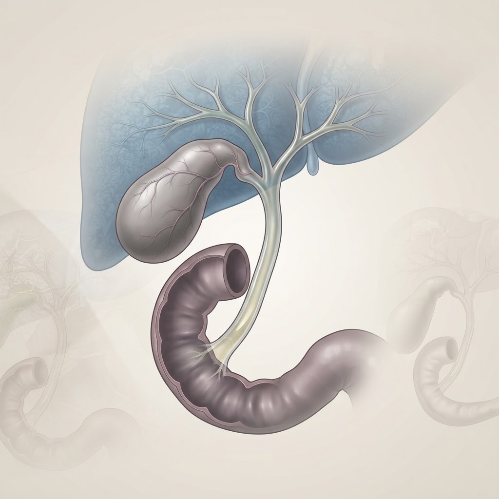
    At its core, gallbladder dyskinesia is a functional disorder of the biliary tract. To understand this condition, one must first look at the mechanics of the gallbladder. This pear-shaped organ acts as a reservoir for bile, a digestive fluid produced by the liver that is essential for breaking down dietary fats. When you consume a meal, the gallbladder receives a hormonal signal—typically from cholecystokinin—to contract and squeeze that concentrated bile into the small intestine. In a healthy system, this happens efficiently and without significant discomfort. In individuals with gallbladder dyskinesia, the motility or rhythmic contractions of the gallbladder are impaired, leading to incomplete emptying or disorganized movement of bile.

    The term “dyskinesia” literally translates to “abnormal movement,” and in this context, it refers to the biliary tree’s inability to coordinate with the sphincter of Oddi, the valve that controls the flow of bile into the intestine. When the gallbladder cannot contract with enough force or frequency, bile becomes stagnant. This stagnation does not just hinder digestion; it can lead to a buildup of pressure, discomfort, and irritation of the gallbladder wall. Because this condition is functional rather than structural, it is often missed by routine ultrasounds, which are excellent at detecting physical obstructions like stones or polyps but are blind to the gallbladder’s internal muscular performance.

    This dysfunction is frequently associated with a low ejection fraction, a metric used to quantify how much bile the gallbladder releases during a stimulated event. When this fraction drops below a certain threshold—often considered by clinicians to be lower than 35-40%—the gallbladder is effectively labeled as “hypokinetic.” It is not broken in the traditional, mechanical sense, but it is underperforming significantly enough to impact the quality of life. For the patient, this means the body struggles to emulsify fats, which leads to a cascade of downstream issues, including nutrient malabsorption and chronic digestive distress. Recognizing this as a dynamic, muscular issue rather than a static hardware problem is vital for shifting toward more effective management strategies.

    Common Symptoms of Biliary Dysfunction

    The symptoms of biliary dysfunction are notoriously non-specific, which is why many individuals spend months or even years visiting various specialists before landing on the correct diagnosis. The most hallmark sign of gallbladder dyskinesia is biliary colic, which manifests as a sharp, cramping, or aching pain in the right upper quadrant of the abdomen, just below the ribs. This pain is often episodic and triggered by the ingestion of fatty or fried foods, which force the gallbladder to contract. Because the organ is malfunctioning, the effort to squeeze results in a painful spasming sensation rather than a smooth release of bile.

    However, the clinical picture is rarely limited to pain alone. Many people report a range of systemic digestive issues that occur shortly after eating. Nausea is extremely common, often accompanied by a feeling of fullness or bloating that lingers long after a meal has ended. Some individuals describe a “gnawing” sensation in the center of their chest or upper abdomen, which they may mistakenly attribute to heartburn or acid reflux. When the gallbladder fails to deliver sufficient bile, the fat-digesting process is compromised, leading to symptoms like light-colored or “floating” stools, gas, and excessive belching.

    It is important to note that these symptoms can sometimes radiate to other parts of the body. A classic sign of biliary issues is referred pain, which is typically felt in the right shoulder blade or the space between the shoulder blades. This happens because the gallbladder shares nerve pathways with other structures in the upper torso. Furthermore, the chronic stress of poor fat digestion can lead to fatigue, skin issues, or a general sense of malaise. Because the body is struggling to absorb fat-soluble vitamins like A, D, E, and K, long-term dysfunction may even show up as brittle nails, dry skin, or weakened immune responses. Understanding these biliary dyskinesia symptoms as a collective pattern, rather than isolated events, helps in identifying the root cause of the discomfort.

    Management Approach Focus Area Best For
    Dietary Modification Reducing fat load Immediate symptom mitigation
    Natural Bile Support Enhancing liver-gallbladder flow Long-term functional improvement
    Surgical Intervention Removal of the gallbladder Cases of severe, chronic tissue damage
    Stress Management Vagus nerve regulation Patients with tension-related triggers

    Understanding the Role of the HIDA Scan

    When standard ultrasound and blood panels return normal results, the HIDA scan, or cholescintigraphy, serves as the investigative gold standard. This specialized nuclear medicine test is designed to evaluate the functional health of your biliary system. During the procedure, a technician injects a safe, radioactive tracer into the bloodstream. This tracer is taken up by the liver and excreted into the bile ducts, which then leads it into the gallbladder. By using a specialized camera, the medical team can track the flow of this tracer in real-time, observing how quickly it fills the gallbladder and, more importantly, how effectively it empties.

    The most critical part of the HIDA scan is the administration of a synthetic hormone called cholecystokinin (CCK). After the gallbladder has filled with the tracer, CCK is introduced to simulate the physiological response to a fatty meal. In a healthy gallbladder, this hormone triggers a robust contraction, flushing the tracer into the small intestine. In cases of gallbladder dyskinesia, the scan will often reveal a failure to empty the tracer properly. This is quantified as the Gallbladder Ejection Fraction (GBEF). If the GBEF is low, it provides objective evidence that the gallbladder is not performing its mechanical duties, validating the patient’s reports of pain and discomfort.

    It is worth noting that while the HIDA scan is powerful, it is not without nuance. A “low ejection fraction” result is a strong indicator of dyskinesia, but clinicians generally view it alongside the patient’s full clinical profile. There is a debate in the medical community regarding exactly what the “cutoff” for a normal ejection fraction should be, as individual biology can vary significantly. Some people may have a lower-than-average ejection fraction but experience zero pain, while others may have slightly better numbers but suffer significantly. Consequently, a HIDA scan should be viewed as one piece of a puzzle rather than a final verdict. It is an tool for measuring function, not for defining the entirety of the patient’s experience, and it is most effective when interpreted by a specialist who understands the broader context of digestive motility.

    Primary Causes and Potential Triggers

    Identifying the exact origin of gallbladder dyskinesia is often difficult, as it is rarely the result of a single event. Instead, it is usually the culmination of chronic stressors that interfere with the gallbladder’s ability to contract. Inflammation is perhaps the most significant contributor. When the gallbladder wall is subjected to chronic, low-grade inflammation—sometimes due to systemic oxidative stress or imbalances in the gut microbiome—it becomes “boggy” and less responsive to the hormonal signals that should trigger contraction. This inflammation can be driven by a poor diet, specifically one high in processed inflammatory fats and refined sugars, which places a heavy, constant demand on the biliary system.

    Another major factor is the composition of the bile itself. Bile is a complex mixture of cholesterol, bile salts, and bilirubin. If this ratio becomes imbalanced—often referred to as “biliary sludge”—the bile becomes thicker and more viscous. While this is not yet a stone, the thick sludge is physically more difficult to move through the narrow cystic duct. As the gallbladder works harder to push this thick, sludgy bile, the muscular wall begins to tire, eventually leading to a reduction in its ejection fraction. Factors like rapid weight loss, hormonal fluctuations (especially regarding estrogen levels), and certain medications can all alter the consistency of bile, making it more prone to stagnation.

    Neurological triggers also play an under-recognized role. The gallbladder is governed by the autonomic nervous system, specifically the vagus nerve, which manages the “rest and digest” state. When we are chronically stressed, our bodies remain in a sympathetic “fight or flight” mode, which actively inhibits digestive processes, including gallbladder contraction. Furthermore, dysbiosis—an imbalance of bacteria in the small intestine—can interfere with the signaling pathways that tell the gallbladder when to release its contents. If the gut environment is unhealthy, the complex signaling loop between the duodenum and the gallbladder may be interrupted. By identifying these triggers, whether they are dietary, hormonal, or neurological, individuals can begin to address the root causes rather than simply managing the downstream symptoms.

    The Connection Between Bile Flow and Digestion

    Bile is far more than just a waste product; it is a critical digestive enzyme booster. Without a consistent and efficient flow of bile, the entire process of nutrient assimilation is compromised. Think of bile as a natural detergent. When you eat fat, your body needs to emulsify that fat—breaking large lipid droplets into tiny particles—so that pancreatic enzymes have enough surface area to work on them. If the gallbladder is not contracting well, the amount of bile entering the small intestine is insufficient. This leaves large fat globules undigested, which pass into the lower intestine, where they can cause gas, bloating, and diarrhea. This is the mechanism behind the classic “post-meal distress” associated with biliary dysfunction.

    Furthermore, bile plays a crucial role in maintaining a healthy environment in the small intestine. Bile salts have mild antimicrobial properties, acting as a natural regulator of bacterial populations. When bile flow is sluggish, the small intestine may experience an overgrowth of bacteria, a condition known as SIBO (Small Intestinal Bacterial Overgrowth). This overgrowth can further complicate digestion by fermenting food prematurely, producing gas and toxins that irritate the gut lining. This creates a vicious cycle: the poor bile flow leads to bacterial imbalance, and the resulting inflammation from that imbalance makes it even harder for the biliary system to function correctly.

    The health of your gallbladder also impacts your ability to absorb fat-soluble vitamins A, D, E, and K. These vitamins are essential for everything from hormone production and skin health to bone density and immune function. If you are chronically low in these nutrients despite a healthy diet, your biliary health may be the missing link. Additionally, bile is the body’s primary mechanism for excreting excess cholesterol and toxins that the liver has processed. When bile flow is impaired, the body’s detoxification pathways are essentially backed up, which can put a significant burden on the liver. By optimizing bile flow through a proper biliary dyskinesia diet and lifestyle changes, you aren’t just fixing a “digestive pain” issue; you are supporting your liver, improving your nutrient status, and reducing the toxic load on your entire system. This interconnectedness is why addressing gallbladder function is often the missing key in holistic wellness journeys.

    Dietary Changes to Manage Gallbladder Pain

    When dealing with gallbladder dyskinesia, the primary goal of your dietary strategy is to reduce the workload on your gallbladder. Since your organ is struggling to contract efficiently, high-fat meals act as a major trigger, forcing the gallbladder to work harder than it is capable of, which often leads to the sharp, cramping pain associated with biliary dyskinesia symptoms. By shifting toward a gallbladder-friendly diet, many individuals notice a significant decrease in the frequency and intensity of their episodes.

    The cornerstone of a biliary dyskinesia diet is portion control and fat management. Instead of eating three large meals, many experts recommend moving to five or six smaller, nutrient-dense meals throughout the day. This approach prevents the gallbladder from having to release a massive bolus of bile at once, which is often when dysfunction causes the most distress. Focus on increasing your intake of fiber-rich foods like vegetables, legumes, and whole grains. Fiber helps bind bile acids and promotes healthy digestion, ensuring that your bowels move regularly, which prevents the stagnation of bile.

    Avoid highly processed foods, trans fats, and hydrogenated oils, as these are notoriously difficult for the liver and gallbladder to process. Even “healthy” fats can be problematic if consumed in large quantities. For example, while avocado and olive oil are generally considered heart-healthy, consuming them in high concentrations may trigger gallbladder pain if the gallbladder is already inflamed or weak. Opt for steaming, poaching, or baking your foods rather than frying, as high-heat frying can create inflammatory compounds that further stress the biliary system.

    Hydration is another frequently overlooked component of dietary management. Bile is composed largely of water; if you are dehydrated, your bile can become thick and viscous, making it harder for a poorly functioning gallbladder to eject it. Aim for consistent water intake throughout the day, perhaps flavored with a squeeze of fresh lemon, which some traditional wellness practices believe can help stimulate the flow of digestive juices.

    Natural Supplements to Support Gallbladder Function

    While diet is the foundation, natural gallbladder support can provide an extra layer of assistance for those dealing with a low ejection fraction gallbladder. Supplements are generally used to thin the bile, soothe inflammation, or support the liver’s ability to process fats more effectively. It is vital to approach supplementation cautiously, as some herbs that stimulate bile flow (choleretics) could theoretically cause discomfort if your biliary ducts are obstructed.

    Commonly discussed supplements include ox bile, which provides the gallbladder with a “rest” by supplying the enzymes necessary for fat digestion exogenously. Many people with gallbladder dysfunction report better comfort during meals when supplementing with digestive enzymes that include lipase. Another popular option is milk thistle, known for its liver-protective properties. Because the liver produces the bile that the gallbladder stores, maintaining liver health is inherently tied to gallbladder performance.

    Peppermint oil is also highly regarded for its antispasmodic properties, which can help relax the smooth muscle of the gallbladder and bile ducts, potentially reducing the sensation of cramping. Turmeric, specifically the compound curcumin, is frequently utilized for its powerful anti-inflammatory benefits, which may help mitigate the localized irritation caused by poor gallbladder function.

    Supplement Mechanism of Action Best For
    Ox Bile Provides supplemental bile salts Those with low fat digestion capability
    Milk Thistle Promotes liver detoxification Overall liver and biliary health
    Peppermint Oil Antispasmodic effect on smooth muscle Relieving cramping and bloating
    Lipase Enzymes Breaks down dietary fats Immediate mealtime relief

    Lifestyle Habits for Better Bile Production

    Managing gallbladder dysfunction goes beyond what you put on your plate. Your nervous system plays a massive role in digestion; specifically, the vagus nerve coordinates the release of bile. When you are chronically stressed, your body enters a “fight or flight” state, which effectively shuts down digestive processes like bile secretion. Incorporating stress-reduction techniques—such as deep breathing exercises, meditation, or gentle yoga—can help shift your body into a “rest and digest” state, which is the physiological environment where the gallbladder functions best.

    Physical movement is also crucial. The gallbladder is not an autonomous pump; it relies on the movement of the diaphragm and the abdominal muscles to help assist in the healthy flow of bile. Sedentary behavior can lead to bile stagnation. Regular, low-to-moderate intensity exercise, such as brisk walking, swimming, or cycling, encourages abdominal circulation and promotes systemic digestive health. However, avoid high-intensity maneuvers that place excessive stress on the upper right quadrant of the abdomen if you are currently experiencing a flare-up.

    Sleep hygiene also correlates with biliary health. Many traditional health systems suggest that the liver and gallbladder perform their most intense “cleansing” and processing work during the night. By prioritizing a consistent sleep schedule and ensuring you are not eating heavy meals right before bed, you allow your body the necessary resources to manage bile metabolism overnight. Furthermore, managing your weight is essential. Obesity is a well-known risk factor for biliary issues, as it increases the saturation of cholesterol in the bile, which can exacerbate the symptoms of an already poorly functioning gallbladder.

    When Is Surgical Intervention Necessary?

    While many individuals successfully manage biliary dyskinesia symptoms through dietary and lifestyle modifications, there are instances where surgical intervention, typically a cholecystectomy (gallbladder removal), becomes the necessary path forward. Medical experts generally consider surgery when the quality of life is severely impacted by chronic, recurring pain that does not respond to conservative management.

    If your gallbladder dysfunction is causing frequent hospitalizations, systemic infection (such as cholangitis), or if testing confirms that your gallbladder is essentially non-functional (a very low ejection fraction), your medical team may advise removal. The gallbladder is a storage organ; the body can adapt to its absence by secreting bile directly from the liver into the small intestine. While most people recover well, the transition period often involves a heightened need for the dietary changes mentioned earlier, as the constant drip of bile into the digestive tract requires a more consistent, moderate intake of healthy fats.

    Ultimately, the decision for surgery is a personal one, usually made in consultation with a gastroenterologist or surgeon. If you are experiencing symptoms like jaundice, fever, or intense, radiating pain, these are considered “red flags” that require immediate medical evaluation rather than home-based natural support. In these cases, the gallbladder may be at risk of rupture or blockage, and prompt medical intervention is vital to prevent serious complications.

    Frequently Asked Questions

    Can biliary dyskinesia cause pain in the back or shoulder?

    Yes, it is very common for gallbladder-related pain to be “referred.” The gallbladder shares nerve pathways with other structures, meaning that inflammation or dysfunction in the gallbladder can often be felt as sharp, stabbing pain between the shoulder blades or even in the right shoulder area.

    Is it possible to improve a low ejection fraction gallbladder naturally?

    While some people report symptomatic improvement by optimizing their diet, reducing stress, and using supportive supplements to thin their bile, the physical ejection fraction percentage is a structural measurement. Whether this number can be permanently increased is debated, but many individuals find they can manage the condition effectively without surgery if they remain disciplined with their lifestyle.

    How long should I follow a low-fat diet for gallbladder issues?

    There is no set time frame. Most people find that they need to maintain a gallbladder-friendly, moderate-fat diet long-term to keep their symptoms at bay. If you reintroduce high amounts of saturated fats too quickly, the gallbladder will be forced to overwork again, which often leads to the return of biliary symptoms.

    Does gallbladder dysfunction always lead to gallstones?

    Not necessarily. While they are related, gallbladder dyskinesia (functional disorder) is distinct from cholelithiasis (stones). However, a sluggish gallbladder that does not empty bile efficiently may create an environment where the bile becomes supersaturated, which can increase the risk of stone formation over time.

    Are there specific drinks that help with gallbladder pain?

    Staying well-hydrated with plain water is the most important factor. Some people find relief with warm water mixed with lemon juice or diluted apple cider vinegar, as these may help stimulate digestive secretions. Avoid sugary sodas, alcohol, and excessive caffeine, as these can irritate the digestive tract or contribute to inflammation.

    What does a “biliary attack” feel like?

    A biliary attack, or gallbladder attack, is typically characterized by a sudden onset of intense pain in the upper right quadrant of the abdomen, just below the rib cage. The pain may last from thirty minutes to several hours, is often described as a “cramping” or “boring” sensation, and may be accompanied by nausea, vomiting, or bloating.

    Conclusion

    Navigating gallbladder dyskinesia requires patience, self-awareness, and a proactive approach to your daily habits. By understanding that your gallbladder is struggling to perform its storage and ejection duties, you can make the necessary adjustments to your diet, stress management, and lifestyle to minimize discomfort. While the path to relief often involves strict dietary discipline and the use of targeted natural support, many people successfully lead full, symptom-free lives by honoring their body’s limitations and choosing supportive, whole-food-based habits.

    Remember that your digestive health is deeply interconnected. Taking care of your gallbladder through consistent meal timing, stress reduction, and healthy fats is an investment in your long-term vitality. Listen to your body, monitor your triggers, and maintain regular communication with your healthcare provider to ensure you are on the right track for your unique needs. If you are ready to take control, start today by evaluating your next meal and reducing the burden on your biliary system—your body will thank you for the extra support.

    By healthauthoritylife Editorial Team

    This article is for informational purposes only and is not a substitute for professional medical advice. Always consult your doctor before making changes to your diet, exercise routine, or treatment plan.


  • Occipital Neuralgia: Causes, Symptoms, and Natural Relief Tips

    Key Takeaways

    • Occipital neuralgia is a distinct condition involving irritation of the nerves running from the spine to the scalp, often causing sharp, electric-shock sensations.
    • Identifying the difference between a standard tension headache and occipital nerve pain is essential for seeking the correct treatment.
    • Poor posture, particularly “tech neck,” is a leading mechanical factor in compressing the nerves at the base of the skull.
    • Craniocervical instability can lead to chronic nerve irritation, requiring a focus on stabilizing the upper neck muscles.
    • Natural relief for occipital neuralgia often centers on a multi-faceted approach, including physical therapy, ergonomic adjustments, and inflammation management.

    The sensation of a sudden, sharp jolt of electricity racing up the back of your head is an experience that many who suffer from occipital neuralgia describe as both jarring and debilitating. Unlike the dull, throbbing pressure of a typical tension headache, this condition arises from a specific pathway of nerve irritation that requires a unique understanding of anatomy and mechanical health. As part of a broader spectrum of cervical spinal issues, including craniocervical instability, understanding how your neck alignment influences these nerves is the first step toward reclaiming your comfort. At the healthauthoritylife Editorial Team, we are dedicated to peeling back the layers of this often-misunderstood condition to help you navigate your journey toward nerve pain relief.

    What Is Occipital Neuralgia and How Does It Feel?

    Occipital neuralgia is a specific neurological condition characterized by chronic, paroxysmal pain in the upper neck, the back of the head, and behind the ears. This pain originates from the occipital nerves, two pairs of nerves that start at the top of the spinal cord and weave their way through the muscles of the scalp to the skin. When these nerves become compressed, inflamed, or injured, they send distorted signals to the brain, which are interpreted as intense, stabbing, or burning pain.

    For those living with the condition, the pain often comes in distinct waves. Imagine a flickering lightbulb or a frayed electrical wire; the sensation is similarly unpredictable. Many patients describe the pain as “electric,” “shooting,” or “piercing,” radiating from the very base of the skull—the junction where the neck meets the head—up toward the crown or even behind the eyes. It is rarely a dull ache in the traditional sense; instead, it is a high-intensity disruption that can make basic activities, such as brushing your hair or even resting your head on a pillow, feel agonizing.

    The anatomy here is critical. The occipital nerves must pass through dense muscular structures, including the trapezius, the splenius capitis, and the semispinalis capitis. If these muscles are chronically tight or if the vertebrae beneath them are shifting due to craniocervical instability, the nerves can become trapped, much like a garden hose being kinked under a heavy object. This mechanical obstruction is a fundamental aspect of the causes of occipital neuralgia. Because the nerves also provide sensation to the skin of the scalp, a person might experience extreme sensitivity in these areas, a phenomenon known as allodynia, where even a gentle breeze or a soft touch can trigger a painful response.

    Understanding the “how it feels” aspect is vital because it helps distinguish this condition from other forms of cranial discomfort. While migraine sufferers often experience a throbbing sensation associated with light sensitivity, the pain of occipital neuralgia is typically localized to the path of the nerves themselves. It is a structural and neurological issue rather than a chemical or vascular one. Consequently, treatments that focus solely on traditional headache medication often fall short, leading many to search for natural relief for occipital neuralgia that addresses the underlying mechanical environment of the neck and upper cervical spine.

    Common Symptoms of Occipital Nerve Pain

    Recognizing the hallmark occipital neuralgia symptoms is the cornerstone of early management. Because the occipital nerves are responsible for transmitting sensory information from the skin covering the upper neck, back of the head, and behind the eyes, the symptoms are almost always concentrated in these specific regions. The most prominent symptom is a sudden, sharp, or shooting pain that feels like a bolt of electricity. These attacks can last for a few seconds to several minutes and are often interspersed with a lingering, aching sensation in the scalp or base of the skull.

    Another common symptom is significant tenderness. If you run your fingers along the base of your skull, you may find specific “trigger points”—small, hardened knots of muscle—that, when pressed, radiate pain into your head. This sensitivity is often accompanied by scalp paresthesia, a tingling or “pins and needles” sensation. Some individuals report a feeling of numbness in the scalp, as the nerve signal is being muffled by constant compression. This sensory fluctuation can be highly unsettling, leading many to believe they are experiencing an issue with their skin rather than the nerves beneath it.

    Visual disturbances or sensitivity to light are also frequently reported. Because the occipital nerves share pathways with nerves that branch toward the eyes, it is not uncommon for someone suffering from an attack to feel pain behind one or both eyes. This often causes sufferers to retreat to dark, quiet rooms, not necessarily because they have a migraine, but because the nerve irritation is so severe that sensory input becomes overwhelming.

    The following table illustrates common approaches to managing these symptoms and how they serve different needs in your wellness routine:

    Approach Mechanism of Relief Best For
    Targeted Myofascial Release Relaxes tight muscles compressing the nerve Chronic muscle-based nerve entrapment
    Cervical Ergonomic Correction Reduces strain on the upper cervical spine Preventing flare-ups during sedentary work
    Thermal Therapy (Alternating) Improves blood flow and reduces inflammation Immediate, acute nerve pain relief
    Upper Cervical Stabilization Supports the base of the skull Addressing underlying craniocervical instability

    It is important to note that symptoms can be unilateral (one-sided) or bilateral. Unilateral pain is frequently cited in clinical observations, often correlating with the side of the body that carries the most habitual postural tension. Tracking your symptoms in a journal—noting the time of day, your activities prior to the pain, and the nature of the sensation—can provide invaluable data for you and your health practitioners to identify triggers. Consistent tracking turns an invisible, subjective experience into a concrete map of your nerve health.

    Primary Causes and Triggers of Nerve Irritation

    To address the root of the problem, one must investigate the causes of occipital neuralgia, which are rarely singular. Instead, they are usually a result of cumulative stress on the upper cervical anatomy. At the center of this is the relationship between the skull and the first few vertebrae of the neck (the C1 and C2 vertebrae). When the ligaments that stabilize this junction are weakened or the muscles are overcompensated, a state of craniocervical instability can occur. This instability allows for micro-movements that irritate the nerves residing in the suboccipital region.

    Trauma is a frequent catalyst. A history of whiplash, even a minor fender-bender from years ago, can leave the cervical muscles in a chronic state of guarded spasm. Over time, these muscles lose their flexibility and become dense, fibrous bands that no longer cushion the nerves but instead act as vices. This is why many people who suffer from nerve pain do not experience relief until they address the muscular integrity of their neck.

    Another major trigger is inflammation. Systemic inflammation—often linked to dietary choices, chronic stress, or underlying health conditions like arthritis—can cause the tissues surrounding the nerves to swell. When the space between the vertebrae and the muscles is already tight, even slight swelling can trigger an aggressive response from the occipital nerves. This is often why patients find their pain worsens during periods of high stress; cortisol and other inflammatory hormones can exacerbate the body’s sensitivity to nerve compression.

    Repetitive strain also plays a significant role. Today’s digital landscape forces many of us to maintain a forward-head posture for hours at a time. This position shifts the weight of the head significantly forward, placing an immense load on the muscles at the base of the skull. These muscles are forced to stay in a state of constant contraction to keep the head from falling forward, inevitably leading to the entrapment of the nerves passing through them. Furthermore, conditions such as diabetes or gout, which can cause peripheral neuropathy, are known to make the occipital nerves more susceptible to irritation. In such cases, managing systemic health is just as important as physical neck stabilization.

    Ultimately, identify your specific trigger. Is it a stressful week at work? Is it the chair you sit in? Or is it a lack of movement? By identifying the “why,” you transition from passive sufferer to active manager of your health, allowing you to intercept a flare-up before it reaches its peak intensity.

    How Posture Impacts Occipital Nerve Health

    The phrase “posture is everything” is particularly true when it comes to the health of the occipital nerves. The human head weighs approximately 10 to 12 pounds, and it is balanced precariously atop the cervical spine. When your posture is aligned—with your ears positioned directly over your shoulders—the weight is distributed evenly across the muscles and joints. However, when the head shifts forward, even by just two or three inches, the gravitational load on the muscles of the upper neck increases exponentially.

    This forward-head posture is the arch-nemesis of occipital nerve health. As the head migrates forward, the suboccipital muscles (the tiny, deep muscles at the base of the skull) have to work overtime to act as a counterweight. These muscles were designed for stabilization and fine-tuned movement, not for holding the heavy, bowling-ball weight of the skull in a forward-leaning position for eight hours a day. As they grow fatigued and tight, they compress the occipital nerves, leading to that characteristic nerve pain relief cycle that so many individuals struggle to break.

    This mechanical stress is further exacerbated by the rise of craniocervical instability. If the connective tissues in the neck have been compromised, the body often attempts to “splint” the area by inducing muscle spasms. While this is an evolutionary survival mechanism, it is counterproductive in a modern context. You end up with a neck that feels stiff and guarded, which effectively pinches the nerves every time you turn your head or look down. Maintaining a neutral spine is not just about aesthetics; it is about creating enough “breathing room” for your nerves to function without being compressed by overly tight, defensive musculature.

    Practical postural interventions start with your environment. Consider the height of your computer monitor; if you are looking down, you are likely putting yourself at risk. Your eyes should naturally hit the top third of your screen without the need to tilt your chin down. Similarly, avoid the “text neck” phenomenon by bringing your phone up to eye level rather than dropping your head to meet your device. Incorporating regular “posture breaks”—where you reset your shoulders, retract your chin slightly (a movement known as a chin tuck), and realign your head over your shoulders—can physically unload the pressure on your occipital nerves. These small, frequent adjustments serve as a vital form of natural relief for occipital neuralgia, preventing the buildup of tension that eventually leads to a painful episode.

    Diagnostic Signs: Distinguishing From Common Headaches

    Distinguishing occipital neuralgia from a garden-variety headache is essential because the two require vastly different management strategies. A common tension headache is often described as a tight band around the forehead or a generalized feeling of pressure. It is typically symmetrical, affecting both sides of the head equally, and is often related to dehydration, stress, or eye strain. In contrast, the occipital neuralgia symptoms are distinctly localized, often starting from the base of the skull and moving upward like a directional arrow, following the anatomy of the nerves themselves.

    One of the most reliable diagnostic markers is the “tineal sign” or localized tenderness upon palpation. If a practitioner or a knowledgeable caregiver applies firm pressure to the base of your skull and you experience a “lightning bolt” of pain that travels up to your ear or eye, it is a strong indicator of nerve involvement rather than a typical muscular tension headache. Furthermore, a tension headache usually responds to rest, hydration, or over-the-counter pain relievers, while occipital neuralgia often proves resistant to these standard measures.

    Consider the timing of your pain. Tension headaches often develop gradually throughout the day as stress accumulates. Occipital nerve pain, however, is frequently paroxysmal. It hits in sudden, intense bursts. You might be perfectly fine one moment, and then a specific movement—like turning your head to reverse your car—triggers a stabbing pain that makes you gasp. This sensitivity to movement is a hallmark of an entrapment syndrome.

    It is also worth noting that migraine headaches are distinct from occipital neuralgia. While migraines can involve the base of the skull, they are generally accompanied by systemic symptoms like nausea, extreme sensitivity to light and sound, and occasionally visual auras. Occipital neuralgia, as a mechanical and neurological phenomenon, typically does not present with nausea unless the pain is so intense that it triggers a vasovagal response. If you find that your pain is strictly localized to the back of the head, is triggered by touch, and feels electrical, you are likely looking at an issue that requires a specialized approach to nerve pain relief. Understanding these nuances empowers you to speak more clearly with healthcare providers, ensuring that you receive the most accurate guidance for your specific situation.

    Natural Anti-Inflammatory Approaches for Nerve Pain

    When addressing the root of nerve irritation, systemic inflammation often acts as an accelerant. While conventional medicine frequently turns to non-steroidal anti-inflammatory drugs (NSAIDs) for quick relief, these can lead to long-term gastrointestinal or renal challenges. Many individuals seeking natural relief for occipital neuralgia turn to botanical and physical methods designed to modulate the body’s inflammatory response at the cellular level.

    Turmeric, specifically its active compound curcumin, remains one of the most researched natural interventions for neuropathic pain. Curcumin works by inhibiting NF-kB, a molecule that travels into the nuclei of your cells and turns on genes related to inflammation. Because curcumin has low bioavailability on its own, it is typically recommended to consume it alongside black pepper (piperine) or in a lipid-based (fat-soluble) delivery system to ensure the compound reaches the affected nerves in the suboccipital region effectively.

    Topical botanical applications can also provide localized nerve pain relief without the systemic side effects of oral medications. Capsaicin, derived from chili peppers, works by depleting Substance P—a neurotransmitter involved in pain signaling—from the nerve endings. When applied to the base of the skull or the upper neck, it may desensitize the occipital nerves over time. Similarly, magnesium oil or transdermal magnesium chloride is frequently utilized to relax the tight musculature at the craniocervical junction, which often compresses the nerves in the first place.

    Furthermore, cold and heat therapy remain cornerstone conservative treatments. Cryotherapy helps to constrict blood vessels and reduce swelling in the acute phase of a flare-up, while heat therapy, specifically via a heating pad or warm hydrotherapy, promotes circulation and relaxes muscles that may be triggering the neuralgic response. Alternating these modalities can help reset the autonomic nervous system’s pain response.

    Intervention Mechanism of Action Best For
    Curcumin (with Piperine) Inhibits systemic inflammatory pathways (NF-kB) Long-term reduction of chronic nerve irritation
    Topical Magnesium Promotes muscle relaxation and nerve conduction Acute muscular tension causing nerve entrapment
    Capsaicin Cream Depletes Substance P at local nerve endings Localized burning, shooting pain
    Cold/Heat Contrast Vasoconstriction vs. Vasodilation Immediate, episodic pain management

    The Role of Physical Therapy and Cervical Alignment

    In cases of craniocervical instability, the primary goal of physical therapy is not just symptom management, but mechanical restoration. The occipital nerves pass through several layers of muscle before reaching the scalp. When the neck is misaligned—often due to “tech neck,” poor posture, or ligamentous laxity—these muscles become chronically shortened or hypertonic, creating a “pincer effect” on the nerve.

    A specialized physical therapist often focuses on the deep cervical flexors. These muscles are the “core” of your neck, and when they are weak, the larger, superficial muscles (like the trapezius and splenius capitis) take over the workload. These superficial muscles are prone to cramping and spasm, which directly exacerbates occipital neuralgia symptoms. Strengthening the deep cervical flexors through gentle chin-tuck exercises can significantly offload the tension from the suboccipital triangle.

    Cervical alignment also involves postural retraining. Ergonomic modifications to one’s workspace, such as raising monitors to eye level and ensuring lumbar support, are essential to preventing the forward-head posture that compresses the occipital nerve. Furthermore, gentle manual therapy—specifically myofascial release—can help break up adhesions in the fascia surrounding the atlas and axis vertebrae. By improving the mobility of these top two vertebrae, the nerves have more space to glide freely, preventing the irritation that leads to electric-shock sensations.

    Nutritional Support to Calm Inflamed Nerves

    Your nerves are highly metabolic structures, and their health is intrinsically linked to your nutritional status. When nerves are inflamed, they require specific substrates for repair and signal transmission. Vitamin B-complex, particularly B12, B6, and folate, are non-negotiable for nerve health. Deficiency in these vitamins is well-documented as a potential cause of neuropathic pain. B12, in particular, is essential for the maintenance of the myelin sheath—the fatty coating that insulates your nerves and ensures efficient electrical signal conduction.

    Beyond B-vitamins, Omega-3 fatty acids play a critical role. EPA and DHA, found abundantly in wild-caught fatty fish or high-quality algae oil, act as precursors to anti-inflammatory mediators called resolvins. These resolvins are responsible for “turning off” the inflammatory signal once the body has attempted to heal an injury. For individuals with chronic nerve pain, maintaining a high intake of Omega-3s can dampen the sensitivity of pain receptors.

    Alpha-lipoic acid (ALA) is another key nutrient for nerve health. It is a potent antioxidant that works in both water and fat-soluble environments. ALA has shown promise in various studies for its ability to improve blood flow to the nerves and improve the speed of nerve conduction. By reducing oxidative stress around the occipital nerves, ALA provides a supportive environment for nerve regeneration and stability.

    Lifestyle Changes to Prevent Recurrent Nerve Flare-ups

    Preventing recurrent flare-ups requires a multi-faceted approach to daily living. Stress management is perhaps the most overlooked component of occipital neuralgia treatment. When the body is under chronic stress, it produces cortisol, which can heighten the perception of pain and increase muscular tension in the trapezius and neck. Techniques such as diaphragmatic breathing, meditation, or progressive muscle relaxation can downregulate the sympathetic nervous system, preventing the “fight or flight” muscle bracing that triggers neuralgia.

    Sleep hygiene is equally vital. The position in which you sleep can exacerbate craniocervical issues. Many people with occipital nerve involvement find that sleeping on their stomachs puts too much torque on the neck, causing the vertebrae to shift during the night. Switching to a back-sleeping position with a cervical-support pillow that maintains the natural curve of the neck can prevent morning flare-ups and reduce pressure on the nerve roots.

    Hydration also plays an understated role in joint and disc health. The intervertebral discs and the connective tissue ligaments that stabilize the neck are largely composed of water. Dehydration can lead to a loss of disc height and increased ligamentous laxity, both of which contribute to cervical instability. Staying well-hydrated ensures that your spinal column maintains the necessary cushioning to protect the sensitive nerves exiting the brainstem.

    When to Seek Professional Medical Evaluation

    While natural relief for occipital neuralgia is often effective for mild to moderate cases, there are specific “red flags” that mandate an immediate consultation with a neurologist or spine specialist. If you experience a sudden “thunderclap” headache, which is an intense, explosive pain that reaches peak intensity within seconds, you must seek emergency care. This can sometimes indicate underlying vascular issues that require urgent diagnostics.

    Furthermore, if your occipital neuralgia symptoms are accompanied by neurological deficits—such as weakness in the arms or legs, persistent numbness in the hands, balance issues (ataxia), or a sudden change in vision—it is essential to rule out more serious complications like structural damage to the spinal cord or brainstem. If your pain does not respond to conservative lifestyle adjustments and physical therapy after several weeks, or if the intensity of the pain prevents you from participating in daily life, a doctor may evaluate you for interventional procedures. These may include nerve blocks, diagnostic imaging (such as an upright MRI to visualize craniocervical instability), or medications designed to stabilize nerve membranes.

    Frequently Asked Questions

    Can occipital neuralgia cause vision problems?

    While occipital neuralgia primarily affects the nerves at the back of the head, many individuals report light sensitivity (photophobia) or blurred vision during a flare-up. This is often because the trigeminal nerve and the occipital nerves share pain-processing pathways in the brainstem, a phenomenon known as central sensitization.

    Is craniocervical instability considered a primary cause of occipital neuralgia?

    Craniocervical instability is increasingly recognized as a root cause for many chronic cases. When the ligaments holding the skull to the spine are too loose, the vertebrae can shift, irritating the occipital nerves. Addressing the underlying mechanical stability is often the key to long-term resolution.

    How long does a typical flare-up of occipital neuralgia last?

    The duration of a flare-up is highly individual. It can range from a few minutes of sharp, electric-like pain to a chronic, aching discomfort that lasts for days or even weeks. Consistent application of anti-inflammatory and postural strategies can often shorten the duration and frequency of these episodes.

    Are there specific foods that trigger nerve pain?

    Yes, highly processed foods, excess sugar, and refined carbohydrates are known to spike systemic inflammation, which can lower your threshold for nerve pain. For many, limiting inflammatory oils (such as soybean, corn, and canola oil) and focusing on an anti-inflammatory diet is a helpful step in managing chronic neuralgic conditions.

    Can stress really make my neck pain worse?

    Absolutely. Stress leads to unconscious muscle clenching, particularly in the shoulders and neck. This increased muscular tension can physically entrap the occipital nerves, turning a minor sensitivity into a full-blown nerve pain episode. Managing the stress response is a critical part of a holistic treatment plan.

    What is the most effective sleeping position for nerve relief?

    Sleeping on your back is generally considered the most supportive position for the neck. Using a cervical pillow that supports the natural inward curve of the neck helps keep the spine in a neutral position, reducing the likelihood of nerve compression during the night.

    Conclusion

    Managing occipital neuralgia and craniocervical instability is a journey that requires patience, consistency, and a comprehensive look at your overall health. By focusing on mechanical alignment, reducing systemic inflammation, and nourishing your nervous system, you can effectively manage symptoms and significantly improve your quality of life. Remember that the body has a remarkable capacity for healing when provided with the right tools and an environment conducive to repair. Start by implementing one or two of the strategies mentioned above—such as improving your posture or adding a high-quality Omega-3 supplement to your daily routine—and monitor your progress closely. You do not have to settle for a life defined by pain; take the first step toward reclaiming your well-being today.

    By healthauthoritylife Editorial Team

    This article is for informational purposes only and is not a substitute for professional medical advice. Always consult your doctor before making changes to your diet, exercise routine, or treatment plan.

  • Adult Sleep Apnea: Symptoms, Causes, and Natural Relief Tips

    Adult Sleep Apnea: Symptoms, Causes, and Natural Relief Tips

    Key Takeaways

    • Sleep apnea is a serious medical condition characterized by repeated breathing pauses that can severely impact cardiovascular health.
    • Recognizing the difference between simple snoring and chronic sleep apnea is crucial for early intervention.
    • Weight management and structural factors are primary causes of sleep apnea in adults.
    • Chronic fatigue is often a secondary symptom of fragmented sleep, leading to reduced productivity and cognitive decline.
    • Positional therapy and lifestyle modifications serve as accessible starting points for those seeking to improve sleep apnea naturally.

    Waking up feeling as though you never slept at all is a frustration shared by millions, yet for many, the culprit is far more complex than a mere lack of hours in bed. If your nights are punctuated by restlessness, gasping, or a partner’s complaints about your loud snoring, you may be navigating the silent, exhausting world of adult sleep apnea. While often dismissed as a social nuisance, this condition represents a significant disruption to your body’s ability to repair itself during the night. Understanding the underlying mechanisms of sleep apnea is the first step toward reclaiming your energy, improving your long-term health, and exploring effective natural sleep apnea remedies that can supplement traditional treatments. In this comprehensive guide, the healthauthoritylife Editorial Team explores the intricacies of this condition, from the physiological triggers to actionable, natural strategies for nightly restoration.

    Understanding Adult Sleep Apnea and Its Health Impact

    At its core, sleep apnea is a sleep disorder where breathing repeatedly stops and starts throughout the night. The most common form, obstructive sleep apnea (OSA), occurs when the muscles in the back of the throat fail to keep the airway open during sleep. When you breathe, air travels through the nose and mouth into the throat. If the structures in your throat—such as the tongue, tonsils, or soft palate—collapse or become too relaxed, the airway narrows or closes completely. This collapse prevents oxygen from reaching your lungs, triggering the brain to send an urgent wake-up signal. This cycle can happen dozens, sometimes hundreds, of times in a single night.

    The health impact of these recurring pauses is profound. Because the body is deprived of consistent oxygen flow, it undergoes a systemic stress response. Each time a breathing pause occurs, the heart rate fluctuates and blood pressure may spike, forcing the cardiovascular system to work overtime while it should be at rest. Over time, this constant state of physiological alert can contribute to an increased risk of hypertension, irregular heart rhythms, and other cardiovascular challenges. Experts generally agree that untreated sleep apnea places a significant burden on the body, acting as a catalyst for systemic inflammation.

    Furthermore, the cumulative effect of these micro-arousals—the moments when your brain forces you out of deep sleep to restore breathing—prevents you from achieving the restorative cycles of REM and deep sleep. These stages are vital for memory consolidation, hormonal regulation, and cellular repair. When these cycles are broken, the body fails to flush out waste products in the brain efficiently, which may explain why individuals with sleep apnea often report “brain fog” and difficulty concentrating. The impact extends beyond physical wellness, influencing emotional stability and mood regulation. By viewing sleep apnea as a systemic health issue rather than just a nightly inconvenience, you can begin to prioritize the structural and lifestyle changes necessary for long-term health. Whether you are currently using a CPAP machine or are just beginning to investigate why you feel constantly drained, understanding the mechanics of your airway is the foundation of recovery.

    Common Symptoms of Sleep Apnea You Should Not Ignore

    Identifying the symptoms of this condition can be challenging because many occur while you are unconscious. However, the signs are often evident to those around you or manifest as persistent patterns during your waking hours. The most frequently reported indicator is chronic, loud snoring. It is important to distinguish between social snoring and clinical sleep apnea. While standard snoring is often caused by nasal congestion or the vibration of soft tissues due to mouth breathing, snoring linked to sleep apnea is typically louder, more rhythmic, and often punctuated by silence followed by a sharp gasp or choking sound. If your snoring sounds like a struggle for air, it is a significant red flag.

    Beyond audible cues, daytime sleepiness is perhaps the most reliable indicator of a compromised night. If you find yourself drifting off during quiet meetings, while driving, or while watching television, your sleep quality is likely severely degraded. This is not the same as standard tiredness; it is a pervasive, heavy fog that persists regardless of how many hours you “spent” in bed. Morning headaches are another common symptom. These occur because the drop in oxygen levels throughout the night can cause blood vessels in the brain to dilate, leading to a dull, throbbing pain upon waking that usually subsides within a few hours of rising.

    Mood changes, including irritability, anxiety, and depressive symptoms, are frequently overlooked. When your brain is starved of oxygen and deprived of deep sleep cycles, it struggles to manage stress and regulate emotions. You might notice that your patience is thin, your short-term memory is failing, or that you have lost interest in activities you once enjoyed. Physical indicators also include a dry mouth or sore throat upon waking, as the mouth-breathing associated with airway obstruction leads to rapid dehydration of the oral tissues. To help you differentiate between common issues and potential apnea, consider the following comparisons:

    Symptom/Behavior Occasional Snoring Sleep Apnea Best For
    Sound Quality Steady, rhythmic vibration Erratic, gasping, choking Distinguishing the severity
    Daytime Alertness Generally alert Excessive, uncontrollable sleepiness Assessing functional impact
    Morning Status Clear-headed Headache, dry mouth, irritability Identifying physical aftermath
    Breathing Pattern Uninterrupted Apneic pauses/gasping Clinical diagnosis indicators

    Ignoring these symptoms can lead to a long-term decline in metabolic and cardiovascular health. If you recognize these patterns in yourself or a loved one, it is time to move beyond self-observation and seek guidance to explore potential treatments, including various CPAP alternatives or lifestyle-driven solutions.

    Primary Causes and Risk Factors for Obstructive Sleep Apnea

    The causes of sleep apnea in adults are rarely tied to a single factor; rather, they usually stem from a combination of anatomical, genetic, and lifestyle elements. Understanding these drivers is essential, as many of these factors can be managed or modified to improve the quality of your airway function. Anatomically, many individuals have a narrow throat or a tongue that is relatively large compared to their oral cavity. If your tonsils or adenoids are enlarged, or if you have a recessed jaw, these structural characteristics can physically crowd the airway, making a collapse much more likely when the muscles relax during sleep.

    Weight is perhaps the most widely recognized risk factor, yet it is often misunderstood. Excess weight, particularly around the neck area, can add significant pressure to the airway. When you lie down, the weight of the neck tissues can cause the airway to collapse more easily. However, it is a mistake to assume only those who are overweight suffer from this condition. Many individuals with a healthy body mass index (BMI) still experience sleep apnea due to genetics—specifically, the shape and size of their throat and facial structure. Furthermore, fat distribution plays a role; individuals who carry weight around the upper body or neck are statistically more prone to airway obstruction than those who carry weight elsewhere.

    Age and gender also contribute significantly. As we age, the throat muscles naturally lose some of their tone, which can increase the likelihood of obstruction. Men are generally considered to be at a higher risk than women, although the risk for women increases significantly post-menopause, likely due to hormonal shifts that affect muscle tension and soft tissue support. Lifestyle choices such as alcohol consumption and smoking are major contributors as well. Alcohol acts as a muscle relaxant; if you consume it close to bedtime, your throat muscles are significantly more likely to collapse. Smoking causes inflammation and fluid retention in the upper airway, which restricts the amount of space available for air to pass through during the night.

    Lastly, nasal congestion and chronic allergies can exacerbate the issue. If you are forced to breathe through your mouth because your nasal passages are blocked, you are inherently altering the mechanics of your airway. This shift makes it easier for the tongue to fall back and block the throat. By addressing these primary causes—such as managing allergies, reducing alcohol intake, and maintaining a healthy weight—you are not just treating a symptom; you are addressing the foundational issues that contribute to the severity of sleep apnea. Recognizing these risk factors is the first step in deciding which strategies, from medical interventions to natural lifestyle shifts, will provide the most effective relief for your unique situation.

    The Connection Between Sleep Apnea and Chronic Fatigue

    Chronic fatigue is often dismissed as a byproduct of a busy modern lifestyle, but for the person suffering from sleep apnea, it is a constant, debilitating reality. The fatigue associated with this condition is not simply being “tired.” It is a state of deep, systemic exhaustion that affects every facet of life. Because the body is forced into a state of semi-arousal hundreds of times per night, the individual never reaches the deepest, most restorative stages of sleep. This cycle of fragmented sleep is the primary driver behind the cognitive and physical weariness that marks the day.

    When sleep is fragmented, the brain is deprived of the neurochemical “reset” it requires. Research suggests that during deep sleep, the glymphatic system—a waste clearance system in the brain—is highly active. It flushes out metabolic byproducts that accumulate during the day. In an individual with untreated sleep apnea, this process is frequently interrupted. Consequently, these waste products linger, leading to the hallmark “brain fog” that makes complex decision-making, memory recall, and focus incredibly difficult. This is why individuals with sleep apnea often feel like they are working harder than their peers, yet achieving less, simply because their cognitive engine is running on a depleted battery.

    Physically, the fatigue is compounded by the body’s attempt to survive. Each time you stop breathing, your sympathetic nervous system—the “fight or flight” system—is activated. This releases a surge of adrenaline and cortisol, hormones that are designed to deal with immediate physical threats. Having these hormones flooding your system all night is exhausting. By the time you wake up, your body has effectively been through a marathon, despite having spent eight hours in bed. This hormonal imbalance also impacts your metabolism. Elevated cortisol levels are linked to insulin resistance and weight gain, creating a vicious cycle: the fatigue makes you crave energy-dense foods, while the hormonal fallout makes it harder to maintain a healthy weight, which in turn worsens the sleep apnea.

    Improving this exhaustion requires more than just trying to sleep longer; it requires improving the quality of the sleep you do get. Strategies to manage this fatigue often involve a multi-pronged approach. While you might be looking for how to improve sleep apnea naturally, it is vital to understand that patience is required. Restoring energy levels takes time as the body recovers from months or years of chronic sleep deprivation. By addressing the apnea, you allow your nervous system to return to a parasympathetic, or “rest and digest,” state. This transition is crucial for healing the body, reducing inflammation, and finally breaking the cycle of persistent daytime weariness.

    How Positional Therapy Can Improve Your Nightly Rest

    For many individuals, the severity of sleep apnea is dictated by the position in which they sleep. This is known as positional sleep apnea, where the airway obstruction is significantly worse when lying on the back (supine position) compared to the side (lateral position). When you sleep on your back, gravity encourages the tongue and soft tissues in the throat to collapse backward toward the pharynx, which can block the airway and trigger the apneic events mentioned earlier. Positional therapy is a highly effective, non-invasive method that aims to prevent you from rolling onto your back during sleep, effectively reducing the frequency and duration of airway obstruction.

    The logic behind positional therapy is simple: when you sleep on your side, the forces of gravity are redistributed. Instead of the tongue falling directly into the throat, it tends to fall to the side of the mouth, keeping the primary airway passage clearer. For those with mild to moderate sleep apnea, this can lead to a dramatic reduction in snoring and a significant increase in oxygen saturation levels throughout the night. Many people report that simply training themselves to stay in a side-lying position results in fewer middle-of-the-night awakenings and a more refreshed feeling upon waking.

    There are several ways to implement positional therapy. Traditionally, some have used a “tennis ball technique,” where a ball is sewn into a pocket on the back of a pajama top to discourage rolling onto the back. While effective for some, modern solutions are often more comfortable and sophisticated. Today, there are specialized pillows designed to support the head and neck while promoting a side-sleeping position, as well as electronic vibrating sensors that you wear on your chest or neck. These devices are programmed to emit a gentle vibration if they detect that you have transitioned into a supine position, nudging you to shift back to your side without fully waking you up.

    It is important to note that while positional therapy is one of the most accessible natural sleep apnea remedies, it may not be a complete cure for everyone, especially those with severe anatomical obstructions. However, it can serve as a vital component of a broader strategy, potentially allowing for a lower pressure setting if you use a CPAP machine or serving as a helpful habit to pair with other lifestyle adjustments. When combined with throat exercises, weight management, and healthy sleep hygiene, positional therapy empowers you to take an active role in managing your own rest. By changing your relationship with your sleeping environment, you remove the physical leverage that gravity holds over your breathing, allowing your body to settle into the uninterrupted cycles it needs to thrive.

    Dietary Changes and Weight Management for Airway Health

    The relationship between body composition and airway patency is one of the most critical factors in managing sleep apnea naturally. Excess weight, particularly fat deposits around the neck and throat area, can exert significant pressure on the upper airway during sleep. This structural crowding makes the tissues more prone to collapsing when muscles relax at night. Addressing these physical factors is often the first step in reducing the frequency of apneic events.

    Adopting an anti-inflammatory diet is a cornerstone of this process. Chronic inflammation can cause the tissues of the throat and nasal passages to swell, further narrowing the airway. By shifting toward whole, nutrient-dense foods—such as leafy greens, fatty fish rich in omega-3 fatty acids, berries, and nuts—individuals may reduce systemic inflammation. Limiting processed sugars, refined carbohydrates, and heavy late-night meals is equally essential. Eating large meals shortly before bedtime can trigger acid reflux, which is known to cause airway inflammation and increase the likelihood of choking sensations or disruptions in breathing during sleep.

    Weight management should not be viewed as a crash-diet solution but as a lifestyle shift. Even modest, gradual reductions in total body mass have been associated with improvements in airway stability. Engaging in consistent physical activity, such as brisk walking, swimming, or resistance training, supports metabolic health and helps maintain muscle tone throughout the body. While weight loss is a powerful tool, it is important to remember that airway anatomy is complex. Some individuals have “thin” obstructive sleep apnea due to narrow jaw structures or tonsillar hypertrophy, meaning that weight management alone may not be a total cure, but it remains a vital component of holistic airway health.

    Hydration also plays a subtle yet significant role. Keeping the mucous membranes of the nose and throat properly hydrated helps prevent the stickiness and inflammation that can exacerbate snoring and blockages. Avoiding alcohol is perhaps the most critical dietary modification for sleep apnea patients. Alcohol is a potent muscle relaxant; it significantly decreases the tone of the muscles that keep the airway open, leading to longer and more frequent pauses in breathing. For those looking to improve their sleep quality, eliminating alcohol intake for several hours before bed is one of the most effective natural interventions.

    The Role of Oral Myofunctional Therapy in Airway Strengthening

    Many people are surprised to learn that the muscles of the mouth, tongue, and throat can be trained just like any other muscle group in the body. Oral Myofunctional Therapy (OMT) is a specialized series of exercises designed to correct “orofacial myofunctional disorders”—basically, improper patterns of muscle function in the face and neck. If the tongue has poor resting posture or the muscles of the soft palate are weak, the airway is much more likely to collapse during sleep.

    In a healthy state, the tongue should rest against the roof of the mouth, not the floor, and the lips should be sealed. OMT helps patients retrain these habits. Exercises might include specific tongue-pushing drills against the hard palate, stretching the frenulum, or performing rhythmic movements with the soft palate to increase its stiffness and reduce its tendency to vibrate or sag. These exercises are often performed under the guidance of a trained myofunctional therapist, but they can be integrated into daily routines once the techniques are learned.

    The goal of OMT is to transform the airway from a “passive” tube that collapses easily into an “active” structure that maintains its shape even when the brain is in deep REM sleep. While it requires time and consistency—often several months of dedicated daily practice—the long-term benefits include improved breathing patterns, reduced snoring, and better success rates for individuals using other CPAP alternatives. It is particularly effective for those with mild to moderate obstructive sleep apnea who are looking for ways to strengthen their physiology rather than just relying on mechanical devices.

    Supplements and Herbs That May Support Better Sleep Hygiene

    While no supplement is a direct replacement for medical treatment, certain natural compounds may support the biological systems involved in sleep quality and respiratory health. When considering supplements, the focus should be on substances that reduce inflammation, promote nasal patency, or assist in muscle relaxation without causing excessive lethargy.

    Magnesium is frequently cited as a foundational mineral for sleep. Because it helps regulate the nervous system and encourages muscle relaxation, it can assist in creating a state of physical readiness for rest. Many people find that magnesium glycinate is the most easily absorbed form. Additionally, anti-inflammatory herbs like turmeric (specifically when combined with black pepper for bioavailability) may help manage the general inflammation of the upper respiratory tract. Quercetin, a flavonoid found in various plants, is often used by those who suffer from nasal congestion to help stabilize cells that release histamine, potentially making it easier to breathe through the nose during the night.

    It is important to approach herbal remedies with caution. Some natural sleep aids, such as valerian root or high doses of melatonin, can be sedating. For a person with sleep apnea, over-sedation is a risk factor because it can further depress the respiratory drive and reduce muscle tone. Always consult with a healthcare professional to ensure that any supplement does not conflict with existing health conditions or medications.

    Natural Strategy Primary Mechanism Best For
    Oral Myofunctional Therapy Muscle tone improvement Strengthening the tongue and throat
    Anti-inflammatory Diet Reducing tissue swelling Long-term airway health
    Nasal Strips/Dilators Physical airway opening Reducing nasal resistance
    Magnesium Supplementation Nervous system relaxation General sleep hygiene and muscle health

    When to See a Specialist for Sleep Apnea Testing

    While natural interventions are excellent for preventative care and managing mild symptoms, sleep apnea is a medical condition that can have serious systemic consequences if left unmanaged. It is vital to recognize when the situation has progressed beyond the point of self-care. If you experience excessive daytime sleepiness that impacts your ability to drive or perform job duties, you should prioritize a medical consultation.

    Other “red flags” include waking up gasping for air, suffering from chronic morning headaches, noticing a significant decline in memory or focus, or experiencing mood swings and irritability. If a bed partner reports that you have long pauses in breathing or loud, disruptive snoring that ceases only to start again with a gasp, this is a clear indication that a professional sleep study is necessary. A sleep study, whether conducted in a clinical facility or via a home-testing kit, provides a formal diagnosis and an “apnea-hypopnea index” (AHI) score. This score quantifies the severity of the condition, allowing you and your doctor to determine if natural methods are sufficient or if medical intervention, such as oral appliances or specialized therapy, is required for your safety.

    Frequently Asked Questions

    Can you cure sleep apnea completely with natural remedies?

    For many, particularly those with mild obstructive sleep apnea caused by lifestyle factors, natural remedies can significantly reduce symptoms and, in some cases, bring AHI levels into a normal range. However, for those with anatomical obstructions—such as large tonsils, a deviated septum, or specific jaw structures—natural remedies may be insufficient as a standalone cure, and they should be used as part of a comprehensive management plan under medical supervision.

    Is snoring the same thing as sleep apnea?

    No, snoring is a symptom, but it is not synonymous with sleep apnea. Snoring occurs when tissues in the throat vibrate as air passes through a narrow space. While almost everyone with obstructive sleep apnea snores, not everyone who snores has sleep apnea. Sleep apnea is specifically defined by the cessation of breathing for 10 seconds or longer, which is not required for simple snoring.

    Do mouth guards really stop sleep apnea?

    Custom-made oral appliances, often referred to as mandibular advancement devices, are effective for many people. They work by slightly shifting the lower jaw forward, which prevents the tongue from collapsing backward and obstructing the airway. They are generally considered an excellent alternative to CPAP for those with mild to moderate cases, though they must be fitted by a qualified dentist to avoid jaw pain.

    How does sleeping on your side help with sleep apnea?

    When you sleep on your back, gravity causes the tongue and soft tissues in the throat to fall backward, narrowing the airway. Sleeping on your side uses gravity to pull these tissues away from the airway, often resulting in significantly reduced obstruction and quieter, more consistent breathing patterns.

    Can childhood sleep apnea carry over into adulthood?

    Yes, if the underlying structural issues—such as narrow dental arches or enlarged tonsils—are not addressed in childhood, the breathing patterns established during those developmental years can persist. However, many children outgrow their symptoms as their airways grow larger and their facial structures mature.

    What is the most common sign of sleep apnea that people ignore?

    The most commonly ignored sign is “chronic fatigue.” Many people mistakenly attribute their exhaustion to a busy lifestyle, aging, or stress, failing to realize that their sleep quality is being fragmented by hundreds of micro-arousals throughout the night. If you feel tired regardless of how many hours you “spend” in bed, it is often a sign of poor sleep quality rather than sleep quantity.

    Conclusion

    Managing sleep apnea is a journey that requires a proactive approach to your health. By understanding the causes of sleep apnea in adults and recognizing the early sleep apnea symptoms, you are already taking a significant step toward reclaiming your vitality. Whether through dietary changes, weight management, or the dedicated practice of myofunctional therapy, there are many tools available to improve your airway health and the quality of your rest.

    Remember that your sleep quality dictates your life quality. If you suspect you are struggling with sleep-disordered breathing, do not wait for the situation to worsen. Schedule a consultation with a healthcare professional to discuss your concerns, consider a sleep study, and explore the best path forward for your specific needs. Your journey to deeper, more restorative sleep starts with a commitment to addressing the root causes of your symptoms.

    By healthauthoritylife Editorial Team

    This article is for informational purposes only and is not a substitute for professional medical advice. Always consult your doctor before making changes to your diet, exercise routine, or treatment plan.
  • Iron Deficiency During Pregnancy: Symptoms and Natural Support

    Iron Deficiency During Pregnancy: Symptoms and Natural Support

    Key Takeaways

    • Pregnancy significantly increases your body’s demand for iron to support both maternal blood volume expansion and fetal development.
    • Recognizing anemia in pregnancy symptoms early, such as persistent fatigue and dizziness, is critical for timely intervention.
    • Beyond dietary intake, factors like short intervals between pregnancies and malabsorption issues can contribute to low iron levels pregnancy.
    • Left unaddressed, iron deficiency can impact birth weight and increase the risk of postpartum recovery challenges.
    • Integrating iron-rich whole foods with vitamin C-rich pairings is a foundational approach to natural pregnancy anemia relief.

    The journey toward motherhood is a physical feat like no other, placing unprecedented demands on your body’s internal resources. Among the most critical of these is the physiological requirement for iron, a mineral essential for crafting the hemoglobin that carries oxygen to every cell in your body—and to your growing baby. While feeling somewhat tired is often expected during the trimesters, persistent exhaustion, shortness of breath, and pale skin may be indicators that your body is struggling to keep pace with these elevated needs. Addressing iron deficiency during pregnancy requires a nuanced understanding of why your requirements shift, how to spot the warning signs, and how to utilize diet and support strategies to maintain optimal levels. In this guide, the healthauthoritylife Editorial Team explores the landscape of iron health during gestation, offering evidence-based insights into managing your levels safely and effectively.

    Why Iron Needs Increase During Pregnancy

    To understand why iron deficiency during pregnancy is so prevalent, one must first look at the extraordinary physiological overhaul the maternal body undergoes. During gestation, your body significantly expands its total blood volume—sometimes by as much as fifty percent. This expansion is necessary to support the perfusion of the placenta, provide adequate blood flow to the uterus, and ensure the fetus receives a constant supply of oxygenated blood. Because iron is the central component of hemoglobin, the protein in red blood cells that transports oxygen, the body needs a substantial influx of this mineral to create the sheer volume of new red blood cells required for this expansion.

    Beyond the needs of the maternal circulatory system, the developing fetus acts as an incredibly efficient “iron sink.” As the baby grows, they begin to build their own stores of iron, which will be essential for their survival and neurological development during the first six months of life after birth. This transfer of iron from mother to fetus is non-negotiable from a biological perspective; the placenta will prioritize the needs of the fetus, often leaving the mother’s own stores depleted if intake does not keep up. This “priority access” is why many expectant mothers find their ferritin levels—the protein that stores iron in the body—steadily declining as they move into the second and third trimesters.

    Furthermore, the physical changes of pregnancy also involve an increase in heart rate and cardiac output. The heart must pump more blood to distribute these nutrients, and an iron-deficient state forces the heart to work even harder to deliver the same amount of oxygen to the tissues. This increased cardiac workload, combined with the normal, steady decline in hemoglobin concentration that naturally occurs as plasma volume increases faster than red cell mass, creates a “dilutional” effect. While this is a normal part of the physiological adjustment, if it crosses into the territory of true deficiency, it can jeopardize the mother’s energy levels and resilience.

    Experts often point out that the daily requirement for iron can nearly double during the later stages of pregnancy compared to pre-pregnancy levels. Most women enter pregnancy with modest iron stores, and without a deliberate strategy to replenish these through consistent dietary intake or high-quality prenatal iron support, the cumulative effect of these demands can quickly outstrip the body’s reserves. It is a biological balancing act: the body is essentially building an entirely new organ—the placenta—while simultaneously supporting the rapid growth of a human being and managing a massive expansion of its own liquid tissue. Recognizing that this process is taxing is the first step toward proactive management.

    Common Symptoms of Anemia in Expectant Mothers

    Identifying anemia in pregnancy symptoms can be challenging, largely because many of the signs of iron deficiency overlap significantly with the general, expected discomforts of pregnancy. It is very common for a pregnant person to feel tired, experience a racing heart, or feel slightly short of breath simply because they are carrying extra weight and navigating hormonal shifts. However, when these symptoms become persistent, intense, or interfere with daily functioning, they warrant a closer look by your prenatal healthcare provider.

    Fatigue is undoubtedly the hallmark symptom. While most expectant mothers report feeling “wiped out” during the first trimester due to progesterone increases, iron-deficiency fatigue often feels different. It is frequently described as a bone-deep exhaustion that does not resolve after a full night of sleep. If you find yourself struggling to perform even basic daily tasks, or if you notice your energy crashing significantly by mid-afternoon, it may be a sign that your cells are not getting the oxygen they need to produce energy efficiently.

    Another classic indicator is the presence of physical pale skin, specifically noting the inside of the lower eyelids, the nail beds, and the palms of the hands. Hemoglobin is responsible for the rosy, pinkish hue of our skin. When hemoglobin levels drop, the skin can appear notably paler or even yellowish. This is often accompanied by dizziness or a feeling of lightheadedness, particularly when standing up quickly. This happens because the brain may not be receiving a sufficient supply of oxygenated blood, causing a brief “drop” in blood pressure that leads to that familiar, fleeting wave of wooziness.

    Shortness of breath is another common symptom that warrants careful observation. While it is normal for the expanding uterus to push against the diaphragm in the third trimester, if you feel winded while doing something as simple as walking to the mailbox or even at rest, your blood’s oxygen-carrying capacity may be compromised. Heart palpitations or a feeling of a “thumping” heart are also frequently reported. Because the heart is trying to compensate for fewer red blood cells by beating faster or more forcefully, you might feel a fluttering sensation in your chest.

    Finally, some women experience more peculiar symptoms, such as pica—an intense craving for non-food items like ice, starch, or even dirt. While the exact physiological mechanism behind pica remains a subject of debate, it has long been associated with severe iron deficiency. Additionally, some women notice brittle nails, hair thinning, or recurring headaches that do not respond to standard hydration efforts. Because these symptoms are subjective, it is vital to keep a record of how often they occur and share these patterns with your provider, rather than assuming they are just “part of being pregnant.”

    Root Causes of Iron Deficiency Beyond Diet

    While a lack of sufficient dietary iron is the most common reason for low iron levels pregnancy, it is certainly not the only one. Many factors can influence how well your body absorbs, stores, and utilizes iron. Understanding these root causes can help you and your provider determine whether simple dietary adjustments will be enough, or if a more targeted therapeutic approach is necessary to restore your balance.

    One major factor is the interval between pregnancies. If a woman conceives again shortly after a previous birth—often defined as less than 18 months apart—the body has had very little time to recover the iron stores depleted by the previous pregnancy and the blood loss associated with delivery. Each pregnancy and birth experience is a significant “withdrawal” from the body’s iron bank. When the body is forced to start a new pregnancy before it has had a chance to restock those reserves, it begins from a deficit, making it much harder to maintain healthy levels throughout the subsequent trimesters.

    Malabsorption issues also play a significant role. Even if you are eating a diet rich in iron, your body may struggle to actually pull that iron into the bloodstream. This can occur due to underlying gastrointestinal sensitivities, such as celiac disease or inflammatory bowel conditions, which can alter the lining of the small intestine where iron is absorbed. Furthermore, the use of certain medications or supplements can interfere with iron absorption. For instance, calcium-rich foods, dairy products, and certain antacids taken at the same time as an iron-rich meal or supplement can drastically reduce the amount of iron your body successfully extracts.

    Another often-overlooked factor is the presence of heavy menstrual bleeding prior to conception. Women who struggle with chronic, heavy periods often enter pregnancy already teetering on the edge of iron deficiency. Because they begin their pregnancy with an “overdrawn” account, even a healthy, iron-rich diet may not be enough to bridge the gap as the physiological demands of the fetus escalate. In these cases, prenatal iron support is frequently required earlier than the second trimester to ensure the mother’s stores are sufficient.

    Finally, chronic stress and inflammation can impact how the body manages iron. When the body is under stress, it produces a hormone called hepcidin, which acts as the “gatekeeper” of iron metabolism. High levels of hepcidin can effectively block the absorption of iron from the digestive tract and trap iron inside the cells, preventing it from being utilized to make new red blood cells. By identifying these non-dietary root causes, you can take a more holistic approach to managing your health, addressing the blockers that might be standing between you and optimal vitality.

    How Low Iron Affects Maternal and Fetal Health

    The implications of persistent low iron levels pregnancy extend far beyond the mother’s own comfort. While pregnancy is a resilient state, it is also a period where the margin for error is narrower. When iron stores remain chronically low, the body struggles to maintain the homeostasis required for optimal fetal growth and maternal recovery. Addressing these issues early is a cornerstone of prenatal care for a reason.

    For the mother, the most immediate impact of unaddressed iron deficiency is a significantly increased risk of postpartum depression and anxiety. Emerging research suggests that the brain is highly sensitive to iron levels, and the exhaustion that accompanies anemia can make the transition to motherhood—already a period of profound sleep deprivation—much more physically and emotionally taxing. When you are operating in a state of chronic oxygen deprivation due to low hemoglobin, your ability to recover from the physical trauma of childbirth, such as blood loss during delivery, is also hindered. Women with lower iron levels are often more prone to excessive bleeding during and after birth, creating a cycle that can delay healing in the weeks following delivery.

    The impact on the fetus is equally significant, as the baby depends entirely on maternal iron stores for the development of their own red blood cells and for vital neurological processes. Iron is a key nutrient involved in the development of the fetal brain, particularly in the areas responsible for memory and motor function. While the body does its best to prioritize the baby, severe or prolonged deficiency can eventually result in lower iron stores in the newborn. This can manifest as increased risks of low birth weight or, in more serious instances, preterm birth.

    These outcomes are not intended to induce fear, but rather to highlight the biological imperative of iron status during these nine months. The goal is to maintain a “steady state” where both the mother’s cardiovascular health and the infant’s developmental environment are supported. When mothers prioritize their iron status, they are effectively building a reserve that will help them navigate the immediate postpartum period with greater energy and resilience. Ensuring that you are regularly screened for anemia during your routine prenatal visits allows your healthcare team to intervene before these potential complications have the chance to manifest, providing peace of mind and protecting the long-term well-being of both mother and child.

    Approach Focus Best For
    Iron-Rich Whole Foods Bioavailability and nutrient synergy Early pregnancy maintenance and baseline support
    Liquid Iron Supplements Digestive comfort and rapid absorption Mothers sensitive to traditional iron pills
    Vitamin C Pairing Chemical enhancement of absorption Maximizing iron intake from plant-based sources
    Prescription Support High-dose intervention Documented, symptomatic iron-deficiency anemia

    Best Dietary Sources of Iron for Pregnancy

    When searching for pregnancy anemia natural relief, the plate is your most powerful tool. However, it is important to understand the two forms of dietary iron: heme and non-heme. Heme iron, found in animal products, is significantly more bioavailable, meaning your body can absorb and utilize it with much greater efficiency. Non-heme iron is found in plant-based sources, and while it is an excellent way to boost your intake, it requires a little extra “help” to be absorbed effectively. For an optimal pregnancy diet, a blend of both sources is generally recommended to ensure a consistent supply of this vital mineral.

    Heme iron sources include lean red meats, poultry, and fish. Beef, in particular, is one of the richest sources of heme iron. For those who enjoy seafood, small amounts of cooked, low-mercury fish like salmon or sardines can provide both iron and the essential omega-3 fatty acids that support fetal brain development. If you are incorporating animal proteins, try to choose high-quality, pasture-raised options whenever possible to maximize the nutrient density of your meals.

    Plant-based, or non-heme, sources are plentiful and provide a wealth of other phytonutrients that support general pregnancy health. Lentils, chickpeas, black beans, and kidney beans are all nutritional powerhouses. A single serving of cooked lentils can provide a significant portion of your daily recommended intake. Dark, leafy greens like spinach, kale, and Swiss chard are also excellent, though they contain compounds that can slightly inhibit absorption, so they are best served cooked and paired with other enhancers. Other notable sources include pumpkin seeds, sunflower seeds, quinoa, and fortified cereals or grains.

    The secret to mastering non-heme iron is the “vitamin C bridge.” Consuming vitamin C alongside your plant-based iron sources can dramatically increase the rate of absorption. For example, squeeze fresh lemon juice over your lentil salad, toss some bell peppers into your spinach scramble, or enjoy a side of strawberries or orange slices with your iron-fortified oatmeal. The vitamin C helps convert the non-heme iron into a form that is more easily processed by the digestive tract, effectively “unlocking” the nutrients in your food.

    Equally important is knowing what to avoid during iron-rich meals. As mentioned previously, calcium and tannins can act as inhibitors. Try to space your consumption of dairy (milk, cheese, yogurt) or tea and coffee at least two hours away from your iron-rich meals or any iron supplements you might be taking. By strategically timing your meals to include absorption enhancers and avoiding inhibitors, you can naturally optimize your iron levels, providing your body with the sustained support it needs to thrive throughout your pregnancy journey.

    The Role of Vitamin C in Enhancing Iron Absorption

    While consuming an iron-rich diet for pregnancy is a foundational step, the bioavailability of the iron you consume matters just as much as the quantity. Non-heme iron—the type found in plant-based sources like lentils, spinach, and fortified cereals—is naturally more difficult for the human body to absorb than heme iron found in animal products. Fortunately, nature provides a powerful facilitator: Vitamin C (ascorbic acid).

    When you consume Vitamin C alongside non-heme iron sources, it acts as a chemical bridge. It helps convert the iron into a more soluble form that the gut lining can easily recognize and transport into the bloodstream. This process is essential for women experiencing low iron levels in pregnancy who may be relying heavily on plant-based dietary shifts to boost their stores.

    To maximize this synergy, aim to pair your iron sources with high-Vitamin C foods in the same meal. For example, add sliced bell peppers to a black bean salad, squeeze fresh lemon juice over steamed broccoli, or include a side of strawberries with your morning oatmeal. Even small additions, such as a tablespoon of tomato sauce or a handful of citrus, can significantly increase the percentage of iron absorbed from a plant-based meal. It is a simple yet profoundly effective adjustment that requires no extra cost or complex preparation, making it an ideal strategy for busy, expecting mothers.

    Foods and Habits That Inhibit Iron Uptake

    Just as certain nutrients help, others actively work against your body’s ability to pull iron from food. Being aware of these “iron blockers” is critical when managing anemia in pregnancy symptoms. The most common culprits are tannins and polyphenols, compounds found in beverages that many people consume daily.

    Coffee and tea (both black and green) are the most significant inhibitors. When consumed during or immediately after a meal, the tannins bind to iron in the digestive tract, creating a complex that the body cannot absorb. Calcium-rich foods and supplements also pose a challenge. Calcium and iron compete for the same absorption pathways in the gut; if you take your prenatal vitamin with a glass of milk or a calcium-rich yogurt, you may inadvertently reduce the iron uptake by a notable margin.

    Common Inhibitors and How to Manage Them:

    • Coffee and Tea: Try to wait at least one to two hours before or after an iron-rich meal to enjoy your favorite brew.
    • Calcium-Rich Foods: If you are focusing on iron intake, consider eating calcium-rich foods at separate meal times rather than pairing them with your primary iron source.
    • Antacids: Medications used to manage the common pregnancy symptom of heartburn can lower stomach acidity, which is necessary for breaking down and absorbing iron. Consult your healthcare provider about timing these medications if you are also taking iron supplements.
    Food/Substance Impact on Iron Best For
    Black/Green Tea High Inhibition Consuming between meals only
    Coffee High Inhibition Morning consumption, separate from iron-rich meals
    Dairy/Calcium Supplements Moderate Competition Snacking, separate from iron-dense meals
    Citrus/Vitamin C High Enhancement Pairing with every plant-based iron meal

    When to Consult Your Doctor About Iron Supplements

    Dietary adjustments are always the first line of defense, but there are instances where food alone is insufficient to address low iron levels during pregnancy. If your blood tests reveal that your ferritin or hemoglobin markers have dropped below the safety thresholds established by your healthcare provider, they will likely recommend a targeted prenatal iron support plan.

    It is important to contact your doctor if you experience persistent symptoms despite following a nutrient-dense diet. Warning signs include extreme shortness of breath upon minimal exertion, heart palpitations, or severe dizziness that interferes with daily life. Additionally, some women find that standard prenatal vitamins contain forms of iron that are difficult on the digestive system, causing constipation or nausea. Do not simply stop taking your supplement if these side effects occur. Instead, consult your midwife or OB-GYN; they can often switch you to a different formulation, such as a chelated iron supplement, which is frequently gentler on the stomach, or adjust the dosage to be more manageable.

    Always have a professional oversee your supplementation. Taking iron without a confirmed deficiency can lead to iron overload, which carries its own set of risks during gestation. Your doctor will determine the exact dosage based on your lab results and medical history.

    Natural Lifestyle Strategies to Manage Fatigue

    Pregnancy anemia natural relief goes beyond the plate. Fatigue is one of the most common complaints during pregnancy, and while low iron is a major contributor, it is rarely the only factor. Managing your overall energy expenditure is key to maintaining your health and wellbeing.

    Prioritize “active rest.” If you are feeling depleted, avoid the temptation to push through. Even 15 minutes of lying down with your feet elevated can help circulate blood flow and reduce the strain on your cardiovascular system. Gentle, restorative movement—such as prenatal yoga or slow walking—can improve circulation and boost oxygen delivery to your tissues, which may help mitigate that “heavy” feeling associated with anemia.

    Furthermore, focus on blood sugar stability. Frequent, smaller meals help prevent the glucose dips that can exacerbate feelings of weakness. Hydration is equally vital; dehydration reduces blood volume, which can make anemia symptoms feel significantly more intense. Staying hydrated ensures your heart does not have to work harder than necessary to circulate blood to the placenta.

    Monitoring Your Iron Levels Throughout Trimesters

    Iron needs are not static throughout pregnancy. In the first trimester, your body is undergoing massive hormonal changes, and while your blood volume begins to expand, the demand for iron is relatively manageable. However, as you move into the second and third trimesters, your body experiences a significant increase in red blood cell production to support the growing fetus. This “dilutional effect” can often cause a natural dip in hemoglobin, even if your actual iron stores are sufficient.

    Experts generally agree that routine screening at specific intervals is the gold standard for care. Most practitioners conduct a baseline blood count during your initial prenatal visit to identify any existing deficiency before the high-demand stages of pregnancy begin. A follow-up test, typically occurring between 24 and 28 weeks, is crucial, as this is often when the physiological expansion of blood volume is at its peak. If you have a history of anemia or are carrying multiples, your doctor may suggest more frequent testing. Do not hesitate to ask for a copy of your lab results and inquire about the trend. Understanding your own numbers empowers you to make informed decisions about your dietary habits and lifestyle, ensuring that you and your baby remain adequately supported throughout the entire journey.

    Frequently Asked Questions

    Can I get enough iron during pregnancy by eating only plant-based foods?

    Yes, it is possible, but it requires careful planning. Because plant-based (non-heme) iron is less easily absorbed than animal-based iron, you must focus on consuming a higher total volume of iron-rich plants—such as lentils, beans, dark leafy greens, and fortified grains—while consistently pairing these with Vitamin C to boost absorption. It is best to track your intake and work closely with a healthcare provider to ensure your blood levels remain in the healthy range.

    What are the first signs of iron deficiency to look out for?

    The earliest signs are often subtle and easily mistaken for standard pregnancy fatigue. You might notice an unusual level of exhaustion, pale skin (especially in the nail beds or inner eyelids), frequent headaches, or a general feeling of being “run down.” Some women also experience cold hands and feet or a rapid heartbeat after mild physical exertion.

    Is it safe to take iron supplements if I am not diagnosed with anemia?

    It is generally not recommended to take iron supplements unless a blood test confirms you have a deficiency or your doctor specifically advises it as a preventative measure. Excess iron can cause digestive distress and may lead to complications. Always rely on diagnostic testing rather than self-diagnosis when considering supplementation.

    How long does it take for iron levels to improve once I start a supplement?

    Individual results vary significantly depending on the severity of the deficiency and the type of supplement prescribed. Many women begin to feel a boost in energy levels within two to four weeks of consistent supplementation, though your doctor will typically wait several months before re-testing your blood levels to ensure the iron is being effectively stored in your system.

    Do pregnancy symptoms like heartburn interfere with iron absorption?

    Yes, they can. Frequent use of antacids or H2 blockers for heartburn reduces the acidity in your stomach. Iron absorption is highly dependent on stomach acid, which helps break down the iron into a form the body can absorb. If you rely heavily on antacids, talk to your doctor about the timing of your iron supplements to ensure they are not being neutralized.

    Why do iron supplements often cause constipation during pregnancy?

    Iron supplements, particularly those that are not chelated or time-released, can be difficult for the digestive system to process. The excess iron that is not absorbed remains in the intestines, which can disrupt the natural balance of your gut bacteria and slow down digestive transit. Staying well-hydrated, increasing dietary fiber, and asking your doctor for a “gentle” or chelated iron form can often resolve these issues.

    Conclusion

    Maintaining optimal iron levels is a cornerstone of a healthy pregnancy, directly impacting your energy levels, your cardiovascular health, and the wellbeing of your developing baby. By understanding the critical role of heme and non-heme iron, mastering the art of food pairings, and staying vigilant about monitoring your blood work, you can take an active, confident role in your health journey. Remember that your body is performing an incredible feat of biological engineering, and small, consistent habits—like reaching for that extra serving of spinach with a squeeze of lemon or strategically spacing your tea and your supplements—can yield substantial long-term benefits.

    Do not be discouraged if you find yourself struggling with anemia symptoms; it is a common condition that, with the right guidance, is highly manageable. Empower yourself by communicating openly with your healthcare team, advocating for the testing you need, and prioritizing nutrient-dense, whole-food nourishment. Start today by reviewing your daily meals and identifying where you can naturally boost your iron intake, and ensure your next prenatal check-up includes a conversation about your current iron status. You have the tools to support a vibrant, healthy pregnancy.

    By healthauthoritylife Editorial Team

    This article is for informational purposes only and is not a substitute for professional medical advice. Always consult your doctor before making changes to your diet, exercise routine, or treatment plan.

  • Restless Arm Syndrome: Causes, Symptoms, and Natural Relief

    Restless Arm Syndrome: Causes, Symptoms, and Natural Relief

    Key Takeaways

    • Restless arm syndrome is a sensory-motor condition characterized by an irresistible urge to move the arms, often worsening during periods of inactivity.
    • Common arm twitching symptoms frequently overlap with nerve compression issues and electrolyte imbalances.
    • Chronic arm pain relief often begins with identifying systemic triggers like iron deficiency or neurological stress.
    • Upper body restlessness is distinct from traditional Restless Legs Syndrome, requiring a more nuanced approach to posture and ergonomics.
    • Natural muscle relaxation techniques, including magnesium supplementation and targeted stretching, can significantly mitigate nightly discomfort.

    For many individuals, the quiet of the evening brings an unwelcome companion: a persistent, agonizing urge to move their arms. While Restless Legs Syndrome (RLS) is a widely recognized medical condition, its upper-body counterpart—often referred to as restless arm syndrome—is frequently misunderstood, misdiagnosed, or dismissed as mere nervousness. This sensation manifests as a deep-seated discomfort that defies simple scratching or shifting; it is a neurological and muscular demand for movement that can disrupt sleep, hinder productivity, and diminish overall quality of life. Understanding the complexities of this condition is the first step toward reclaiming your comfort and finding lasting relief.

    What Is Restless Arm Syndrome?

    Restless arm syndrome represents a cluster of sensations that share a kinship with traditional restless limb disorders. At its core, it is a sensorimotor affliction. The primary hallmark is an overwhelming, often painful urge to move the arms, which is usually triggered or exacerbated by periods of rest, such as lying in bed or sitting at a desk for extended periods. Unlike a simple muscle cramp, which has a clear, localized origin, the discomfort associated with this condition is often described as a creeping, crawling, or pulling sensation that runs deep within the bicep, forearm, or shoulder.

    Health experts generally agree that this condition is likely a variant of the broader Restless Legs Syndrome spectrum, though it presents unique challenges due to the functional anatomy of the upper body. While our legs are built for repetitive, rhythmic movement throughout the day, our arms are tasked with complex, fine-motor dexterity. When the sensory nerves in the arms become hyper-stimulated or the brain’s signaling pathways become agitated, the resulting upper body restlessness can feel significantly more intrusive than symptoms occurring in the legs.

    Many people find that the condition fluctuates in intensity. You might go weeks with only minor, intermittent twitching, followed by a flare-up that makes it impossible to fall asleep for several nights in a row. This cyclic nature is common in sensory-motor disorders. It is also important to distinguish between restless arm syndrome and other neurological conditions such as essential tremors or focal dystonia. In restless arm syndrome, the movement is a voluntary, conscious response to an involuntary sensory urge. You move your arm because you must to alleviate the sensation, not because the arm is jerking without your permission. Identifying this distinction is crucial for pursuing the right path toward chronic arm pain relief.

    Common Symptoms and Physical Sensations

    Recognizing the symptoms of this condition early can prevent the cycle of sleep deprivation and anxiety that often follows. The physical sensations are notoriously difficult to describe, which is perhaps why patients often struggle to explain their experience to healthcare providers. Common descriptors include a sense of “electric buzzing,” “fluid flowing under the skin,” or an intense “internal vibration” that feels as though it originates in the marrow rather than the muscle tissue.

    Arm twitching symptoms are rarely isolated to a single muscle group. Instead, they typically radiate from the shoulder down to the fingertips. You might experience the sensation traveling through the triceps, creating a focal point of irritation that shifts when you change position. These sensations are almost exclusively tied to circadian rhythm; they are nearly non-existent during the busy hours of the morning and afternoon but ramp up aggressively as you try to wind down. This specific timing is a primary diagnostic clue for practitioners.

    Beyond the internal sensation, there are observable behaviors associated with the condition. These may include:

    • The constant need to rotate the wrists or shrug the shoulders to “release” tension.
    • An inability to keep arms pinned to the sides, necessitating frequent readjustment of pillows or blankets.
    • Heightened sensitivity to touch, where even the weight of a bedsheet can feel irritating or overwhelming.
    • A paradoxical increase in symptoms during relaxation, such as watching a movie or reading a book.

    To better understand how these symptoms compare to other common approaches for muscular discomfort, the following table outlines common management strategies:

    Strategy Mechanism Best For
    Magnesium Topical Sprays Transdermal muscle relaxation Immediate soothing of tight arm muscles
    Ergonomic Posture Correction Reducing nerve compression Individuals with desk-related restlessness
    Iron-Rich Diet/Supplementation Addressing neurotransmitter health Long-term systemic correction of symptoms

    The progression of these symptoms often mirrors stress levels. During periods of high psychological tension, many report that their arm restlessness intensifies, suggesting a strong interplay between the central nervous system and the musculoskeletal system. Recognizing these individual patterns is key to developing a personal relief plan.

    Underlying Causes and Potential Triggers

    Uncovering the root cause of restless arm syndrome is akin to solving a puzzle. Rarely is there a single “smoking gun.” Instead, the condition is typically the result of a convergence of factors. Experts generally agree that dopamine regulation plays a significant role, as dopamine is the primary neurotransmitter responsible for smooth, fluid muscle movement. If the brain’s dopamine signaling is interrupted or fluctuates, the body may experience the “false signal” of muscle irritation that we perceive as the urge to move.

    However, beyond neurochemistry, there are environmental and physiological triggers. Lifestyle habits often serve as the biggest catalysts for flare-ups. Caffeine, for example, is a known central nervous system stimulant. While it may help you power through your workday, excessive intake late in the day can over-sensitize your nerves, making them prone to twitching once you lie down. Similarly, alcohol, which acts as a depressant, may initially seem to relax the body but often results in “rebound restlessness” as the substance metabolizes, leading to more intense sensations in the middle of the night.

    Another major contributor is chronic stress. When we are stressed, our bodies enter a state of heightened “fight or flight,” which naturally increases muscle tension, particularly in the shoulders and neck. This tension can constrict the nerves and blood vessels supplying the arms, leading to the feeling of upper body restlessness. This explains why people who work high-stress jobs are often the most affected. If you find your arms twitching at night, consider the activities of your day: did you carry significant tension in your traps or shoulders? Were you hunched over a laptop for eight hours? These physical stresses often manifest as neurological restlessness in the quiet evening hours.

    Finally, we must consider the impact of medication. Many common pharmaceuticals—including some antihistamines, antidepressants, and anti-nausea medications—have been known to aggravate restless limb symptoms. Always check with a health professional if you suspect a new medication might be interfering with your nerve function. By keeping a detailed symptom diary, you can begin to identify which of these triggers are most responsible for your specific discomfort, allowing you to make informed lifestyle modifications.

    The Connection Between Nerve Compression and Arm Restlessness

    One of the most overlooked aspects of this condition is the role of the cervical spine and the brachial plexus. The brachial plexus is a network of nerves that sends signals from your spinal cord to your shoulder, arm, and hand. When this pathway is compromised, the brain may receive erratic signals that are interpreted as a need to move the limb. Nerve irritation causes are more prevalent than many realize, especially in our modern, tech-heavy world.

    The “tech neck” phenomenon is a prime example. When we look down at smartphones or hunch over tablets, we shift the alignment of the cervical spine. This can place subtle, sustained pressure on the nerves exiting the neck, which then radiate down the entire arm. You may not feel acute pain or numbness—which are typical signs of severe injury—but rather a low-level, persistent annoyance. This is the hallmark of sub-clinical nerve compression. The nerves are not being “pinched” enough to cause damage, but they are irritated enough to create the sensory demand for movement.

    Furthermore, poor posture during sleep can exacerbate these issues. Many people sleep with their arms tucked under their pillows or bent at sharp angles near their heads. This position can compress the ulnar nerve or restrict blood flow to the distal muscles. Over several hours, this creates a hypoxic environment for the nerve tissue, which responds by sending “wake up” signals to the brain. This is often why the restless sensation becomes most unbearable exactly when you are trying to drift into deep sleep. By focusing on neutral spinal alignment and using supportive pillows, many find significant improvements in their symptoms.

    Physical therapies that focus on thoracic outlet decompression and cervical mobility can be transformative. Exercises that encourage the chest to open and the shoulders to retract help relieve the pressure on the brachial plexus, providing natural, non-invasive relief. If you spend significant time at a desk, integrating frequent “micro-breaks” to perform gentle neck tilts and shoulder rolls can prevent the buildup of that evening restlessness before it even begins. Addressing the physical structure of your upper body is just as important as addressing the internal neurochemistry.

    Nutritional Deficiencies That Impact Muscle Function

    When the body lacks the raw materials required for nerve signaling and muscle contraction, it often communicates this deficiency through restlessness. The nervous system is highly sensitive to electrolyte balance, and specific minerals are required for the “reset” phase of a muscle contraction. Without these, muscles remain in a state of semi-excitement, leading to the twitching and urgency associated with restless arm syndrome.

    Magnesium is perhaps the most vital player in this process. It is often referred to as the “relaxation mineral” because it helps regulate the neurotransmitters that calm the nervous system. A deficiency in magnesium can lead to increased neuronal excitability, where nerves fire more easily than they should. Many people consume diets high in processed foods, which are notoriously devoid of bioavailable magnesium. Supplementing with high-quality magnesium glycinate or utilizing transdermal magnesium oil can offer profound natural muscle relaxation, helping to quell the nightly agitation of the arms.

    Iron also plays a critical, and often misunderstood, role. While we typically associate iron with blood health and energy levels, it is also a cofactor in the production of dopamine. Because dopamine is essential for regulating limb movement, low iron levels—even when they do not reach the threshold of clinical anemia—can directly contribute to the sensation of restlessness. It is common for health practitioners to check ferritin levels in those struggling with restless limb issues. If levels are low, even on the lower end of the “normal” range, increasing iron intake through diet (such as leafy greens, lentils, and lean proteins) or guided supplementation can sometimes eliminate the syndrome entirely.

    Finally, we cannot ignore the role of hydration and B-vitamins. B12 and folate are essential for the maintenance of the myelin sheath, the protective covering around your nerves. If the myelin is damaged or poorly maintained due to a deficiency, nerve signaling becomes erratic, resulting in the “buzzing” sensations characteristic of this condition. Hydration acts as the medium for all these chemical reactions. Even mild dehydration can lead to electrolyte imbalances that render muscles more prone to involuntary twitching. Prioritizing a balanced intake of these micronutrients is not just general health advice; it is a targeted intervention for those suffering from chronic restless arm symptoms. By nourishing the nerves and muscles from the inside out, you provide the structural foundation for lasting comfort and better sleep quality.

    The Role of Stress and Nervous System Regulation

    When discussing chronic restless arm syndrome, it is impossible to overlook the profound connection between the mind and the peripheral nervous system. Stress is not merely a psychological state; it is a physiological event that triggers the release of cortisol and adrenaline. In a state of chronic sympathetic nervous system dominance—commonly referred to as “fight or flight”—the body remains in a heightened state of alert. This systemic tension can exacerbate upper body restlessness, as muscles are held in a state of subconscious bracing, and nerve pathways become sensitized to internal stimuli.

    Nervous system regulation involves shifting the body back into a parasympathetic state, or “rest and digest” mode. When the nervous system is dysregulated, the brain may misinterpret minor sensory signals from the arms—such as tingling, temperature fluctuations, or minor muscle fibers firing—as significant disturbances. This sensory amplification is a core component of the “itch” or “crawling” sensation associated with restless arm syndrome. By prioritizing nervous system regulation, individuals may reduce the overall sensitivity of their nerves, thereby decreasing the frequency and intensity of arm twitching symptoms.

    Practices like vagus nerve stimulation, diaphragmatic breathing, and progressive muscle relaxation are essential tools for those suffering from chronic arm discomfort. Diaphragmatic breathing, in particular, signals to the brain that the body is safe, effectively lowering heart rate and reducing the sympathetic output that keeps muscles tight. Furthermore, mindfulness-based stress reduction (MBSR) techniques have been shown to help patients decouple the physical sensation from the emotional reaction, preventing the “vicious cycle” of anxiety about the restlessness, which only serves to trigger further nerve irritation.

    Lifestyle Changes to Soothe Arm Discomfort

    Managing restless arm syndrome through lifestyle adjustments requires a holistic approach that targets both the immediate physical triggers and the underlying environmental factors. Because this condition is often linked to nerve irritation causes, minimizing systemic inflammation and physical strain is paramount.

    Ergonomics play a critical role, especially for those who spend significant time at a desk. Poor posture, particularly the rounding of the shoulders or a forward-head position, compresses the nerves traveling from the cervical spine down into the arms. Implementing a workstation audit can be transformative. Ensure that your elbows are supported, your wrists remain in a neutral position, and your screen is at eye level to prevent the compensatory tension that travels down the limbs. Taking “micro-breaks” every thirty minutes to mobilize the shoulders and upper back can prevent the buildup of stagnant muscular tension that often precedes an episode of restlessness.

    Sleep hygiene is equally vital. Since restless symptoms often peak during periods of inactivity or at night, creating a pre-sleep routine that promotes physical cooling and relaxation can mitigate the severity of symptoms. Avoid stimulants like caffeine and nicotine in the late afternoon, as these are known nervous system agitators that can heighten twitching. Consider the temperature of your sleeping environment; many individuals report that cool temperatures help reduce the “burning” or “crawling” sensations that accompany restless arm episodes. Maintaining a consistent circadian rhythm also helps regulate the neurotransmitters, such as dopamine and serotonin, which play a role in sensory processing and nerve calm.

    Natural Supplements for Nerve and Muscle Relaxation

    When dietary intake is insufficient or when specific deficiencies exist, targeted supplementation may provide the support needed to calm an overactive nervous system. While these supplements should never replace a balanced diet, they can be valuable allies in managing chronic arm pain relief.

    Supplement Primary Mechanism Best For
    Magnesium Glycinate Muscle relaxation and nervous system stabilization Chronic muscle tension and nighttime twitching
    Vitamin B12 Nerve sheath (myelin) health and nerve signaling Numbness, tingling, and nerve irritation
    Omega-3 Fatty Acids Reduction of systemic neuro-inflammation Inflammation-related nerve sensitivity
    L-Theanine Promotes relaxation without sedation Stress-induced upper body restlessness

    Magnesium is frequently cited by health experts as a cornerstone for muscle health. Specifically, magnesium glycinate is often preferred due to its high bioavailability and gentle effect on the digestive system. It acts as a natural calcium channel blocker, which helps to prevent excessive muscle contraction. When nerves are sensitized, the “gatekeeper” function of magnesium helps ensure that muscles do not twitch or spasm in response to minor environmental triggers.

    Vitamin B12 is essential for the maintenance of the myelin sheath—the protective coating around nerves. If there is a deficiency, nerve conduction can become erratic, leading to the sensations associated with restless arm syndrome. Similarly, the anti-inflammatory properties of high-quality Omega-3 fatty acids, particularly EPA and DHA, help to soothe irritated nerve endings. By dampening the inflammatory response in the surrounding tissues, these nutrients can create a more hospitable environment for nerves to recover. Always remember to prioritize high-quality, third-party tested supplements to ensure purity and potency.

    Stretches and Exercises to Reduce Tension

    Physical movement, when performed correctly, acts as a reset button for the muscles and nerves of the upper body. The goal of these exercises is not high-intensity exertion, but rather the facilitation of blood flow and the release of chronic tension patterns that compress nerve pathways.

    1. The Pectoral Doorway Stretch: Tight chest muscles often pull the shoulders forward, restricting blood flow to the arms. Stand in a doorway, place your forearms on the door frame with elbows at shoulder height, and gently lean forward until a stretch is felt in the chest. Hold for 30 seconds while practicing deep, rhythmic breathing.

    2. Nerve Gliding Exercises: Nerve glides are designed to help nerves move freely through the surrounding muscle and connective tissue. One effective move: extend your arm out to the side with your palm facing the ceiling. Gently tilt your head toward the opposite shoulder. You should feel a mild, non-painful stretch. Slowly tilt your head back to center while maintaining the arm position. This gentle movement encourages “sliding” of the nerves without placing them under excessive tension.

    3. Thoracic Spine Mobility: Restricted movement in the upper back often forces the arms and shoulders to do extra work. Use a foam roller or simply practice “cat-cow” stretches on all fours to encourage flexibility in the thoracic spine. When the mid-back moves fluidly, the stress on the nerves branching out toward the arms is significantly reduced.

    When to See a Professional for Chronic Arm Issues

    While many cases of restless arm syndrome can be managed through lifestyle and home care, it is essential to recognize when symptoms warrant a professional evaluation. Chronic arm pain or restlessness can occasionally be a symptom of an underlying medical condition, such as cervical radiculopathy, peripheral neuropathy, or metabolic imbalances that require diagnostic testing.

    Seek medical advice if you experience “red flag” symptoms. These include, but are not limited to, sudden or progressive loss of muscle strength, unexplained muscle wasting, intense burning or shooting pains that radiate into the fingers, or symptoms that are accompanied by significant swelling or discoloration. Furthermore, if the restlessness is impacting your mental health or significantly disrupting your sleep over a long duration, a healthcare provider can help rule out secondary causes.

    A professional may recommend blood panels to check for vitamin deficiencies, nerve conduction studies to assess the integrity of the nerve pathways, or imaging of the cervical spine to ensure there is no impingement. Receiving a formal diagnosis not only provides peace of mind but also allows for a targeted treatment plan that addresses the specific root cause of your symptoms.

    Frequently Asked Questions

    Is restless arm syndrome the same as restless leg syndrome?

    While they are closely related and share a similar underlying pathophysiology, they manifest in different parts of the body. Both are neurological movement disorders characterized by an overwhelming urge to move the limbs due to uncomfortable sensations. In many cases, patients who experience symptoms in their legs may eventually notice them in their arms as well, though arm-only presentation is less common.

    Can caffeine actually make my arm twitching worse?

    Yes, caffeine is a central nervous system stimulant that can increase nerve sensitivity and muscle excitability. For individuals already experiencing restless arm symptoms, caffeine can exacerbate the “crawling” sensation and increase the frequency of involuntary twitches. Reducing or eliminating caffeine, especially in the afternoon, is often a recommended first step in natural management.

    Are there specific minerals I should be consuming for nerve health?

    Yes, minerals such as magnesium, potassium, and calcium are essential for proper nerve conduction and muscle relaxation. Magnesium, in particular, is vital for preventing muscle spasms and calming the nervous system. Ensuring you have adequate levels of these electrolytes through a nutrient-dense diet is a foundational approach to managing chronic restlessness.

    How long does it take for lifestyle changes to show results?

    The timeline varies greatly from person to person. While some individuals report subtle improvements within a few days of consistent stretching and stress reduction, it may take several weeks for the nervous system to recalibrate and for inflammation to subside. Patience and consistency are required, as natural interventions often work cumulatively rather than overnight.

    Can poor posture really lead to restless arms?

    Absolutely. The nerves that supply the arms originate in the cervical spine (neck). When you maintain a “forward head” or “slumped shoulder” posture, you physically narrow the spaces where these nerves exit the spine and travel down the arm. This mechanical restriction can lead to nerve irritation, which the brain often interprets as the restless or crawling sensation associated with the syndrome.

    Should I use heat or cold for arm restlessness?

    The choice between heat and cold depends on the underlying trigger. If the restlessness is due to muscle tension, heat therapy (like a warm compress or Epsom salt bath) is often superior as it increases blood flow and relaxes tight fibers. If the sensation is burning or nerve-related, some find brief applications of cold helpful to “numb” the sensory irritation, though this should be used cautiously to avoid further stiffening the muscles.

    Conclusion

    Restless arm syndrome can be a deeply disruptive condition, affecting not only your physical comfort but also your ability to rest, work, and engage in daily life. However, by understanding the complex interplay between nerve irritation, muscular tension, and nervous system health, you can move from a state of frustration to one of control. Through a combination of intentional lifestyle changes, targeted nutrition, consistent mobility work, and proper stress management, you can create the optimal conditions for your nerves and muscles to thrive.

    The journey to relief is rarely linear, but by systematically addressing the stressors and physical imbalances that contribute to your symptoms, you can reclaim your comfort. Remember to listen to your body, proceed with patience, and consult with a professional if symptoms persist. Take the first step today by auditing your workstation or beginning a daily nervous system regulation practice—your arms deserve the relief.

    By healthauthoritylife Editorial Team

    This article is for informational purposes only and is not a substitute for professional medical advice. Always consult your doctor before making changes to your diet, exercise routine, or treatment plan.

  • Uric Acid Stones: Symptoms, Causes, and Natural Relief Tips

    Uric Acid Stones: Symptoms, Causes, and Natural Relief Tips

    Key Takeaways

    • Uric acid stones form when urine becomes overly acidic, preventing the proper dissolution of waste products.
    • Common kidney stones symptoms include intense flank pain, blood in the urine, and persistent nausea.
    • Dietary choices, specifically high intake of purine-rich animal proteins, are primary causes of uric acid stones.
    • Chronic dehydration significantly reduces urine volume, creating a concentrated environment where crystals easily bond.
    • Natural relief for kidney stones focuses on long-term lifestyle modifications, including pH balance and hydration strategies.

    Experiencing the sudden, sharp agony of a kidney stone is a moment many patients never forget. Among the various types of stones that can develop within the urinary tract, uric acid stones represent a unique challenge because they are heavily influenced by the internal chemical environment of your body. Unlike calcium-based stones that often get the most attention in medical literature, uric acid stones thrive in acidic conditions, making them a direct reflection of metabolic health and dietary habits. Understanding how these jagged deposits form is the first essential step in regaining control over your renal wellness and preventing the recurring cycles of discomfort that characterize this condition. In this guide, the healthauthoritylife Editorial Team explores the mechanics of stone formation, the warning signs that indicate a problem, and the foundational lifestyle shifts required to support optimal kidney health.

    What Are Uric Acid Stones and Why Do They Form

    To understand uric acid stones, one must first look at the body’s metabolic waste management system. Uric acid is a byproduct of the breakdown of purines, which are chemical compounds found in many foods and produced naturally by our cells. Under normal physiological conditions, this waste product travels through the bloodstream, is processed by the kidneys, and is eventually excreted in the urine. However, when the delicate pH balance of the urine shifts toward the acidic side, uric acid ceases to remain in a dissolved, liquid state. Instead, it begins to precipitate out of the solution, forming tiny, microscopic crystals that can gradually grow into larger, obstructive stones.

    The transition from dissolved waste to solid stone is a process known as crystallization. When urine is consistently too acidic—often defined by a pH level below 5.5—the kidneys struggle to keep uric acid soluble. This is not merely a matter of having “too much” uric acid in the system, but rather a failure of the urine environment to keep that acid in suspension. When these crystals remain in the kidney, they can bond with other waste particles or other crystals, slowly accumulating mass over months or even years. This is why many people are unaware they have a problem until a stone shifts into the ureter, the narrow tube connecting the kidney to the bladder, causing a blockage.

    Several physiological factors influence this acidification. For many individuals, metabolic syndrome or insulin resistance can lower the amount of ammonia excreted by the kidneys, which normally helps buffer the urine’s acidity. When this buffer system is compromised, the urine stays acidic, favoring the environment required for uric acid stones. Furthermore, certain genetic predispositions and underlying health conditions can influence how the body handles waste, making some individuals significantly more prone to stone development than others. It is important to view the formation of these stones not as an isolated incident, but as a chronic manifestation of an internal environment that is currently out of balance.

    For those seeking to prevent these stones, the focus must shift from simply treating the pain to modifying the urinary chemistry. By understanding that these stones are a chemical reaction to an acidic environment, one can better appreciate why interventions such as dietary adjustments and hydration are not just “suggestions” but foundational requirements for long-term health. The goal is to keep the urine closer to a neutral pH, creating a “solubility zone” where uric acid can exit the body safely before it ever has a chance to coalesce into a painful, solid mass.

    Common Symptoms of Uric Acid Kidney Stones

    Recognizing the early warning signs of kidney stones is vital, as early detection can often prevent the development of a full-blown obstruction. While the experience of a stone passing through the urinary tract is famously agonizing, the process often begins with subtle symptoms that are easy to overlook. The most characteristic indicator of uric acid stones is a deep, dull ache in the flank—the area on either side of the spine between the ribs and the hip. This pain is caused by the stone causing a buildup of pressure within the kidney, often fluctuating in intensity as the stone shifts positions.

    As a stone begins to move into the ureter, the pain typically transforms into a sharp, spasmodic sensation known as renal colic. This is often described as some of the most intense pain a person can experience. It typically comes in waves, lasting for several minutes to an hour, and then subsiding before returning with increased ferocity. This pain is not localized solely to the back; it frequently radiates downward toward the groin, the lower abdomen, or the inner thigh. The shifting location of the pain often correlates with the movement of the stone along the ureter, providing a physical map of the blockage as it progresses.

    Beyond pain, there are several systemic signs that point toward the presence of crystals or stones. Changes in urination habits are among the most common. You may experience a frequent, persistent urge to urinate, even when your bladder contains very little fluid. When you do urinate, the stream may be intermittent or painful, accompanied by a burning sensation. It is also quite common to notice visible blood in the urine, a condition known as hematuria. This occurs because the jagged edges of the uric acid stone scrape the delicate lining of the urinary tract, causing minor lacerations as they travel.

    Other symptoms are systemic, reflecting how the body reacts to the stress of an obstruction. Many people report significant nausea and vomiting, which are common autonomic responses to the severe pain of renal colic. You may also experience a general feeling of malaise, chills, or a low-grade fever if the stone is causing a localized infection or significant inflammation. If these symptoms are accompanied by a high fever or an inability to pass any urine at all, this is considered a medical priority that requires immediate clinical evaluation to ensure there is no damage to the renal function or severe systemic infection.

    Approach Mechanism Best For
    Hydration Strategy Dilutes concentration of minerals General stone prevention
    Alkalinizing Diet Raises urine pH to dissolve uric acid Chronic uric acid stones
    Purine Restriction Lowers total uric acid load High-protein consumers

    Primary Causes and Risk Factors for Crystal Formation

    The formation of uric acid stones is rarely the result of a single factor; rather, it is usually a “perfect storm” of biological and environmental contributors. Understanding these risk factors is essential for anyone interested in kidney health support, as it allows for a targeted approach to risk reduction. One of the primary drivers is the composition of the urine itself. If your body consistently produces urine with a low pH, the chemical stability of uric acid is compromised. This acidic environment is often tied to metabolic health, including conditions like obesity, type 2 diabetes, and metabolic syndrome. In these states, the kidney’s ability to secrete substances that would otherwise neutralize the urine is impaired, leading to a chronically acidic environment.

    Dietary habits play a massive role, perhaps more so for uric acid stones than for any other type of kidney stone. The ingestion of high quantities of animal-based proteins—such as beef, pork, poultry, and certain types of shellfish—increases the metabolic load of uric acid. When you digest these proteins, your body produces uric acid as a waste product. If you consume these foods in excess, your kidneys must work harder to process the surplus. If your body is already predisposed to a lower urine pH, this excess uric acid will quickly reach the saturation point, initiating the process of crystallization.

    Genetics also contribute to the risk profile. Some individuals have a hereditary predisposition that affects how their kidneys process purines or manage pH levels. If you have a family history of kidney stones, your likelihood of developing them increases. Furthermore, certain medications can exacerbate the problem. For example, some diuretics used to treat hypertension can sometimes alter the urinary environment in ways that favor crystal formation. Additionally, those who suffer from conditions like gout—where uric acid crystals form in the joints—are inherently more likely to see similar crystals form in the kidneys. These conditions are linked by the same underlying metabolic struggle: an excess of uric acid in the system that the body cannot effectively clear.

    Environmental factors cannot be ignored. Climate and lifestyle also influence risk. People living in hot, dry climates who are physically active are at a higher risk of developing stones simply because they lose more fluid through perspiration, resulting in lower urine volume. When combined with a diet high in processed foods or proteins, the risk compounds. By identifying which of these risk factors apply to you—whether it is an underlying metabolic condition, a dietary pattern, or a lifestyle factor like dehydration—you can create a more effective, personalized strategy for long-term stone prevention.

    The Role of Purine-Rich Foods in Stone Development

    Purines are natural compounds that exist in every cell of the human body and in virtually all foods. When the body breaks down these compounds, it generates uric acid. In a healthy system, this is a routine part of metabolism. However, for those susceptible to uric acid stones, the volume of purine intake is a critical lever that can be adjusted to prevent stone recurrence. Not all purines are created equal, and understanding the difference between high-purine and low-purine foods is a cornerstone of the uric acid diet.

    Animal organs, such as liver, kidney, and sweetbreads, are among the most concentrated sources of purines. Regular consumption of these items significantly elevates the internal production of uric acid, placing a heavy burden on the kidneys. Similarly, certain types of game meats and red meats contribute significantly more to the uric acid pool than plant-based protein sources. It is not necessarily about eliminating protein entirely—which would be dangerous—but about shifting the *source* of that protein. Experts generally agree that replacing a portion of animal-derived protein with plant-based alternatives like legumes, nuts, and soy products can effectively reduce the purine load, thereby lowering the risk of crystal formation.

    Furthermore, it is important to look at the role of alcohol and certain types of sugar. Beer, specifically, is a double-threat for those prone to uric acid stones. It is high in purines and can also inhibit the excretion of uric acid by the kidneys, effectively “trapping” the waste in the body. Similarly, the consumption of high-fructose corn syrup, frequently found in sodas and processed snacks, has been linked to increased uric acid production in the liver. These dietary triggers can act as a catalyst, rapidly shifting the chemical balance of your urine and creating an environment where crystals can bond and grow.

    For those looking for natural relief for kidney stones or prevention strategies, adopting a modified diet is usually the most impactful first step. This does not mean you must immediately switch to a restrictive regimen, but rather that you should aim for a “purine-aware” diet. This involves limiting high-purine foods, opting for leaner cuts of meat in smaller portions, and prioritizing hydration. By monitoring your intake of these triggers and focusing on a diet rich in vegetables, fruits, and whole grains, you help maintain a more neutral, balanced urine pH. This approach provides the kidneys with the environment they need to function efficiently without being overwhelmed by excess metabolic waste.

    How Dehydration Contributes to Kidney Stone Growth

    Dehydration is arguably the most common and easily corrected factor in the development of kidney stones. When you do not consume enough fluids throughout the day, your kidneys must conserve water to maintain blood volume and cellular health. This means the urine produced is highly concentrated, containing a high density of waste products in a very small volume of liquid. In this “supersaturated” environment, the chemicals that are usually dissolved—like uric acid—have nowhere to go. They begin to clump together, forming the microscopic crystals that eventually become stones.

    Hydration acts as a natural solvent. When you drink adequate water, you increase the total volume of urine, which physically dilutes the concentration of uric acid and other stone-forming minerals. Think of it like adding water to a cup of tea with too much sugar; if the water is insufficient, the sugar settles at the bottom. By increasing your fluid intake, you ensure that the waste products have enough volume to remain in a liquid state as they travel through the kidneys, ureters, and bladder. This is the simplest and most effective form of natural relief for kidney stones, and it is a practice that should be maintained daily, not just when symptoms appear.

    It is important to note that not all fluids have the same effect. While water is the ideal choice, other beverages contribute differently to your renal health. Caffeinated drinks and alcohol can sometimes act as diuretics, leading to further fluid loss and potential dehydration. Conversely, water infused with a squeeze of fresh lemon or lime can be beneficial; the citrate in these fruits may actually help inhibit crystal formation in some individuals. Staying well-hydrated throughout the entire 24-hour cycle is essential, as many people experience a dangerous dip in hydration during the night, which is when the body is most likely to produce highly concentrated, acidic urine.

    The practical application of this knowledge is simple: prioritize consistent water intake. Many experts suggest aiming for a urine output that is pale and clear, which typically requires drinking enough water to produce roughly two liters of urine per day. This might mean keeping a water bottle nearby at all times or setting reminders. For those who struggle to drink enough plain water, incorporating hydrating, water-rich foods like cucumbers, watermelon, and celery can also contribute to your total daily intake. By consistently preventing the concentration of your urine, you provide your kidneys with the best possible defense against the formation of new crystals, supporting long-term kidney health throughout every season of life.

    The Connection Between Metabolic Health and Acidic Urine

    The formation of uric acid stones is rarely an isolated event; it is frequently a secondary symptom of broader metabolic dysfunction. To understand why your kidneys begin to struggle with uric acid clearance, one must look at the body’s overall acid-base balance. Metabolic syndrome, characterized by insulin resistance, elevated blood pressure, and abdominal obesity, is a primary driver of chronically acidic urine. When cells become resistant to insulin, the kidneys’ ability to transport ammonia—a key buffer that helps neutralize urine acidity—is compromised. Consequently, the pH of the urine drops, creating a highly acidic environment that serves as the perfect incubator for uric acid crystals to precipitate.

    Furthermore, chronic inflammation often seen in metabolic disorders can impair renal filtration. When the body remains in a state of low-grade systemic inflammation, the chemical composition of urine shifts. This shift means that even if a person consumes a moderate amount of purine-rich foods, their body may be incapable of processing the resulting uric acid effectively because the “alkalization” mechanisms of the kidney are suppressed. Addressing kidney health support requires more than just avoiding red meat; it necessitates a comprehensive look at improving insulin sensitivity, which in turn helps the kidneys perform their essential task of pH regulation.

    Natural Dietary Strategies to Lower Uric Acid Levels

    A successful uric acid diet is not merely about restriction; it is about strategic inclusion. While the traditional advice involves avoiding organ meats and excessive shellfish, the modern approach focuses on creating an alkaline-forming environment within the body. Plants are your greatest allies here. Dark, leafy greens, root vegetables, and most fruits—despite being acidic in their raw state—leave an alkaline ash in the body after metabolism. This helps push urine pH toward a more neutral or slightly alkaline range, which is essential for keeping uric acid soluble and preventing it from solidifying into stones.

    Hydration is the cornerstone of dietary strategy. It is not enough to drink water; you must drink enough to ensure your urine remains pale and dilute throughout the entire 24-hour cycle. Incorporating citrus-infused water, particularly lemon or lime, can be beneficial. These fruits contain citrates, which bind to calcium and interfere with crystal aggregation, while also helping to neutralize acidity. Additionally, limiting fructose is vital. High-fructose corn syrup, found in many processed snacks and sodas, is uniquely linked to increased uric acid production in the liver, making it a “double threat” for those predisposed to stone formation.

    Dietary Strategy Primary Mechanism Best For
    Alkaline-Rich Vegetables Raises urinary pH to increase uric acid solubility Long-term prevention
    Lemon/Citrus Infusion Provides natural citrates to inhibit crystal growth Acute flushing support
    Low-Fructose Intake Reduces liver-based uric acid synthesis Metabolic-driven stones
    High Fluid Volume Mechanically reduces concentration of solutes Immediate stone passing

    Lifestyle Habits to Support Kidney Function and Flush Crystals

    Lifestyle habits act as the foundation upon which your diet rests. If you are eating a clean diet but remain sedentary, your circulation and lymphatic drainage may be sluggish, impacting how efficiently the kidneys filter blood. Regular, moderate physical activity encourages proper systemic circulation, which ensures that minerals and waste products are transported to the kidneys for excretion rather than settling in the urinary tract. However, it is vital to avoid extreme dehydration during exercise. Always replace electrolytes lost through sweat, as chronic dehydration is the single greatest risk factor for the recurrence of kidney stones.

    Sleep hygiene also plays an underrated role in kidney health. During sleep, the body performs a “maintenance mode” on the renal system. Disruptions in sleep cycles or chronic stress can elevate cortisol, which has been linked to metabolic shifts that may negatively impact urine pH. Practicing stress management techniques—such as deep breathing or meditative walks—helps maintain the autonomic nervous system balance, which keeps the body’s detoxification pathways running smoothly. Consistency is the secret; the kidneys prefer a predictable rhythm of hydration and rest over sporadic health efforts.

    Essential Supplements to Prevent Uric Acid Accumulation

    While food should always be the priority, targeted supplementation can provide the extra support needed to manage uric acid levels. When looking for kidney health support, it is important to choose supplements that work with your body’s natural chemistry rather than against it. Vitamin C, in moderate amounts, has been shown in some studies to help lower uric acid levels by increasing the rate at which the kidneys excrete them. However, caution is advised, as excessive doses can sometimes have the opposite effect or contribute to different types of stones, so moderation is key.

    Magnesium is another vital mineral. Many individuals are deficient in magnesium, and this deficiency can lead to increased calcium and uric acid retention in the kidneys. Magnesium helps relax the smooth muscles of the urinary tract, which may assist in the passage of smaller, gravel-like deposits. Additionally, botanical extracts like Tart Cherry have gained popularity for their ability to support the body’s inflammatory response, which is crucial for those experiencing recurring discomfort related to uric acid levels. Always remember that supplements should never replace a balanced diet, and their efficacy is maximized when combined with adequate hydration and metabolic management.

    When to Seek Medical Attention for Kidney Pain

    Understanding kidney stones symptoms is essential for knowing when to transition from natural relief to clinical intervention. While many small stones pass on their own with increased hydration, there are specific “red flag” symptoms that necessitate immediate medical evaluation. If you experience intense, unbearable pain that radiates into the groin or back, you should seek professional assessment immediately. This pain is often described as cyclical, coming in waves that can be incapacitating.

    Furthermore, if you notice blood in your urine (hematuria), it is a clear sign that a stone may be damaging the lining of the ureter. Perhaps most critically, any symptoms of systemic infection must be treated as a medical emergency. If you develop a high fever, chills, persistent nausea, or vomiting alongside kidney pain, this may indicate that the obstruction is preventing the kidney from draining properly, leading to a dangerous backup of toxins and potential infection (uropathy). Never ignore signs of urinary retention; if you feel the urge to urinate but cannot produce anything, head to an emergency department promptly.

    Frequently Asked Questions

    What are the first signs that I might have uric acid stones?

    Common kidney stones symptoms often begin as a dull ache in the flank area or lower back. As the stone moves, this pain can escalate into sharp, severe cramping that radiates toward the abdomen or groin. Other frequent indicators include blood in the urine, cloudy or foul-smelling urine, a persistent urge to urinate, and burning sensations during urination.

    Can I dissolve uric acid stones naturally?

    While some smaller uric acid crystals can be successfully passed through lifestyle changes, hydration, and alkalizing diets, larger stones typically require medical intervention. Natural strategies focus on preventing the formation of new stones and flushing out smaller gravel, rather than dissolving large, mature stones that may be stuck in the ureter.

    How does a uric acid diet differ from other stone-prevention diets?

    Unlike calcium oxalate stones, which require limiting oxalates, a uric acid diet focuses heavily on reducing animal-based purines, limiting fructose, and increasing the consumption of alkaline-forming vegetables. The primary goal is to change the pH of the urine from acidic to neutral or slightly alkaline to prevent uric acid from settling.

    Is coffee or tea bad for people with uric acid stones?

    Generally, moderate consumption of coffee and tea is not considered problematic and, in some cases, may even offer a protective effect due to their diuretic properties. However, it is essential to ensure these drinks do not replace plain water, as the goal is to keep the urine as dilute as possible throughout the day.

    Do genetics play a role in developing chronic uric acid stones?

    Yes, genetics can influence how your body metabolizes purines and how your kidneys regulate pH levels. If you have a strong family history of kidney stones, you may be genetically predisposed to producing more acidic urine. While you cannot change your genes, you can significantly mitigate these risks through proactive lifestyle and dietary management.

    How long does it take for natural relief strategies to show results?

    The timeline for results varies depending on the severity of the condition and your overall metabolic health. While hydration can offer immediate support for flushing the system, long-term shifts in urine pH and uric acid levels usually require several weeks to months of consistent dietary and lifestyle changes to become measurable and effective.

    Conclusion

    Managing uric acid stones is a journey toward restoring metabolic balance and supporting your body’s natural detoxification pathways. By understanding the causes of uric acid stones and adopting a proactive approach that includes an alkaline-rich diet, consistent hydration, and stress management, you can significantly reduce the likelihood of recurrence. Remember that kidney health support is a long-term commitment, not a short-term fix. Prioritize your fluid intake, watch your intake of processed sugars, and keep a close eye on your body’s signals. Taking control of your health today will pay dividends in your comfort and vitality for years to come. If you have ongoing concerns, prioritize a conversation with your healthcare provider to create a personalized, safe, and effective management plan.

    By healthauthoritylife Editorial Team

    This article is for informational purposes only and is not a substitute for professional medical advice. Always consult your doctor before making changes to your diet, exercise routine, or treatment plan.

  • Hyperuricemia: Symptoms, Causes, and Natural Relief Strategies

    Hyperuricemia: Symptoms, Causes, and Natural Relief Strategies

    Key Takeaways

    • Hyperuricemia occurs when the body produces too much uric acid or fails to excrete it efficiently, often leading to crystal formation in joints and tissues.
    • While not everyone with high uric acid levels develops symptoms, untreated levels can lead to gout, kidney stones, and long-term metabolic complications.
    • Dietary choices—specifically high-purine foods and fructose intake—are major contributors to uric acid buildup, alongside genetic and lifestyle factors.
    • Maintaining optimal kidney health is the most critical defense against hyperuricemia, as the kidneys serve as the primary filtration system for excess acid.
    • Adopting a proactive diet for high uric acid involves focusing on hydration, fiber-rich vegetables, and moderate protein intake to help lower uric acid naturally.

    For many, the first time they encounter the term hyperuricemia is during a routine blood panel or, more unpleasantly, during the sharp, debilitating pain of a gout flare. While often misunderstood as a simple dietary mishap, high uric acid levels represent a complex metabolic imbalance that affects much more than just the big toe. When the body’s natural waste management systems struggle to keep pace with the production or filtration of uric acid, the resulting buildup can create a cascade of inflammatory responses throughout the body. Understanding the science behind this condition is the first step toward effective management and long-term relief. Whether you are currently experiencing symptoms or are simply looking to optimize your metabolic health, this guide from the healthauthoritylife Editorial Team explores the root causes of uric acid excess and provides actionable, science-backed strategies to help you lower uric acid naturally and protect your long-term wellness.

    1. What is Hyperuricemia and Why Does It Matter?

    Hyperuricemia is a clinical term for a state where there is an excess of uric acid in the blood. To understand why this matters, one must first understand what uric acid is. Uric acid is a waste product generated when your body breaks down substances called purines. Purines are found naturally in your body, and they are also present in many of the foods and drinks we consume. In a healthy physiological state, uric acid dissolves in the blood, travels through the kidneys, and is eventually excreted from the body via urine. However, when this system encounters a bottleneck—either because the body is producing too much uric acid or the kidneys are struggling to flush it out—the levels begin to rise, creating a state of hyperuricemia.

    The primary reason this condition matters is that uric acid, when present in high enough concentrations, loses its ability to remain fully dissolved in the blood. Instead, it begins to crystallize, forming microscopic, needle-like urate crystals. These crystals have a tendency to settle in cooler parts of the body, particularly the joints and the kidneys. Once these crystals form, the immune system often identifies them as foreign invaders, triggering an intense inflammatory response. This is the mechanism behind gout, a painful form of inflammatory arthritis that has plagued individuals for centuries. Beyond the immediate pain of gout, chronic hyperuricemia is increasingly being scrutinized by researchers for its potential role in broader metabolic disturbances, including systemic inflammation and cardiovascular concerns.

    Furthermore, it is essential to distinguish between asymptomatic hyperuricemia and clinical disease. Many people may have high uric acid levels for years without ever experiencing a gout flare or kidney stone. This “silent” stage of the condition is often when intervention is most effective. By addressing the imbalance early, it is possible to prevent the crystallization process before it causes tissue damage. Because the body relies heavily on the kidneys for this filtration process, hyperuricemia can also be both a cause and a symptom of declining kidney function. Therefore, managing uric acid is not just about avoiding a painful flare-up; it is about preserving the functional integrity of your renal system and supporting your overall metabolic stability. By focusing on systemic health rather than just symptom suppression, you can take control of your uric acid levels before they lead to more complex clinical challenges.

    2. Common Symptoms of High Uric Acid Levels

    One of the most frustrating aspects of hyperuricemia is that it is often asymptomatic until a threshold is crossed and crystallization occurs. Many individuals are entirely unaware that their uric acid levels are trending upward until they experience a sudden, acute episode. The most classic presentation of symptomatic hyperuricemia is a gout attack. These episodes typically arrive suddenly, often at night, and are characterized by intense, unbearable joint pain. The affected joint—most frequently the base of the big toe, but also the ankles, knees, wrists, or elbows—becomes red, swollen, hot to the touch, and extremely sensitive, to the point where even the weight of a bedsheet can feel excruciating.

    While gout is the most recognizable symptom, there are other manifestations of high uric acid levels that often go unnoticed or are attributed to other causes. For some, the buildup of urate crystals can lead to the formation of tophi. Tophi are visible, firm lumps of urate crystals that deposit under the skin, often around the fingers, toes, or ears. These are a sign of chronic, long-term high uric acid and indicate that the condition has moved past the initial inflammatory stage into a more persistent, structural phase. If you notice persistent swelling or bumps around your joints, this is a clear signal that your body’s uric acid metabolism requires professional attention.

    Beyond joint-related symptoms, individuals with elevated uric acid often report subtle issues related to the renal system. Because uric acid is filtered through the kidneys, excessive buildup can lead to the formation of uric acid stones. Symptoms of these stones include sharp, cramping pain in the back, side, or lower abdomen, blood in the urine, and frequent, painful urination. Additionally, many people report general feelings of malaise, fatigue, or lingering joint stiffness that doesn’t quite reach the level of a full-blown gout attack but certainly impacts quality of life. The following table provides a breakdown of different approaches to monitoring and managing these symptoms, contrasting their utility based on the stage of the condition.

    Management Approach Focus Area Best For
    Blood Chemistry Panels Serum Urate Monitoring Early detection and tracking
    Anti-Inflammatory Nutrition Purine Reduction Long-term prevention of flares
    Hydration Protocols Renal Clearance Acute and chronic kidney support
    Targeted Supplements Antioxidant Support Reducing oxidative stress

    It is important to remember that because hyperuricemia symptoms can overlap with other conditions like osteoarthritis or rheumatoid arthritis, you should never attempt a self-diagnosis. A healthcare provider can utilize simple blood tests to confirm your uric acid levels and help you differentiate between gout and other forms of joint distress. If you are experiencing unexplained joint pain, keep a log of your symptoms—noting the time of day, your diet in the preceding 24 hours, and the duration of the pain. This information is invaluable for identifying specific triggers and tailoring your natural relief strategies.

    3. Primary Causes and Risk Factors for Uric Acid Buildup

    Understanding uric acid causes requires a look at both the internal metabolic machine and the external inputs we provide it. Broadly speaking, hyperuricemia stems from two main issues: either the body is manufacturing an excess of uric acid, or it is unable to dispose of it quickly enough. The production of uric acid is a standard byproduct of purine metabolism. Purines are nitrogen-containing compounds that exist in every cell of your body. When cells die and are replaced, or when you consume purine-rich foods, your body breaks them down. A certain amount of uric acid is normal and expected; the problem arises when this “input” exceeds the “output” capacity of the body.

    Diet is perhaps the most discussed factor, and for good reason. High-purine foods, such as organ meats (liver, kidneys), game meats, certain seafood (like sardines, anchovies, and shellfish), and alcohol—particularly beer—can significantly increase the workload on your metabolic system. However, it is not just purines that matter. Fructose, a sugar found in high-fructose corn syrup, sweetened beverages, and even large quantities of fruit juice, has been shown by many experts to accelerate the production of uric acid. Unlike glucose, fructose is processed primarily in the liver, where it can cause a rapid degradation of ATP, leading to a spike in uric acid production. This makes sugary, processed foods a significant, often overlooked, driver of hyperuricemia.

    Beyond what you eat, your genetic makeup plays a significant role. Some individuals are genetically predisposed to produce more uric acid or have kidneys that are slightly less efficient at filtering it. If you have a family history of gout, your risk of developing hyperuricemia is statistically higher. Lifestyle factors also carry considerable weight. Obesity, for instance, creates a chronic inflammatory state and places metabolic stress on the body, which can make it harder for the kidneys to clear uric acid effectively. Furthermore, certain medications, such as diuretics (often prescribed for hypertension) or low-dose aspirin, can interfere with the kidneys’ ability to excrete uric acid. This is why individuals managing multiple chronic conditions must be especially vigilant about their uric acid levels.

    Lastly, physical stressors can also influence uric acid levels. Intense, strenuous exercise without adequate hydration can cause the body to break down muscle tissue, releasing more purines and potentially leading to a temporary surge in uric acid. Dehydration, in general, is a major risk factor. If you aren’t drinking enough water, the concentration of uric acid in your blood increases, and the urine becomes more acidic, which promotes the crystallization of uric acid in the kidneys. By addressing these primary causes—moving away from high-fructose and high-purine diets, managing body weight, staying hydrated, and reviewing your medication list with a provider—you can mitigate many of the risks associated with uric acid buildup.

    4. The Connection Between Uric Acid and Kidney Function

    The relationship between kidney health and uric acid is a two-way street, forming a cycle that can be difficult to break without targeted intervention. The kidneys are the primary gatekeepers for uric acid. In a well-functioning system, they filter uric acid from the bloodstream and prepare it for elimination via urine. When the kidneys are healthy, they efficiently maintain a balance, ensuring that serum uric acid stays within a range that prevents crystallization. However, when the kidneys are compromised, or when the load of uric acid becomes too high, the renal system struggles to keep up. This leads to an accumulation in the blood, which eventually spills over into tissues and joints.

    The complication arises because high uric acid levels themselves can be toxic to the kidneys. When concentrations are chronically elevated, uric acid crystals can form directly within the renal tubules, creating blockages or leading to the formation of stones. These stones can cause physical trauma to the urinary tract, increasing the risk of infection and further reducing the kidney’s overall filtration efficiency. Over time, this cycle can contribute to a decrease in the glomerular filtration rate, the standard measure of how well your kidneys are performing their job. This is why addressing hyperuricemia is a fundamental component of protecting your long-term renal health.

    Experts generally agree that supporting the kidneys is the best way to lower uric acid naturally. This involves more than just drinking water; it involves understanding the pH of your environment. Uric acid is less soluble in acidic urine. Therefore, if your urine is consistently acidic, uric acid is more likely to precipitate into crystals. Diet plays a massive role in urine pH. A diet rich in alkaline-forming foods—such as dark, leafy greens, root vegetables, and certain fruits—can help shift the body toward a more balanced state, making it easier for the kidneys to dissolve and excrete uric acid safely. Conversely, diets high in animal proteins and processed sugars tend to produce more acidic urine, exacerbating the problem.

    To support your kidneys, you must also minimize the burden of substances that inhibit renal excretion. Certain chemicals and toxins found in highly processed foods can compete with uric acid for “exit paths” in the kidneys. By simplifying your diet and prioritizing whole, unprocessed foods, you reduce this competition, allowing the kidneys to focus on their primary task of metabolic clearance. Additionally, blood pressure management is critical. Hypertension is a leading cause of kidney damage, and because high blood pressure and hyperuricemia often coexist, controlling your cardiovascular health is essentially the same as protecting your renal health. If you are concerned about your uric acid levels, your first step should be to assess how well your kidneys are currently functioning, as this will determine the urgency and the aggressiveness of your natural relief strategies.

    5. Foods to Avoid When Managing Uric Acid

    When you are looking to lower uric acid naturally, your dietary choices are your most potent tool. The goal is to reduce the “purine load” on your system while simultaneously providing the kidneys with the environment they need to excrete existing acid. While everyone’s body reacts differently to various foods, there is a clear consensus on which items are the most significant culprits for those with high uric acid levels. The primary category to avoid is high-purine organ meats. Liver, kidney, sweetbreads, and heart are exceptionally high in purines, which the body converts into uric acid almost immediately. If you have a history of gout, these items should be moved to the “rare indulgence” or “total avoidance” list.

    Next on the list is a specific group of seafood. While fish is generally considered a healthy protein source, certain varieties contain high levels of purines. Sardines, anchovies, mackerel, herring, and shellfish (like scallops and mussels) are well-documented triggers for gout flares. This doesn’t mean you must abandon fish entirely, as fatty fish like salmon or cod are typically lower in purines and provide beneficial omega-3 fatty acids, but it does mean that you should be selective. Moderating your portion sizes and frequency is key to maintaining a balanced uric acid level while still enjoying the nutritional benefits of seafood.

    Perhaps more dangerous than high-purine foods, however, is the intake of refined sugars, particularly high-fructose corn syrup. We often associate purines with meats, but sugar—especially in sodas, energy drinks, and processed snacks—is a massive driver of hyperuricemia. Fructose bypasses the usual metabolic checkpoints and forces the liver to work harder, directly increasing the amount of uric acid the body produces as a metabolic byproduct. Replacing these sweetened drinks with water, herbal teas, or infused water is perhaps the single most effective dietary change you can make. The goal is to hydrate the kidneys sufficiently to keep them flushing acid effectively, which sugar-heavy drinks simply do not support.

    Finally, alcohol consumption, particularly beer, is a major risk factor. Beer is unique because it is both a source of purines (from the yeast used in brewing) and a promoter of dehydration. Dehydration caused by alcohol consumption makes it harder for the kidneys to filter out the uric acid that has been created. Furthermore, alcohol can temporarily slow down the renal clearance of uric acid, leading to a rapid spike in blood levels. While red wine in strict moderation is often considered less problematic than beer or spirits for some, it is generally safer for those with hyperuricemia to limit or abstain from alcohol during periods where they are attempting to lower uric acid levels. By cutting these high-risk items from your diet, you provide your body with the necessary “breathing room” to process existing waste and regain metabolic balance.

    Best Foods and Herbs to Help Lower Uric Acid Naturally

    Managing high uric acid levels through nutrition is one of the most effective ways to mitigate the risk of crystalline deposits in joints and tissues. While it is impossible to eliminate purines entirely—as they are a natural component of many healthy foods—you can shift your dietary patterns to favor foods that are low in purines and high in natural uric acid-lowering compounds.

    One of the most potent weapons in your pantry is the cherry. Specifically, tart cherries (or montmorency cherries) contain anthocyanins, which are powerful antioxidants known for their anti-inflammatory properties. Many who suffer from gout symptoms incorporate cherry juice or concentrated extracts into their routine to help reduce the frequency of discomfort. Similarly, vitamin C-rich foods, such as citrus fruits, bell peppers, and strawberries, have been observed in numerous studies to potentially assist the kidneys in excreting uric acid more efficiently.

    Incorporating fiber-rich foods is another essential strategy. Soluble fiber, found in oats, flaxseeds, and many vegetables, helps bind to uric acid in the digestive tract, allowing it to be eliminated through waste before it enters the bloodstream. Furthermore, complex carbohydrates like brown rice and quinoa are preferable to refined sugars or high-fructose corn syrup, the latter of which is known to accelerate uric acid production in the liver.

    Herbal support can also play a supplemental role. For example, stinging nettle is often cited in traditional wellness practices for its diuretic properties, which may encourage the kidneys to process waste more quickly. Celery seed extract is another commonly utilized natural remedy, favored by some for its ability to help the body manage excess acid. Below is a comparison table of common dietary choices and their relative efficacy for those looking to lower uric acid naturally.

    Food/Herb Mechanism of Action Best For
    Tart Cherries High concentration of anthocyanins; anti-inflammatory Acute symptom management
    Coffee (Moderation) May increase uric acid excretion Daily metabolic support
    Fiber-rich Vegetables Binds to toxins/acid in the gut Long-term digestive health
    Stinging Nettle Mild diuretic properties Kidney filtration assistance
    Apple Cider Vinegar Maintains systemic pH balance General alkaline support

    When selecting these items, prioritize whole, unprocessed ingredients. Avoid “health” foods that have hidden additives or added sugars, as these can counteract the anti-inflammatory benefits you are seeking. By crowding out purine-dense meats and processed sweets with these supportive foods, you create a physiological environment that is much less hospitable to the crystallization of uric acid.

    Lifestyle Habits to Support Healthy Uric Acid Metabolism

    Diet is only one piece of the puzzle; the way you live your daily life has a profound impact on how your body processes and eliminates waste. One of the most critical lifestyle factors is maintaining a healthy weight. Excess adipose tissue, particularly around the midsection, can increase the production of uric acid and simultaneously make it more difficult for your kidneys to clear it out. However, it is vital to approach weight loss with caution; crash dieting or aggressive fasting can actually spike uric acid levels as the body breaks down stored tissues too rapidly. Gradual, sustainable weight management is the gold standard.

    Stress management is another frequently overlooked component. Chronic stress triggers the release of cortisol, which can alter metabolic pathways and potentially hinder normal detoxification processes. Engaging in low-impact activities like yoga, tai chi, or consistent walking can lower systemic inflammation, which is closely linked to how your body manages metabolic waste like uric acid. Furthermore, these activities improve circulation, ensuring that the kidneys receive adequate blood flow to perform their filtration duties.

    Physical activity also plays a direct role in insulin sensitivity. Research often highlights a connection between insulin resistance and hyperuricemia; when your body does not manage insulin effectively, the kidneys may struggle to dump uric acid. By engaging in moderate aerobic exercise—such as swimming, cycling, or brisk walking—you improve your body’s insulin response, which in turn helps keep uric acid levels within a healthier range. Avoid overly strenuous exercise that leads to extreme dehydration, as concentrated fluids in the body can cause a temporary spike in acid concentration.

    Finally, your sleep environment and quality are essential. Poor sleep patterns have been linked to increased inflammation markers in the body. Ensuring you get seven to nine hours of restorative rest allows your body’s internal repair systems, including the endocrine and renal systems, to function at their peak capacity. Consistency is key; a stable circadian rhythm supports the steady release of hormones that regulate metabolic functions, including the excretion of waste products.

    The Role of Hydration in Flushing Excess Uric Acid

    Hydration is perhaps the most simple, yet often neglected, tool for managing high uric acid levels. Your kidneys function as the body’s primary filtration system, and like any filter, they require adequate fluid flow to remain clean and efficient. When you are dehydrated, your urine becomes concentrated, which makes it far more difficult for the body to dissolve and excrete uric acid crystals.

    The goal is to maintain light-colored, dilute urine throughout the day. This constant fluid turnover ensures that uric acid does not have the opportunity to stagnate in the blood or settle into the joints. While water is the ideal choice, other hydrating liquids can also play a role. Herbal teas, specifically those with mild diuretic properties, can provide additional support, while plain mineral water may help balance the body’s pH levels.

    It is important to understand the concept of “fluid timing.” Rather than consuming a large volume of water at once and then neglecting it for hours, aim for consistent sipping throughout the day. This keeps the filtration process steady. If you live in a hot climate or exercise regularly, your baseline water intake needs to increase significantly to compensate for fluid loss through sweat. If you do not replace these fluids, the remaining uric acid in your blood reaches a higher concentration, significantly increasing the risk of an acute flare-up.

    Limit dehydrating beverages, particularly those containing caffeine or alcohol, if you are currently dealing with elevated uric acid. Alcohol—especially beer—is particularly problematic, as it is high in purines and simultaneously acts as a diuretic, which causes the kidneys to lose fluid rapidly. If you find plain water unappealing, consider infusing it with fresh cucumber, lemon, or berries. These additions not only improve the flavor but can also provide trace electrolytes that support overall cellular hydration.

    When to See a Professional for Uric Acid Management

    While natural strategies are highly effective for prevention and mild maintenance, there are specific scenarios where professional medical intervention is non-negotiable. If you experience sudden, intense pain in a joint—most commonly the big toe, ankle, or knee—accompanied by swelling, redness, and heat, you should seek guidance from a doctor promptly. These are classic indicators of an acute gout attack, which requires medical assessment to rule out infection or other joint conditions.

    You should also consult a healthcare provider if you have tried dietary and lifestyle adjustments for several weeks without seeing a reduction in your symptoms, or if your uric acid blood tests consistently return results above the recommended reference range. Chronic hyperuricemia, even without pain, can lead to the formation of kidney stones or chronic kidney damage over time. A doctor can assess your kidney function through blood tests like creatinine and eGFR (estimated glomerular filtration rate) to ensure your internal organs are not being compromised.

    If you have a family history of gout, kidney disease, or metabolic syndrome, it is wise to be proactive. Discuss your risk profile with a medical professional so they can establish a baseline for your uric acid levels and help you develop a long-term monitoring plan. They may also suggest medications if your levels are dangerously high, as natural methods are sometimes insufficient to prevent damage in individuals with a genetic predisposition to high uric acid.

    Finally, always consult with a professional before adding significant quantities of herbal supplements to your regimen. Many herbs can interact with prescription medications, particularly those meant to lower blood pressure, thin the blood, or manage diabetes. Transparency with your medical team ensures that your holistic approach is both safe and synergistic with any necessary clinical treatments.

    Frequently Asked Questions

    Can stress really cause high uric acid levels?

    While stress itself does not directly produce uric acid, it significantly impacts the body’s metabolic processes. High levels of cortisol can lead to inflammation and insulin resistance, both of which can hinder the kidneys’ ability to efficiently excrete uric acid. Managing stress through mindfulness, rest, and low-impact activity is a valuable part of an overall strategy for uric acid management.

    Is fruit sugar, or fructose, bad for gout?

    Yes, excessive intake of high-fructose corn syrup and processed fruit juices has been linked to increased uric acid production. When the liver breaks down fructose, it releases purines as a byproduct. While whole fruits are generally safe because they contain fiber and antioxidants, highly concentrated fructose sources should be limited or avoided if you are struggling with hyperuricemia.

    How long does it take to lower uric acid naturally?

    The time frame varies from person to person based on their current uric acid levels, kidney health, and consistency in lifestyle changes. Many individuals report noticing a reduction in symptoms or improved blood markers within four to twelve weeks of strictly adhering to a low-purine diet and increased hydration. However, this is a long-term commitment, not a short-term fix.

    Should I avoid all protein if I have high uric acid?

    No, you should not avoid protein entirely. Protein is essential for tissue repair and muscle maintenance. The key is to prioritize “low-purine” protein sources. This includes plant-based proteins like legumes, tofu, and nuts, as well as low-fat dairy, which has been shown in some research to help lower uric acid levels. You should primarily limit red meats, organ meats, and certain shellfish.

    Can drinking coffee help reduce uric acid?

    Several observational studies suggest that coffee consumption may be associated with lower levels of uric acid in the blood. Experts believe this may be due to certain antioxidants in coffee or its mild diuretic effect. However, it should be consumed in moderation and without large amounts of added sugar, and it should not replace water as your primary source of hydration.

    Why do uric acid attacks often happen at night?

    The prevalence of nocturnal flare-ups is largely attributed to lower body temperatures and dehydration. During sleep, your body does not ingest fluids, leading to more concentrated blood. Furthermore, joints are cooler at night, and uric acid is more likely to crystallize at lower temperatures, making it easier for crystals to deposit in the extremities while you are inactive.

    Conclusion

    Chronic uric acid excess is a complex condition, but it is far from an insurmountable challenge. By understanding the causes of hyperuricemia—ranging from dietary purine intake to metabolic efficiency—you can take actionable steps to regain control of your health. Through the intentional selection of anti-inflammatory foods, consistent hydration, and a supportive lifestyle, you can minimize your risk of painful gout attacks and protect your long-term kidney health.

    Remember that change is a journey. Start by integrating one or two of the strategies outlined here, such as increasing your daily water intake or swapping high-purine meats for plant-based alternatives. As you notice improvements in your well-being, you can continue to refine your habits. Take the first step today by reviewing your current diet and ensuring you are staying adequately hydrated; your body will thank you for the extra support.

    By healthauthoritylife Editorial Team

    This article is for informational purposes only and is not a substitute for professional medical advice. Always consult your doctor before making changes to your diet, exercise routine, or treatment plan.

  • Sibutramine and Thyroid: Is It Safe for Your Metabolic Health?

    Sibutramine and Thyroid: Is It Safe for Your Metabolic Health?

    Key Takeaways

    • Sibutramine is a pharmacological appetite suppressant that was removed from the market due to significant cardiovascular risks.
    • Thyroid function is a central regulator of metabolic health, dictating how efficiently the body converts fuel into energy.
    • The drug functions by altering neurotransmitter levels, which can inadvertently stimulate the sympathetic nervous system and place undue stress on the heart.
    • Individuals with pre-existing thyroid disorders may experience exacerbated symptoms if metabolic-altering stimulants are introduced into their system.
    • Current medical standards prioritize lifestyle interventions and safer pharmacological alternatives over historical weight loss drugs like sibutramine.

    For those navigating the complexities of weight management, the search for effective interventions is often clouded by the legacy of older medications. Among these, sibutramine occupies a significant place in medical history, representing a period when potent appetite suppressants were aggressively utilized to combat obesity. However, the intersection of sibutramine and thyroid health remains a subject of concern for many who wonder how past pharmacological choices influence long-term metabolic health. Understanding why this medication was once popular, why it was withdrawn, and how it potentially interacts with the body’s delicate hormonal balance is essential for making informed decisions about contemporary weight loss strategies. In this analysis, the healthauthoritylife Editorial Team explores the physiological mechanics of sibutramine and its implications for those concerned with maintaining a healthy, functioning metabolism.

    What Is Sibutramine and How Does It Work?

    Sibutramine was once a widely prescribed weight loss medication categorized as a serotonin-norepinephrine reuptake inhibitor (SNRI). Its mechanism of action is distinct from common metabolic boosters; rather than directly increasing the caloric burn of muscles or organs, it primarily functions as an appetite suppressant. By inhibiting the reuptake of neurotransmitters like serotonin and norepinephrine within the synaptic cleft, the drug effectively increases the levels of these chemicals in the brain. This creates a state of increased satiety, making the individual feel fuller for longer periods after eating, which theoretically leads to a reduction in caloric intake.

    The drug’s impact on appetite is coupled with a modest influence on thermogenesis, or the body’s heat production process. By elevating norepinephrine levels, sibutramine can slightly increase energy expenditure. While this might sound beneficial for someone struggling with obesity, this effect is generally considered secondary to its primary role as a neurotransmitter modulator. Unlike modern, safer alternatives, the drug does not specifically target thyroid hormone pathways, yet its systemic impact on the nervous system can create ripples throughout the endocrine environment. Because the brain regulates the endocrine system, altering neurotransmitter activity often forces the body to adjust its hormonal outputs in response to perceived stress or heightened alertness.

    Physicians typically prescribe such medications only when lifestyle modifications—such as specific dietary changes and increased physical activity—fail to yield results. However, the pharmacological reality of sibutramine is that it alters the baseline of hunger signaling in the hypothalamus. The challenge with this approach is that the body often compensates for external neurotransmitter manipulation. When the brain is constantly signaled to feel “full” through artificial chemical means, it can become desensitized, and once the medication is withdrawn, the compensatory surge in appetite can be significant. This cycle underscores why modern weight loss drugs risks are viewed with much greater scrutiny today than they were when sibutramine first entered the market. The drug’s reliance on the sympathetic nervous system to manage intake makes it a heavy-handed tool that ignores the underlying hormonal architecture of metabolism, leading to long-term questions regarding how it affects the endocrine system’s delicate balance during and after treatment.

    Understanding the Relationship Between Metabolism and Thyroid Function

    Metabolic health is not merely a measure of how many calories one burns, but rather a complex orchestra of chemical reactions directed largely by the thyroid gland. Often referred to as the body’s “master regulator,” the thyroid produces hormones known as T3 and T4, which act on virtually every cell in the body to control the rate at which cells consume oxygen and nutrients. When this system is in homeostasis, metabolic rate is stable, energy levels are consistent, and body weight is managed efficiently by the brain’s internal feedback loops.

    Thyroid function is inextricably linked to the metabolic rate. A healthy thyroid ensures that the mitochondria—the powerhouses of our cells—are operating at an optimal capacity. If the thyroid is underactive (hypothyroidism), the entire metabolic engine slows down, leading to weight gain, fatigue, and an inability to process glucose efficiently. Conversely, an overactive thyroid (hyperthyroidism) can cause an unnaturally high metabolic rate, often leading to weight loss accompanied by muscle wasting, heart palpitations, and extreme irritability. Because weight loss medications often aim to manipulate these rates to induce weight loss, they frequently collide with the thyroid’s natural regulatory patterns.

    The challenge arises when external factors, such as pharmaceutical interventions, attempt to force the body into a catabolic state (where it breaks down tissues for energy). When a person utilizes a weight loss drug, they are essentially asking the body to operate outside of its natural metabolic set point. If the thyroid is already struggling to maintain balance, adding a pharmacological stressor can exacerbate the existing dysfunction. For example, if a patient has subclinical hypothyroidism, they are already working against a baseline that resists weight loss. Introducing a drug that manipulates the sympathetic nervous system can further confuse the hypothalamus-pituitary-thyroid axis, sometimes masking symptoms of thyroid fatigue until the damage to the patient’s metabolic flexibility becomes more pronounced. Understanding the interplay between thyroid function and metabolism requires acknowledging that weight loss cannot be successfully forced through chemistry without consequences to the endocrine system. The goal of sustainable metabolic health should always be to support the thyroid’s natural rhythm rather than overriding it through stimulants or appetite suppressants that disrupt internal signaling. By focusing on holistic support, individuals can stabilize their metabolism without relying on agents that potentially destabilize this master hormonal controller.

    Approach Mechanism Best For
    Pharmacological Stimulants Increases neurotransmitters to suppress appetite Short-term medically supervised weight management
    Metabolic Optimization Nutrient density, exercise, and hormonal support Long-term metabolic health and hormonal stability
    Endocrine Balancing Targeted support for thyroid hormone pathways Individuals with diagnosed thyroid conditions

    How Sibutramine Impacts the Sympathetic Nervous System

    The sympathetic nervous system, commonly known as the “fight or flight” system, is responsible for the body’s involuntary responses to stress. It regulates blood pressure, heart rate, and the mobilization of energy stores. Because sibutramine acts as a reuptake inhibitor of norepinephrine, it inherently keeps the body in a heightened state of sympathetic activity. This is the physiological “edge” that the drug provides; it keeps the user in a state of alert, which contributes to the suppression of appetite.

    However, this constant activation of the sympathetic nervous system is taxing. In a normal, healthy state, the body fluctuates between sympathetic activity (active) and parasympathetic activity (rest and digest). Sibutramine narrows this window of recovery. When the nervous system is locked into a state of sympathetic dominance, the heart is forced to work harder, often leading to increased blood pressure and a higher baseline heart rate. For many users, this manifests as jitters, insomnia, or a general feeling of agitation. While some may interpret this as “the medication working,” it is actually a reflection of the nervous system being driven by artificial chemical signals rather than organic physiological demand.

    This impact is particularly relevant when considering metabolic health. Chronic sympathetic activation is strongly associated with the release of cortisol, the body’s primary stress hormone. Cortisol is known to be counterproductive to weight loss over time, as it can encourage the storage of visceral fat and lead to insulin resistance. Thus, while the drug might successfully reduce caloric intake in the short term, it creates a systemic environment that is essentially hostile to healthy fat loss. The interplay between the drug’s effect on the sympathetic nervous system and the thyroid gland creates a dual-pressure scenario. If the thyroid is trying to keep the metabolism regulated, but the nervous system is signaling a “stress event,” the endocrine system may prioritize survival mechanisms over metabolic efficiency. This often results in the body “holding on” to weight, leading to a plateau or even weight regain once the stimulant effect of the medication is removed. The long-term risks associated with this nervous system manipulation are why experts generally agree that the cost of such weight loss strategies often outweighs the benefits, especially when compared to modern, more targeted, and less systemic approaches to metabolic management.

    Potential Risks for Patients With Existing Thyroid Disorders

    For patients who already suffer from thyroid disorders such as Hashimoto’s disease or hypothyroidism, the use of stimulant-based weight loss medications presents a uniquely complex set of challenges. When a patient has a compromised thyroid, their body’s ability to self-regulate is already significantly impaired. Their metabolism is often sluggish, and their sensitivity to external stressors, including pharmaceutical stimulants, is heightened. Introducing a substance like sibutramine into this equation is akin to pushing a car with a faulty engine to run at high speeds.

    One of the primary concerns is the risk of “thyroid storm” or the exacerbation of hyperthyroid-like symptoms. While sibutramine does not directly cause an overproduction of thyroid hormones, it acts as a sympathomimetic, which can mimic the symptoms of hyperthyroidism—rapid heart rate, palpitations, and extreme anxiety. For a patient with an existing thyroid imbalance, these sensations can be difficult to distinguish from an acute endocrine crisis. The overlapping symptoms make it notoriously difficult for patients and even medical professionals to determine if the drug is interacting negatively with the patient’s thyroid hormone replacement therapy, or if the drug is simply causing systemic toxicity. This potential for symptom overlap often leads to delays in identifying when a medication is causing harm, which is a major concern regarding weight loss drugs risks.

    Furthermore, thyroid health is heavily dependent on the balance of cortisol and sex hormones. Because stimulant medications like sibutramine can elevate cortisol levels, they can potentially disrupt the conversion of T4 (the inactive thyroid hormone) to T3 (the active form). This conversion process is highly sensitive to stress; when the body is in a state of high physiological demand, it often shifts to producing Reverse T3 (rT3), which effectively blocks the action of active T3. If a patient is taking medication that stimulates the nervous system, they may unintentionally trigger this conversion shift, leading to a state of “cellular hypothyroidism.” Even if blood tests show “normal” TSH levels, the patient may still experience symptoms of a failing metabolism, such as extreme cold intolerance, thinning hair, and persistent weight gain. For these individuals, the use of any medication that forces a state of sympathetic activity is often counterproductive to their primary goal of achieving hormonal balance. The safest approach for this population remains strict adherence to thyroid-supportive care, including diet and mineral supplementation, rather than relying on systemic stimulants that threaten to further destabilize an already delicate hormonal foundation.

    Why Sibutramine Was Withdrawn from Global Markets

    The withdrawal of sibutramine from global markets was not a result of a single isolated incident, but rather the culmination of extensive evidence regarding the cardiovascular risks it posed to the general population. While the drug was effective at suppressing appetite, clinical trials and post-marketing surveillance identified a disturbing trend: an increased incidence of non-fatal myocardial infarction (heart attack) and stroke among users. These events were particularly prevalent in individuals who had a history of cardiovascular disease, though they were also observed in patients with other risk factors, such as high blood pressure and pre-existing obesity-related conditions.

    The decision to pull the drug was driven by the realization that the cardiovascular benefits of weight loss—which are typically significant for long-term health—were being completely negated by the immediate and acute risks introduced by the medication. When a pharmaceutical agent designed to improve health outcomes (weight loss) instead introduces a substantial risk of life-threatening events, its clinical utility is fundamentally undermined. The withdrawal served as a wake-up call for the medical community regarding the hazards of prioritizing short-term weight loss metrics over long-term patient safety, particularly in the context of drugs that work via systemic stimulation.

    Moreover, the withdrawal underscored the need for more rigorous testing of weight loss medications in patients with chronic comorbidities, including thyroid disease. It became clear that the systemic “stress” the drug placed on the cardiovascular system was mirrored by the burden it placed on the endocrine and nervous systems. The lessons learned from sibutramine have changed how regulatory bodies evaluate new obesity drugs today. Modern criteria now require a much longer and more granular safety profile, specifically looking at how these drugs affect the heart, the hormonal axes, and overall metabolic health over the course of several years rather than just a few months. For the health-conscious individual, the story of sibutramine acts as a cautionary tale. It highlights that the fastest route to weight loss is rarely the safest, and that medications that rely on forcing the body into specific states through neurotransmitter manipulation carry risks that can be difficult to manage. Today, the medical consensus has shifted away from generalized stimulants in favor of more specialized, targeted interventions that aim to harmonize the body’s natural regulatory functions rather than overriding them. This shift marks a more mature, evidence-based era of metabolic health that values sustainability over quick, yet potentially dangerous, physiological shortcuts.

    Common Metabolic Side Effects of Stimulant-Based Weight Loss Drugs

    Stimulant-based weight loss medications, such as sibutramine or phentermine-based derivatives, function primarily by influencing neurotransmitters like norepinephrine and serotonin. While these chemical adjustments can effectively suppress appetite, they often come at the cost of metabolic equilibrium. Because the thyroid gland acts as the body’s master metabolic thermostat, introducing potent stimulants into the system can inadvertently force the thyroid to work in ways that are not physiologically sustainable.

    One of the most frequent metabolic side effects is the disruption of heart rate and blood pressure, which can place significant stress on the cardiovascular system. When the body is under a constant state of “fight or flight” due to stimulant activity, it may prioritize the mobilization of glucose and fatty acids for immediate energy. Over time, this chronic activation can deplete glycogen stores and potentially interfere with the hormonal signals that tell the thyroid how much T4 (thyroxine) to convert into the active T3 (triiodothyronine) hormone.

    Furthermore, these drugs often cause an increase in basal body temperature. While this might seem like a benefit for calorie burning, it can mask underlying hormonal imbalances. Many users report that while they lose weight initially, they experience “metabolic stalling” after a few months. This occurs because the body attempts to compensate for the artificial elevation in metabolic rate by slowing down cellular energy production elsewhere, leading to fatigue, cold extremities, and hair thinning—classic hallmarks of a sluggish thyroid.

    Table 1: Comparison of Common Weight Loss Approaches and Metabolic Impact

    Method Mechanism of Action Metabolic Risk Profile Best For
    Stimulant Drugs Norepinephrine/Serotonin inhibition High: Heart rate/Thyroid strain Short-term clinical monitoring
    GLP-1 Receptor Agonists Hormone-mimicking (Incretin) Moderate: GI distress/Muscle loss Patients with insulin resistance
    Nutrient-Dense Diet Nutrient bioavailability Very Low: Supports thyroid Long-term sustainable weight
    Metabolic Supplements Supporting co-factor enzymes Low: Minimal impact Filling micronutrient gaps

    Another overlooked side effect is the impact on sleep architecture. Stimulants can fragment sleep cycles, preventing the deep restorative sleep necessary for the pituitary gland to release growth hormone and regulate thyroid-stimulating hormone (TSH). Since thyroid hormone production is heavily dictated by circadian rhythm, consistent sleep deprivation induced by weight loss medication can lead to a secondary hypothyroid state, essentially fighting against the very weight loss goals the medication was prescribed to achieve.

    Signs That Your Weight Loss Medication Is Affecting Thyroid Health

    Recognizing the warning signs that a weight loss medication is interfering with your thyroid function is critical for preventing long-term metabolic damage. Often, these symptoms are subtle and may be dismissed as simply “part of the weight loss process.” However, if you observe a distinct shift in how your body functions, it is essential to look deeper.

    The first sign is often a paradoxical plateau. If your caloric intake remains low and your activity level high, but your weight loss stalls completely or reverses, this is a major red flag that your thyroid—the driver of your resting metabolic rate—has slowed down in response to the medication. You might feel “wired but tired,” a state where you are physically exhausted but mentally unable to switch off, indicating that your sympathetic nervous system is hyper-aroused while your metabolic engine is stalling.

    Physical markers also provide clear clues. Pay close attention to your hair, skin, and nails. Thyroid hormones regulate cell turnover; therefore, thinning hair, dry or brittle nails, and unusually dry skin despite increased water intake are common clinical signs of reduced thyroid activity. Additionally, notice changes in your temperature regulation. If you find that you are the only one in a room who feels cold, or if your hands and feet feel persistently icy despite the ambient temperature, your thyroid may be struggling to maintain homeostasis.

    Cognitive and emotional symptoms should not be ignored either. “Brain fog,” difficulty concentrating, and a sudden onset of depression or low mood are frequently linked to thyroid dysfunction. Because thyroid hormones are essential for neurotransmitter regulation, a dip in T3 levels often manifests as emotional instability. If you find your mood worsening while taking weight loss drugs, do not assume it is just the stress of dieting. It may be a direct metabolic consequence of the pharmacological intervention.

    Lastly, pay attention to muscle recovery. If you are exercising regularly but find that your muscles take significantly longer to recover than they did previously, or if you experience unexplained muscle aches or weakness, your thyroid might not be supporting the necessary metabolic turnover required for tissue repair. Keeping a symptom diary is one of the most effective ways to track these shifts and provide your healthcare provider with actionable data.

    Safer Alternatives for Supporting Metabolic and Thyroid Function

    Achieving weight loss without the risks associated with stimulant-based drugs involves supporting your metabolic health rather than artificially forcing it. The focus should shift from “burning” calories to “optimizing” the internal environment so that the body naturally releases excess weight.

    Nutrient density is the foundation of thyroid support. The thyroid gland is highly dependent on specific micronutrients, particularly iodine, selenium, zinc, and magnesium. Incorporating foods rich in these minerals—such as Brazil nuts for selenium, wild-caught seafood for iodine, and pumpkin seeds for zinc—can help the thyroid function optimally. Avoiding processed foods that cause inflammation is equally important, as systemic inflammation can block the conversion of T4 to active T3 in the liver and kidneys.

    Blood sugar management is the second pillar of metabolic health. Stable blood sugar prevents the spikes in insulin that can drive weight gain and suppress fat-burning hormones. Replacing simple carbohydrates with fiber-rich vegetables, healthy fats (like avocado and olive oil), and high-quality protein allows the body to reach a metabolic state where it can access stored fat more easily. When insulin levels are kept low and stable, the body is naturally more inclined to support thyroid function rather than diverting energy away from it.

    Gentle movement, such as restorative yoga, walking, or strength training, is often superior to high-intensity cardiovascular exercise for those concerned about metabolic health. High-intensity exercise can sometimes spike cortisol levels, which, if left unchecked, can inhibit thyroid function and encourage the body to store fat around the midsection. By prioritizing lower-stress movement, you signal to your body that it is safe to expend energy and release fat stores.

    Finally, consider the role of lifestyle habits. Stress management techniques, such as mindfulness meditation or deep-breathing exercises, are not just “soft” interventions; they have tangible physiological effects on the HPA (hypothalamic-pituitary-adrenal) axis. By reducing cortisol levels, you remove one of the biggest inhibitors of thyroid function, allowing your metabolic engine to run more efficiently and consistently without the need for high-risk stimulant medications.

    When to Consult Your Doctor Regarding Metabolic Health Concerns

    Deciding when to consult a medical professional about your metabolic health requires a proactive approach. You should never wait until you experience a health crisis to seek guidance. If you are currently taking any weight loss medication and notice persistent changes in your resting heart rate, blood pressure, or body temperature, these are clear indicators that your body is struggling to adapt to the pharmacological load.

    Consult your doctor immediately if you experience “palpitations,” which feel like a fluttering in your chest, or if you experience severe insomnia that does not resolve with standard sleep hygiene adjustments. Furthermore, if you suspect that your metabolic rate has slowed down despite maintaining your diet and exercise program, request a comprehensive thyroid panel. Do not settle for just a TSH test; ask for a full breakdown that includes Free T3, Free T4, and reverse T3, as these markers provide a much clearer picture of how your thyroid hormones are actually working at the cellular level.

    It is also crucial to speak with your doctor if you have a family history of thyroid disorders. Pre-existing vulnerabilities can make you more sensitive to the side effects of weight loss drugs, even if the drugs are within a recommended dosage range. During your consultation, be transparent about all supplements and medications you are taking, as drug-nutrient interactions can significantly complicate metabolic health.

    When you sit down with your physician, be prepared with a list of your symptoms, a log of your diet, and a record of your weight trends. Approach the conversation as a partnership. Ask questions about the long-term impact of your current medication on your metabolic rate and explore what a transition plan might look like if you decide to discontinue the medication in favor of a more holistic approach. Your health is a long-term investment, and having a doctor who understands the nuances of metabolic and thyroid function is essential for your success.

    Frequently Asked Questions

    Can weight loss medication permanently damage my thyroid?

    While stimulant-based medications are generally intended to have temporary effects on the nervous system, chronic use may contribute to persistent metabolic dysregulation in some individuals. Research suggests that by forcing the body to work at an accelerated rate, these drugs can lead to a “down-regulation” of thyroid hormone sensitivity, which may take time and lifestyle interventions to correct after the medication is discontinued.

    Is it possible to take thyroid medication and weight loss pills at the same time?

    Taking thyroid medication alongside weight loss pills is generally discouraged without strict clinical supervision. Many weight loss drugs can interfere with the absorption or efficacy of thyroid hormones, potentially leading to fluctuating hormone levels. Always inform your doctor if you are prescribed both, as frequent blood monitoring will be necessary to ensure you are not entering a state of hyperthyroidism or hypothyroidism.

    What is the link between “brain fog” and thyroid health during dieting?

    The thyroid gland is deeply involved in brain function; specifically, T3 hormones are necessary for healthy neurotransmitter synthesis. When dieting—especially with the use of stimulants—if the thyroid slows down due to stress or nutrient deficiency, the brain often does not receive the fuel or hormonal signaling it needs, resulting in cognitive impairment commonly referred to as “brain fog.”

    Do I need to track my basal body temperature to monitor thyroid health?

    Tracking your resting morning basal body temperature is a common, non-invasive method used to gauge general metabolic function. A consistently low morning temperature (below the standard range) often correlates with lower thyroid activity. While this should not replace clinical lab testing, it can be a useful personal metric to share with your doctor to illustrate how your metabolism is responding to your current diet or medication.

    Are there natural weight loss drugs that do not impact the thyroid?

    Most substances labeled as “natural weight loss boosters” still work via stimulant mechanisms (such as caffeine or green tea extracts in high concentrations), which can stress the endocrine system. True metabolic support involves addressing nutrient deficiencies and blood sugar balance rather than looking for a “magic pill.” Always research the specific active ingredients in supplements to understand if they act as stimulants.

    How long does it take for metabolic health to recover after stopping medication?

    Metabolic recovery time varies significantly from person to person. For some, the body returns to baseline within a few weeks of discontinuing the drug. For others who have used stimulants for an extended period, it may take several months of focused nutritional support, sleep optimization, and stress reduction to help the thyroid and metabolic systems recalibrate and return to efficient function.

    Conclusion

    The journey toward sustainable weight management is rarely a straight line, and the allure of pharmaceutical shortcuts can often obscure the risks posed to our internal health. As we have explored, the thyroid gland serves as a delicate, essential partner in your metabolic health; when stimulants are introduced to force weight loss, that partnership can be disrupted, leading to consequences that affect your energy, mood, and long-term vitality. True metabolic health is built upon the foundation of consistent, nutrient-dense habits that honor your body’s unique physiological needs rather than overriding them.

    If you are currently navigating your weight loss journey, remember that your health is far more valuable than the number on a scale. Prioritize your thyroid function, listen to the subtle signals your body sends you, and do not hesitate to seek the guidance of a trusted healthcare provider to ensure you are moving toward your goals in a way that preserves your wellbeing. Start by focusing on small, actionable changes—like balancing your blood sugar or improving your sleep quality—and watch as your body begins to respond with newfound energy and balance.

    Take control of your health today by scheduling a consultation with a metabolic health specialist or nutritionist to review your current protocol and ensure it aligns with your long-term wellness goals.

    By healthauthoritylife Editorial Team

    This article is for informational purposes only and is not a substitute for professional medical advice. Always consult your doctor before making changes to your diet, exercise routine, or treatment plan.

  • Somatic Symptom Disorder: How Chronic Gut Issues Manifest

    Somatic Symptom Disorder: How Chronic Gut Issues Manifest

    Key Takeaways

    • The gut-brain axis is a bidirectional communication network that directly links emotional health to digestive function.
    • Somatic symptom disorder occurs when chronic physical sensations in the gut are exacerbated by psychological distress.
    • Nervous system dysregulation can cause real, measurable changes in gut motility, even when standard medical tests appear clear.
    • Chronic digestive issues are often a manifestation of stored trauma or prolonged stress responses rather than isolated organ failure.
    • Somatic healing approaches focus on calming the nervous system to improve physical health outcomes rather than just managing symptoms.

    When you feel a knot in your stomach before a big presentation or notice your digestion stalling during a high-stress period, you are experiencing the powerful, unfiltered reality of the mind-body connection. For many, this isn’t just a fleeting feeling; it is a persistent, debilitating cycle of chronic digestive issues that refuse to be explained by traditional blood tests or imaging. When the medical community labels these experiences as functional or stress-related, it can feel dismissive, yet there is a profound biological framework at play: somatic symptom disorder. By exploring how the nervous system dictates digestive function, we can begin to understand why our gut is often the first place to hold onto our deepest internal struggles, and more importantly, how we can begin the process of somatic healing to restore balance.

    Understanding the Gut-Brain Axis and Somatic Response

    The gut-brain axis represents one of the most complex communication highways in the human body. It is a bidirectional system, meaning the brain and the gastrointestinal tract are in constant, real-time dialogue. This system is facilitated by the enteric nervous system—often called the “second brain”—which contains millions of neurons embedded in the lining of the digestive system. Because of this intricate web of nerves, neurotransmitters like serotonin are actually produced in higher concentrations in the gut than in the brain. When the brain perceives a threat, whether that threat is a deadline at work or a deeper, unresolved emotional trauma, it releases chemical messengers that travel directly to the gut, altering its function.

    A somatic response is essentially the physical manifestation of these internal psychological states. When we talk about the gut-brain axis in the context of health, we are acknowledging that the physical health of your stomach and intestines is inseparable from your emotional landscape. If the brain is stuck in a state of high alert—a physiological state of “fight or flight”—the digestive system is typically deprioritized. Blood flow is shunted away from the gut to support the muscles and the heart, and the rhythmic, wave-like contractions that push food through the system, known as peristalsis, become either sluggish or erratic.

    This biological reaction is not “all in your head”; it is a physiological reality of human evolution. In our ancestral past, if we were being chased by a predator, we did not need to be digesting a meal; we needed to run. Today, our bodies often treat emotional stressors—like family conflict, financial pressure, or childhood trauma—with the same biological intensity as those ancestral threats. When this state becomes chronic, the gut begins to experience long-term structural and functional changes. The intestinal lining may become more permeable, the microbiome may shift toward less favorable bacteria, and the nerves in the gut may become hypersensitive. This is the foundation of the somatic response: the body is literally reacting to the state of the mind. By understanding that the gut is an emotional organ, we can begin to shift our perspective from seeing digestive issues as an isolated “broken” part of the body to seeing them as a reflection of the nervous system’s current set point. Healing requires more than just dietary changes; it requires addressing the nervous system’s sensitivity to internal and external stressors.

    What Is Somatic Symptom Disorder in a GI Context?

    Somatic symptom disorder (SSD) is frequently misunderstood, both by patients and by healthcare providers. In a clinical sense, it is defined by the presence of distressing physical symptoms that take a significant toll on a person’s daily life, paired with persistent thoughts, feelings, or behaviors related to those symptoms. In the context of the gastrointestinal system, this often manifests as a preoccupation with bowel habits, constant discomfort, or the fear of a specific, undiagnosed disease. It is important to emphasize that the distress is real and the symptoms are not “imagined.” The diagnosis of SSD suggests that the focus on these symptoms and the emotional reaction to them are what amplify the physical experience, creating a feedback loop that makes the digestive system even more reactive.

    When someone is living with chronic digestive issues that defy simple diagnosis, they often enter a cycle of high-stress hyper-vigilance. They become acutely aware of every “gurgle” or shift in bowel habits. This hyper-focus sends constant signals back to the brain, which may interpret these sensations as signs of catastrophe. The brain then responds by releasing more stress hormones, which further disrupts the gut’s ability to function normally, leading to even more symptoms. This is the core of the somatic loop: the mind-body connection turns a temporary digestive hiccup into a chronic state of pain or discomfort.

    Healthcare providers often utilize a multidisciplinary approach to identify when SSD might be contributing to gastrointestinal distress. The goal is not to discount the patient’s physical suffering but to address the fact that the nervous system has become conditioned to stay in a state of alarm regarding the gut. Addressing this involves moving away from “symptom chasing” and toward a comprehensive strategy that includes nervous system regulation. Patients who find success in managing this disorder often incorporate therapies that teach the body how to feel safe again. By breaking the cycle of preoccupation, the nervous system can begin to downregulate, allowing the gut to return to its natural resting state. This is a critical step in somatic healing, as it empowers the individual to realize that their body is not an enemy to be defeated, but a responsive system that is simply reflecting a high-stress internal environment. Recognizing this disorder is not about labeling a person as “too sensitive”; it is about identifying a specific biological pathway that can be addressed to facilitate real, long-term improvement in digestive health and overall quality of life.

    Approach Focus Area Best For
    Gut-Directed Hypnotherapy Nervous system desensitization Hypersensitive gut nerves
    Somatic Experiencing Trauma release Chronic, unexplained pain
    Cognitive Behavioral Therapy (CBT) Thought-symptom loops Anxiety-driven GI flares
    Vagus Nerve Stimulation Rest-and-digest activation Slowed motility and bloating

    Common Gut Symptoms Linked to Nervous System Dysregulation

    When the nervous system falls out of balance, the digestive system frequently bears the brunt of the physiological fallout. Many patients with functional gastrointestinal disorders report a specific constellation of symptoms that seem to intensify during times of heightened stress. These are rarely random occurrences; they are direct physical expressions of the autonomic nervous system’s inability to switch from its stress-response mode to its rest-and-digest mode. Common symptoms include persistent bloating that occurs regardless of food intake, inconsistent bowel habits that swing between extremes, and a sensation of “fullness” or discomfort that feels disproportionate to the amount of food consumed.

    One of the most common manifestations is visceral hypersensitivity. In a healthy state, the nerves in the gut lining are able to process sensory input—like the stretching of the stomach wall after a meal or the movement of gas—without sending a pain signal to the brain. In a state of nervous system dysregulation, these nerves become over-reactive. They essentially sound a “false alarm,” sending pain signals to the brain for actions that should be entirely painless. This explains why an individual might feel debilitating cramps after eating something perfectly benign. The gut is not necessarily malfunctioning in a structural way; it is hypersensitive due to the constant input of stress signals from the brain.

    Additionally, motility issues are a hallmark of nervous system dysregulation. Digestion is a rhythmic process driven by the parasympathetic nervous system. When the sympathetic system, which controls fight-or-flight, dominates, it inhibits these contractions. This can lead to rapid transit (diarrhea) if the nervous system is in a state of chaotic, panicked signaling, or delayed transit (constipation) if the system is chronically “shut down.” Many people also experience what is commonly called “the knot in the stomach,” which is technically a state of hyper-tonicity in the smooth muscle of the GI tract. Because the gut is muscular, it can tighten just as your shoulders or jaw tighten when you are stressed. Over time, this constant muscular tension leads to fatigue, localized pain, and a feeling of being constantly restricted. Identifying these symptoms as signs of nervous system dysregulation rather than mysterious, incurable illnesses is the first step toward reclaiming digestive comfort. By focusing on calming the signals being sent down the gut-brain axis, patients can often reduce the intensity and frequency of these symptoms significantly, even before addressing external dietary factors.

    The Role of Stress and Trauma in Digestive Health

    The connection between historical trauma—both big “T” trauma and smaller, chronic stressors—and digestive health is increasingly recognized as a foundational aspect of clinical wellness. It is a biological fact that our life experiences are encoded into our nervous systems. When a person experiences prolonged stress or trauma, the nervous system can become “stuck” in a state of defense. This is not a failure of character; it is a sophisticated, biological survival mechanism. However, when the environment becomes safe again, the nervous system often struggles to recalibrate. If you spent years in a high-stress household or a demanding work environment, your body may have learned to prioritize survival over the “non-essential” processes of digestion.

    This history of stress can manifest as chronic digestive issues that seem completely detached from your current life. You might be in a very stable, happy, and safe period of your life, yet your stomach remains perpetually irritated. This is because the body holds onto the *pattern* of stress long after the stressor has passed. Somatic healing experts often describe this as “the body keeping the score.” The gut essentially remembers the physiological state of your past, and it continues to operate within those parameters. This is why standard advice like “eat a balanced diet” or “reduce your stress levels” often fails to solve the problem—it doesn’t address the deep-seated, systemic programming that the nervous system is running.

    In practice, addressing the role of trauma in digestive health involves a gentle, non-pathologizing approach to nervous system regulation. It involves teaching the body, through techniques such as breathwork, grounding, and gentle movement, that it is no longer in danger. As the nervous system begins to feel safer, the chronic, low-grade inflammatory signals that often accompany digestive distress can begin to subside. Many people find that as they process past traumas or learn to release the physiological tension associated with chronic stress, their digestive system begins to “unlock.” This is not a linear process; there will be ups and downs, but it is a highly effective way to treat the root cause rather than just managing symptoms. When we view chronic digestive issues through the lens of trauma and nervous system regulation, we stop looking for a “fix” in the form of a pill and start looking for the capacity of the body to heal itself when given the appropriate safety cues. This paradigm shift is essential for those who have spent years feeling like their bodies are fundamentally broken or uncooperative.

    Why Traditional Medical Tests Often Come Back Normal

    One of the most frustrating experiences for patients with chronic digestive issues is hearing the words “all your tests came back normal” from their healthcare provider. While this is objectively good news—it means there is no cancer, no active infection, and no visible structural damage like an ulcer—it is often received as a invalidation of the patient’s daily reality. The reason for this discrepancy lies in the nature of modern medical testing. Most diagnostic tools, such as endoscopies, colonoscopies, and blood panels, are designed to look for structural pathology. They are excellent at finding tissue damage, inflammatory markers, or tumors. However, they are fundamentally ill-equipped to measure the nuanced, microscopic functionality of the gut-brain axis.

    When you have a functional gastrointestinal disorder, your “hardware”—the organs themselves—is likely perfectly healthy. The problem lies in the “software,” or the signaling patterns of the nervous system. An endoscopy cannot see the way your vagus nerve is struggling to send calming signals to your stomach; a blood test cannot detect the over-reactivity of your intestinal nerves to stress hormones. Because these issues are functional rather than structural, they remain invisible to standard diagnostic imaging. This invisibility is precisely why so many people are left feeling unsupported and confused, often labeled with broad terms like Irritable Bowel Syndrome (IBS), which is effectively a diagnostic “catch-all” for these kinds of invisible, nervous-system-driven symptoms.

    Understanding this limitation is vital for patient advocacy. It does not mean the symptoms are fake; it means that the diagnostic tools were not designed to catch this specific type of physiological imbalance. The medical community is moving toward recognizing that chronic digestive issues often represent a “third category” of disease: one that is neither purely organic nor purely psychiatric, but a hybrid of systemic, nervous system-led dysfunction. When tests come back normal, it should be a signal to pivot toward approaches that target the autonomic nervous system, rather than continuing to seek more invasive structural testing. By accepting that your diagnostic results are a starting point—and not a conclusion—you can shift your focus toward therapies that engage with the mind-body connection. This involves exploring modalities like nervous system regulation, dietary patterns tailored to sensitivity, and psychological frameworks that help de-escalate the brain’s alarm response to gut sensations. Recognizing the gap between what a test can see and what your body is actually experiencing allows you to move beyond the frustration of “nothing is wrong” and into the proactive process of somatic healing, where your actual daily experience is the most important data point in your journey toward wellness.

    Identifying the Warning Signs of Somatic Digestive Issues

    When physical digestive symptoms persist despite clear clinical examinations and normal lab results, it is common to feel frustrated and confused. Somatic symptom disorder often presents in the digestive tract because the gut is exceptionally sensitive to emotional states and nervous system dysregulation. Recognizing the early warning signs that your gut health is being influenced by somatic factors is the first step toward reclaiming your well-being.

    One of the primary indicators is the “context-dependent flare.” This occurs when your digestive distress, such as bloating, cramping, or sudden changes in bowel habits, consistently correlates with periods of high stress, interpersonal conflict, or specific environments, rather than with the consumption of certain trigger foods. While it is natural to blame a specific meal, if your symptoms occur even when adhering to a strict, bland diet, the cause may be rooted in how your body processes internal and external stressors.

    Another warning sign is “visceral hypersensitivity.” This is a physiological condition where the nerves in your gut become over-responsive to normal processes like peristalsis or gas production. Instead of these sensations remaining subconscious, your brain perceives them as painful or threatening. If you find yourself hyper-fixating on these internal sensations, or if the intensity of your pain seems disproportionate to the physical evidence observed by your physician, it is often a sign that the brain-gut interaction is stuck in a loop of alarm.

    Furthermore, many individuals with somatic digestive issues report a “multisystem flare.” This means your digestive symptoms are frequently accompanied by other somatic indicators, such as tension headaches, unexplained fatigue, heart palpitations, or localized muscle pain. Because the nervous system governs all these processes, it rarely malfunctions in isolation. If your GI issues travel in a pack with these other physical complaints, the likelihood of a somatic component is significantly higher.

    Finally, consider your behavioral response to your symptoms. Do you find yourself avoiding social situations, travel, or certain activities for fear of a digestive episode? This cycle of avoidance is a classic marker of somatic symptom disorder. It serves to reinforce the nervous system’s belief that the digestive tract is an unsafe territory, thereby increasing your baseline anxiety and worsening the very physical symptoms you are trying to mitigate.

    How Chronic Inflammation Affects Your Nervous System

    The gut-brain axis functions as a bidirectional superhighway, but when chronic inflammation enters the picture, that highway becomes obstructed. When the gut is inflamed, it releases chemical messengers into the bloodstream that communicate directly with the immune system and the central nervous system. This state of low-grade, systemic inflammation can effectively “re-wire” how your brain interprets signals from the rest of your body.

    Experts often point to the role of the vagus nerve in this process. The vagus nerve is the primary conduit between the gut and the brain, responsible for maintaining the parasympathetic “rest and digest” state. Chronic inflammation can blunt the tone of the vagus nerve, making it harder for your body to transition out of a “fight or flight” response. When this transition is impaired, the gut is left in a state of hyper-arousal, where blood flow is diverted away from digestion and toward the limbs, leading to secondary issues like impaired nutrient absorption and altered motility.

    Furthermore, inflammation can compromise the integrity of the gut lining. When the intestinal barrier becomes permeable, substances can cross into the bloodstream that shouldn’t be there. This triggers an immune response that creates a feedback loop: the immune system signals the brain to feel “sick” or anxious, and the brain, in response to that stress, releases hormones that further increase gut permeability. This cycle is a hallmark of many functional gastrointestinal disorders.

    The long-term impact on the nervous system is profound. Constant exposure to these inflammatory signals can sensitize the amygdala, the brain’s emotional processing center. Over time, you may find that your threshold for physical discomfort decreases. A sensation that might have been perceived as mild fullness a few years ago might now be felt as acute pain. Breaking this cycle requires addressing both the inflammation at the site of the gut and the nervous system’s reactivity to that inflammation.

    Approach Mechanism Best for
    Gut-Directed Hypnotherapy Alters brain-gut sensory processing Reducing visceral hypersensitivity and pain perception
    Vagus Nerve Stimulation Enhances parasympathetic activation Calming the “fight or flight” digestive response
    Somatic Tracking Retrains the brain to neutralize sensations Individuals who fixate on and fear physical symptoms
    Anti-Inflammatory Nutrition Reduces systemic immune activation Creating a physical foundation for nervous system repair

    Healing the Mind-Body Connection for Lasting Gut Relief

    Healing a somatic digestive issue is rarely about finding the one “perfect” diet or medication; it is about building a dialogue of safety between your mind and your gut. The somatic approach assumes that your gut is not “broken” in the traditional mechanical sense, but rather that it is currently functioning under a state of perceived threat. Your goal is to signal to your nervous system that it is safe to return to the calm, restorative functions of digestion.

    The process begins with “interoceptive exposure.” This sounds intimidating, but it is actually a gentle practice of observing your digestive sensations without judgment. Instead of reacting to a cramp with, “Here we go again, something is wrong,” you practice neutral labeling: “I am feeling a sensation of tightness in my lower abdomen.” By removing the emotional layer of fear and frustration, you start to decouple the physical sensation from the panic response. This is a critical step in lowering the “volume” of pain signals sent to the brain.

    Consistency is also vital. The nervous system is a creature of habit, and it learns through repetition. Incorporating small, non-threatening practices into your daily life—such as slow, diaphragmatic breathing while you eat or practicing a body scan before bed—helps to retrain the brain-gut connection. These practices work to gradually shift your baseline from sympathetic dominance to parasympathetic dominance.

    It is also important to address the mental narratives you hold about your digestive system. Many people living with somatic digestive issues develop a relationship of hostility with their gut, viewing it as an enemy that constantly betrays them. Reframing this internal dialogue—acknowledging that your gut is actually trying to protect you by being hyper-vigilant—can foster a sense of partnership. Self-compassion is not just a psychological construct; it is a physiological tool that helps lower cortisol levels and creates the internal environment necessary for actual healing to take place.

    Somatic Techniques to Calm Your Digestive Tract

    When you are in the middle of a somatic digestive flare, you need immediate tools to calm your system. These techniques focus on providing the nervous system with physical feedback that “all is well,” effectively bypassing the brain’s alarm system.

    First, try the “coherency breathing” technique. Because the vagus nerve passes through the diaphragm, moving your diaphragm deliberately can stimulate the nerve to slow down your heart rate and settle your gut. Place one hand on your chest and one on your abdomen. Inhale slowly through your nose for a count of four, ensuring that only the hand on your belly moves. Exhale slowly for a count of six. The key is the longer exhale; the exhalation phase of breathing is directly linked to the activation of the parasympathetic nervous system.

    Second, engage in “gentle grounding.” If you are feeling a surge of digestive anxiety, take your focus away from your stomach and place it entirely on your physical contact with the environment. Feel the weight of your feet against the floor or the texture of the chair against your back. By grounding your sensory focus in the external world, you reduce the brain’s ability to hyper-focus on the internal sensations of the gut, providing it with a much-needed mental break.

    Third, implement “thermal regulation.” Applying a warm compress or a heating pad to the abdomen serves two purposes: it helps to physically relax the smooth muscles of the digestive tract, and it provides a comforting, calming sensory input to the nervous system. The warmth signals safety and can help modulate the intensity of the pain signals being sent from the gut to the brain.

    Finally, practice “pacing.” Often, when we feel bad, we push through tasks, which only heightens our stress levels. If your gut is acting up, take five minutes to drop your shoulders, unclench your jaw, and consciously soften your abdominal wall. Often, we hold our stomachs in, which restricts blood flow and increases tension. Consciously letting your belly be “soft” can send a powerful, non-verbal message to your nervous system that the threat has passed.

    Frequently Asked Questions

    Is somatic symptom disorder “all in my head”?

    Absolutely not. Somatic symptom disorder describes a very real, biological reality where the brain and the body are communicating in a way that generates genuine physical pain and dysfunction. While the symptoms are influenced by the nervous system and emotional states, the pain, cramping, and digestive distress are experienced physically and are not “imagined.”

    Can chronic digestive issues lead to anxiety?

    Yes, and it works both ways. The gut is often called the “second brain” because it contains its own nervous system, the enteric nervous system, which is in constant communication with the brain. Chronic gut issues can lead to persistent discomfort and a sense of losing control over one’s body, which naturally triggers anxiety. In turn, that anxiety further disrupts the gut-brain axis.

    What is the difference between IBS and somatic digestive issues?

    Irritable Bowel Syndrome (IBS) is a functional gastrointestinal disorder, meaning the gut looks normal during tests but does not function properly. Somatic symptom disorder is a broader category that recognizes how psychological factors, such as stress and anxiety, exacerbate or maintain those functional symptoms. Many people with IBS also meet the criteria for somatic symptom disorder because their nervous systems have become highly sensitized to their gut activity.

    Can I heal from somatic digestive issues without medication?

    Many individuals find significant, lasting relief through non-pharmacological approaches. By focusing on nervous system regulation, somatic tracking, dietary adjustments, and stress management, you can retrain the brain-gut axis. However, it is essential to work with a healthcare provider to ensure that any underlying medical conditions have been ruled out and to create a plan that is appropriate for your specific needs.

    How long does it take to see improvement with somatic healing?

    Healing the nervous system is a gradual process. While some people notice a reduction in the intensity of their symptoms within a few weeks of consistent somatic practice, deep-rooted changes in how the brain processes gut signals can take several months. It is important to approach this as a long-term shift in lifestyle and self-care rather than a quick fix.

    Why do my symptoms get worse during stressful events?

    When you experience stress, your body enters a “fight or flight” state, which prioritizes survival functions like muscle tension and alertness over “maintenance” functions like digestion. This leads to reduced blood flow to the gut, altered digestive enzymes, and increased gut sensitivity. Because the brain-gut connection is so tight, the nervous system essentially “borrows” energy from your digestive tract, leading to a temporary flare-up of your usual symptoms.

    Conclusion

    Navigating the complex landscape of somatic symptom disorder and chronic gut issues is a challenging journey, but one that is entirely manageable with the right tools and perspective. By understanding that your digestive system is intricately tied to your nervous system, you can move away from the frustration of “unexplained” symptoms and toward a path of proactive, mind-body healing. Whether you are beginning with simple breathing techniques, exploring somatic tracking, or working with a healthcare professional to address systemic inflammation, you are taking vital steps toward restoring balance.

    The goal is to move from a state of hyper-vigilance and fear to a state of internal safety and resilience. Remember that your gut is capable of healing, and your brain is capable of learning new, calmer ways of communicating with your digestive tract. Be patient with yourself, stay consistent with your practices, and reclaim the comfort and health you deserve.

    By healthauthoritylife Editorial Team

    This article is for informational purposes only and is not a substitute for professional medical advice. Always consult your doctor before making changes to your diet, exercise routine, or treatment plan.

  • Restless Bladder Syndrome: Causes, Symptoms, and Natural Relief

    Restless Bladder Syndrome: Causes, Symptoms, and Natural Relief

    Key Takeaways

    • Restless bladder syndrome represents a constellation of sensations that go beyond simple urinary urgency, often linked to nervous system hypersensitivity.
    • Frequent urination and nocturnal frequency are hallmark symptoms that significantly disrupt sleep architecture and overall quality of life.
    • Pelvic floor health is foundational to bladder control; muscular tension or weakness can both trigger erratic signals between the bladder and the brain.
    • Dietary modifications, specifically identifying common bladder irritants like caffeine and acidic foods, serve as an essential first line of defense in managing symptoms.
    • Nervous system regulation techniques, such as mindful breathing and vagus nerve stimulation, can help dampen the “fight or flight” response that often exacerbates bladder activity.

    The sensation of needing to urinate when your bladder is nearly empty is not merely an inconvenience; for millions, it is a persistent, taxing reality that governs daily choices, sleep patterns, and emotional well-being. Often categorized alongside overactive bladder, the term “restless bladder syndrome” describes a state of chronic sensory discomfort and urgency that seems to pulse with an energy of its own, refusing to settle even after a successful trip to the restroom. This condition is frequently misunderstood, yet it is a deeply physical manifestation of communication breakdown between the bladder muscles, the pelvic floor, and the central nervous system. By shifting the focus from simple symptom management to understanding the underlying triggers of bladder control, we can begin to reclaim peace of mind and bodily autonomy. This guide explores the multifaceted nature of this syndrome, providing you with actionable strategies to calm the bladder and restore balance to your life.

    What Is Restless Bladder Syndrome?

    At its core, restless bladder syndrome is characterized by an insatiable, persistent urge to void that feels involuntary and often disconnected from the actual physical volume of urine in the bladder. While medical literature frequently uses terms like overactive bladder or urinary urgency, “restless bladder” captures the visceral nature of the experience—the constant, low-grade buzzing or tension in the pelvic region that demands attention. Unlike a typical urge that fades once the bladder is emptied, a restless bladder often retains a lingering sense of fullness or hyper-awareness, as if the nerves lining the bladder wall are firing signals at an inappropriate frequency.

    To understand this condition, one must look at the bladder as more than a simple storage tank. It is an organ governed by a complex loop of signals between the detrusor muscle, the autonomic nervous system, and the brain. In a healthy state, the bladder fills comfortably, signaling the brain only when capacity is reached. In a restless state, this sensory signaling becomes dysregulated. Many individuals report that their bladder feels “on edge,” reacting to minor shifts in abdominal pressure, emotional stress, or even environmental temperature changes. This heightened state of alertness within the bladder tissue means that even a few milliliters of urine can trigger a powerful, distracting signal that disrupts focus and comfort.

    The distinction between a standard overactive bladder and what many call a restless bladder often lies in the sensory component. While overactive bladder might be defined by the sudden, strong urge that is difficult to defer, the restless bladder is defined by a chronic inability to feel “at rest.” It is a sensory processing issue where the threshold for signaling “fullness” has dropped significantly. This shift often occurs due to chronic pelvic floor tension, past urinary tract sensitivities, or generalized anxiety that keeps the body in a state of high sympathetic nervous system activity. The “restless” quality implies that the bladder is stuck in a loop of feedback, unable to reset its baseline. Recognizing that this is a nervous system and muscular issue—not just a weakness of the bladder itself—is the first step toward effective management.

    Common Symptoms of a Restless Bladder

    The symptoms of a restless bladder are diverse, ranging from subtle discomfort to severe disruptions in daily living. The most prevalent symptom is, unsurprisingly, frequent urination. Most experts suggest that visiting the restroom more than eight times in a 24-hour period warrants further investigation, but the defining characteristic for restless bladder is the lack of relief felt post-voiding. You may feel as though you have emptied your bladder completely, yet the urge returns almost immediately—a phenomenon often referred to as incomplete emptying sensation or “tenesmus.” This keeps individuals trapped in a cycle of constant monitoring of their bathroom habits.

    Nocturnal frequency, or the need to rise multiple times during the night, is perhaps the most debilitating symptom. It prevents deep, restorative sleep, which in turn spikes cortisol levels and further sensitizes the nervous system, creating a vicious feedback loop. People with this condition often report that the sensation is worse at night when external distractions are minimized, making the internal signal from the bladder feel amplified. This can lead to anxiety surrounding bedtime, where the fear of needing to urinate actually triggers the very urge one is trying to avoid.

    Beyond the frequency, many notice a distinct change in the nature of the urgency. It is rarely a gradual buildup of pressure; instead, it is often described as a sudden “jolt” or a sharp, electric-like sensation in the lower abdomen or perineum. Some people report bladder control issues where the urgency becomes so intense that they experience accidental leakage or a desperate “must-go-now” panic. It is also common to experience referred sensations, such as mild cramping in the lower back or thighs, which occurs because the nerves supplying the bladder share pathways with other structures in the pelvic basin. These symptoms are not merely physical; they create a significant psychological burden, often leading to “map-making”—the habit of constantly scouting for restrooms whenever you leave the house. This behavior, while protective, inadvertently reinforces the brain’s focus on the bladder, keeping the nervous system in a state of hyper-vigilance that only sustains the restless sensation.

    Primary Causes and Underlying Triggers

    Identifying the root cause of a restless bladder requires a detective’s mindset, as the triggers are often multifaceted. One primary suspect is the health of the pelvic floor muscles. The pelvic floor acts as a supportive hammock for the bladder; if these muscles are chronically tight (hypertonic) or weak (hypotonic), they can place unnecessary pressure on the bladder wall or fail to provide the structural support needed to prevent premature signaling. When the pelvic floor is locked in a state of tension, it can irritate the pudendal nerve, which is responsible for sensory input in the pelvic region. This nerve irritation is often misinterpreted by the brain as an urgent need to urinate.

    Another significant trigger is the presence of underlying low-grade inflammation within the bladder lining. Over time, factors such as dietary choices, chronic dehydration, or even recurring subclinical urinary tract imbalances can compromise the GAG (glycosaminoglycan) layer—a protective coating inside the bladder that shields the tissue from the irritants found in urine. When this layer is breached, the acidic or concentrated components of urine come into direct contact with the sensitive bladder wall, causing intense irritation and that characteristic restless sensation.

    Environmental and physiological stressors play an equally critical role. The bladder is highly responsive to the body’s stress hormones. During times of high anxiety, the body’s sympathetic nervous system—responsible for the “fight or flight” response—is activated. This system naturally shifts the body’s priorities; it suppresses digestive processes and alters bladder signaling to prepare for immediate action. For those with a sensitive bladder, this chronic sympathetic state means the bladder is never permitted to truly “relax.” Furthermore, hormonal shifts, such as those experienced during menopause or fluctuations in the menstrual cycle, can impact the elasticity and sensitivity of bladder tissues, often leading to increased urgency. Recognizing these triggers is essential, as managing the condition often requires addressing the tension in the pelvic muscles and the reactivity of the nervous system concurrently, rather than looking for a single “magic pill” cure.

    Approach Mechanism Best For
    Pelvic Floor Physical Therapy Manual release of trigger points and muscle re-education. Those with chronic pelvic pain or muscle tension.
    Bladder Retraining Scheduled voiding to increase bladder capacity over time. Those dealing with habitual frequency and urgency.
    Mindful Nervous System Regulation Vagus nerve stimulation and breathwork to lower cortisol. Stress-induced or anxiety-related restless bladder.
    Dietary Modification Removing acidic, caffeinated, and spicy bladder irritants. Individuals with sensitive bladder wall linings.

    The Link Between Nervous System Regulation and Bladder Control

    The connection between the brain and the bladder is one of the most sensitive pathways in the human body. Often referred to as the “brain-bladder axis,” this communication network relies on a delicate balance between the parasympathetic nervous system (the “rest and digest” mode) and the sympathetic nervous system (the “fight or flight” mode). Under normal conditions, the parasympathetic nervous system allows the bladder muscle to remain relaxed while the bladder fills. However, when we are under constant stress or anxiety, our nervous system remains stuck in a sympathetic state. This state effectively keeps the bladder muscles in a heightened, reactive mode, misinterpreting normal filling as a state of emergency.

    Nervous system regulation is therefore a cornerstone of natural bladder health. When the body perceives stress, it releases hormones like cortisol and adrenaline, which can increase the bladder’s sensitivity to stretch. This is why many people notice their bladder control suffers during periods of professional burnout, grief, or generalized worry. To counteract this, one must intentionally trigger the parasympathetic response. Techniques such as diaphragmatic breathing—where the breath is directed deep into the belly—physically massage the vagus nerve, which runs through the abdomen and plays a key role in slowing down the heart rate and calming the bladder. By practicing these techniques, you are effectively signaling to your autonomic nervous system that the environment is safe, which permits the bladder muscles to soften and extend their capacity.

    In addition to breathing, sensory grounding techniques can be incredibly effective for those who experience the sudden “jolt” of urgency. When an intense urge strikes, rather than rushing to the restroom, which reinforces the “panic-reaction” cycle, one can try “urge surfing.” This involves pausing, taking three slow, deep breaths, and acknowledging the sensation without immediately acting on it. By delaying the void, even by a few minutes, you are retraining the brain to ignore the “false alarm” signals being sent by the bladder. Over time, this helps to shift the set point of the bladder’s urgency threshold. Furthermore, physical movement that focuses on core stabilization without straining the pelvic floor—such as gentle yoga or slow, intentional walking—can help integrate the pelvic region into the rest of the body’s awareness, reducing the sense of isolation and hyper-fixation that often accompanies a restless bladder.

    Dietary Changes to Calm an Overactive Bladder

    What you consume acts as the direct chemical environment for your bladder wall. Because urine is the medium in which your bladder “lives,” the components of your diet have a profound impact on the severity of restlessness and urinary urgency. Many common dietary staples are notorious bladder irritants, meaning they have a chemical structure that can inflame the inner lining of the bladder, making it significantly more sensitive to stretch. Caffeine is perhaps the most significant offender, as it acts as both a diuretic and a stimulant that directly irritates the bladder neck. For those suffering from a restless bladder, caffeine consumption often leads to a rapid increase in the need to void, and the resulting urine is often more concentrated and acidic, causing further discomfort.

    Acidic foods and beverages are another primary culprit. Citrus fruits, tomatoes, and certain spices can alter the pH balance of the urine, leading to irritation of the GAG layer. Alcohol, carbonated beverages (even those without caffeine), and artificial sweeteners like aspartame and saccharin are also frequently cited by health professionals as triggers for overactive bladder symptoms. It is important to note that everyone has a unique threshold; some individuals may be able to tolerate moderate amounts of coffee, while others may find that even a single cup triggers symptoms for the rest of the day. Keeping a detailed food and symptom diary is the most effective way to identify your personal triggers. By recording your intake alongside the frequency and intensity of your bladder sensations, you can uncover patterns that are otherwise invisible.

    Instead of focusing solely on what to remove, consider what you can add to support natural bladder health. Increasing your intake of alkaline-forming foods—such as leafy greens, root vegetables, and mild proteins—can help balance the acidity of your urine, making it less irritating to the bladder lining. Staying consistently hydrated with plain, filtered water is also vital. A common mistake is to reduce water intake in an attempt to curb the urge to go. However, this is counterproductive; when you are dehydrated, your urine becomes highly concentrated, darker, and more acidic, which increases bladder irritation and actually heightens the sensation of urgency. Aim for steady, small sips of water throughout the day rather than large amounts at once. By creating a soothing internal environment, you minimize the irritation and allow the bladder the space to heal and return to a more comfortable, predictable rhythm.

    Herbal Supports and Supplements for Bladder Health

    When addressing the challenges of restless bladder syndrome and frequent urination, many individuals seek support through botanical and nutritional avenues. While the bladder is a muscular organ, its function is deeply intertwined with systemic health, neurological signaling, and hormonal balance. Integrating targeted supplements can provide a gentle, systemic approach to calming the nerves that govern bladder sensation and reducing the inflammation that contributes to urgency.

    One of the most researched botanicals for bladder health is Pumpkin Seed Extract. Often sourced from the Cucurbita pepo variety, these seeds are rich in phytosterols and fatty acids that appear to support the structural integrity of the bladder neck and surrounding pelvic muscles. Studies have suggested that consistent use may help improve nocturnal frequency by supporting a more relaxed state in the bladder wall, allowing for better storage capacity during sleep.

    Another common consideration is Corn Silk. Historically utilized in traditional medicine, corn silk contains soothing compounds that may help calm the urinary tract lining. For those experiencing “restless” sensations or mild irritation, this supplement may act as a demulcent—a substance that forms a protective, soothing film over mucous membranes. By reducing mild irritation within the urethra and bladder, it can help lessen the constant, nagging urge to void that characterizes overactive bladder symptoms.

    Magnesium supplementation is also frequently discussed within the context of bladder control. Because magnesium plays a pivotal role in muscle relaxation, a deficiency can sometimes manifest as muscle spasms or hyper-excitability. If the bladder muscle (the detrusor) is overly sensitive, magnesium may assist in calming the neuromuscular signaling that triggers the urgent need to go. However, it is essential to source magnesium in a form that is bioavailable and gentle on the digestive system, such as magnesium glycinate.

    Supplement Mechanism of Action Best for
    Pumpkin Seed Extract Supports bladder neck tone and muscle health. Reducing nocturnal frequency
    Magnesium Glycinate Promotes neuromuscular relaxation. Calming detrusor spasms
    Corn Silk Provides soothing demulcent properties. Relieving urethral irritation
    Cranberry Extract (PACs) Prevents bacterial adhesion to bladder walls. Preventing irritation-led urgency

    It is vital to approach supplementation with caution. Supplements interact with the body’s physiological processes, and their effects can vary significantly from one person to another. Furthermore, herbal preparations are not one-size-fits-all; an individual with a sensitive gut may react differently to certain concentrated extracts. Always aim for high-quality, third-party tested products to ensure purity and potency, and integrate only one new supplement at a time to monitor how your bladder responds.

    Pelvic Floor Exercises to Improve Bladder Function

    The pelvic floor is a complex “hammock” of muscles that supports the bladder, bowel, and reproductive organs. When these muscles are either too tight (hypertonic) or too weak (hypotonic), they can significantly influence how the bladder stores and releases urine. In the context of restless bladder syndrome, pelvic floor training is not just about strengthening; it is about learning how to release tension effectively.

    For many people suffering from urinary urgency, the default response to a sudden need to urinate is to “hold” by clenching the pelvic floor intensely. While this may prevent immediate leakage, constant clenching can lead to a dysfunctional, high-tension state. Over time, the bladder becomes accustomed to being constantly squeezed, which can actually exacerbate the sensation of urgency. This is often described as a “dysfunctional voiding pattern.”

    Learning how to perform a proper “Kegel” or pelvic floor contraction requires precision. To find the correct muscles, imagine trying to stop the flow of urine or preventing the passage of gas. The sensation should feel like a lift and a squeeze toward the center of your pelvis. It is crucial to avoid “cheating” by using the glutes, thighs, or abdominals; the movement should be internal and isolated.

    Equally important is the “reverse Kegel” or the practice of intentional relaxation. If your pelvic floor is in a state of chronic high-tone, performing strengthening exercises without relaxation may make your bladder symptoms worse. To practice this, place your hand on your lower belly and focus on relaxing your pelvic muscles completely as you inhale. Visualize the muscles expanding and lengthening. This practice helps to reset the neurological pathways, signaling to the bladder that it does not need to be in a constant state of defense.

    Consistency is the cornerstone of pelvic floor health. Most experts recommend starting with sets of short, quick contractions followed by longer, sustained holds. A typical routine might involve three sets of ten repetitions throughout the day. However, if you have a history of pelvic pain, interstitial cystitis, or severe urgency, it is highly recommended to seek guidance from a physical therapist who specializes in pelvic health. They can provide an internal assessment to determine if your specific issue is rooted in weakness or excessive tightness, allowing for a tailored exercise program.

    Lifestyle Habits That Reduce Urinary Urgency

    Managing a restless bladder often requires a holistic look at daily habits. Small, consistent modifications to your routine can significantly lower the threshold of urgency and allow for a more balanced urinary cycle. One of the primary lifestyle areas to audit is fluid intake—not just how much you drink, but when you drink it.

    While staying hydrated is vital, “bladder loading” in the evening can be a primary culprit for nocturnal frequency. Experts generally recommend front-loading your water intake earlier in the day and tapering off about two to three hours before bedtime. This simple shift reduces the volume of fluid the kidneys must process while you are trying to sleep, thereby minimizing the number of middle-of-the-night interruptions.

    Dietary irritants are another frequently overlooked factor. The bladder is highly sensitive to the acidity and chemical composition of what we ingest. Common triggers include caffeine, alcohol, artificial sweeteners, spicy foods, and highly acidic fruits like citrus. Caffeine, in particular, is a known bladder stimulant that can increase the frequency and urgency of contraction. If you struggle with persistent restless bladder symptoms, consider an elimination diet for two weeks: remove these triggers entirely, then reintroduce them one by one to see which specific items aggravate your symptoms.

    Bladder training is another behavior modification technique that can be remarkably effective. Instead of rushing to the bathroom every time you feel a minor urge, try to “delay and distract.” When the feeling hits, take a moment to stand still, perform a few rapid pelvic floor contractions (which can trigger a reflex that tells the bladder to relax), and breathe deeply. By slowly extending the time between voids, you can gradually retrain your bladder to store larger volumes of urine before sending the “full” signal to your brain.

    Lastly, consider the impact of posture and bowel health. Chronic constipation can place significant pressure on the bladder, reducing its capacity and causing the nerves to become hypersensitive. Ensuring a fiber-rich diet and proper hydration to maintain regular bowel movements can indirectly alleviate significant bladder pressure. Furthermore, when sitting on the toilet, using a footstool to elevate your knees above your hips can facilitate a more complete emptying of the bladder, preventing the “double voiding” or residual urine sensations that often accompany an overactive bladder.

    When to See a Specialist for Bladder Issues

    While lifestyle adjustments and natural support strategies provide significant relief for many, they are not a replacement for medical diagnosis. Restless bladder syndrome and frequent urination can sometimes mask underlying conditions that require more targeted medical intervention. Knowing when to escalate your concerns from self-care to professional consultation is critical for your long-term health.

    You should prioritize a visit to a healthcare professional if you experience “red flag” symptoms. These include the presence of blood in your urine (hematuria), which requires immediate evaluation. Additionally, if you experience significant pain or burning during urination, fever, or chills, these may be indicators of a urinary tract infection (UTI) or a more serious kidney issue that necessitates antibiotics or other clinical treatment.

    If your bladder habits are interfering with your quality of life, such as preventing you from sleeping through the night, avoiding social activities, or causing constant anxiety, it is time to seek professional support. A urologist or a urogynecologist can perform diagnostic tests, such as a urinalysis, bladder scan, or urodynamic testing, to determine the exact cause of your frequency or urgency. They can assess whether your symptoms are caused by hormonal shifts (such as those occurring during menopause), prostate enlargement in men, or neurological bladder conditions.

    Even if your symptoms are not severe, they may be progressive. If you find that despite your best efforts at pelvic floor health and dietary changes, your frequency is increasing rather than stabilizing, do not wait for the situation to become unmanageable. A specialist can provide peace of mind by ruling out structural abnormalities or bladder stones. Early intervention often leads to a wider range of treatment options, including physical therapy, specialized medications, or nerve stimulation therapies, which can stop the cycle of restlessness before it becomes a chronic, debilitating condition.

    Frequently Asked Questions

    Can stress alone cause symptoms of a restless bladder?

    Yes, stress and anxiety are major contributors to bladder sensitivity. The “fight or flight” response can cause the bladder muscles to become hyperactive and the pelvic floor muscles to tighten, creating a feeling of urgency even when the bladder is not full.

    Is it normal to wake up once a night to urinate?

    Occasional night-time urination is common for many adults, especially as they age. However, if you are waking up two or more times per night, it is considered nocturia and may indicate that lifestyle adjustments or medical support are needed to help you get better sleep.

    Does caffeine consumption always trigger bladder urgency?

    While caffeine is a well-known bladder irritant, the effect is individual. Some people have very sensitive bladders and notice an immediate increase in urgency after even a small cup of coffee, while others may be less affected. If you suffer from frequent urination, testing a caffeine-free period is a standard recommendation to see if it helps reduce symptoms.

    Can pelvic floor exercises make bladder symptoms worse?

    If your pelvic floor muscles are already chronically tight (a condition known as hypertonicity), performing strengthening exercises without focusing on relaxation can increase tension and exacerbate urgency. In such cases, it is vital to learn how to properly relax the muscles before attempting to strengthen them.

    Are there medications for overactive bladder?

    Yes, there are several classes of prescription medications, including anticholinergics and beta-3 adrenergic agonists, that are designed to help relax the bladder muscle or decrease the number of contractions. These are usually discussed with a doctor after conservative, non-medical strategies have been attempted.

    What is the difference between a restless bladder and a UTI?

    A restless bladder or overactive bladder is typically a chronic, long-term condition involving the frequency and urgency of urination without infection. A urinary tract infection (UTI) is an acute infection that typically presents with pain, burning, potential fever, and sometimes blood in the urine. A UTI requires medical testing and often antibiotics.

    Conclusion

    Managing the challenges of a restless bladder requires a multifaceted approach that honors the connection between your physical, neurological, and lifestyle factors. By understanding that bladder health is dynamic, you empower yourself to make changes that go beyond simple symptom management. Whether you are incorporating targeted herbal supports, committing to pelvic floor balance, or refining your daily dietary and fluid habits, every step you take contributes to restoring control and confidence.

    Remember that your body’s signals are a conversation, not a life sentence. If your bladder is restless, it is asking for attention and care. Start by listening to these cues, being patient with your progress, and knowing when to invite a professional into the process. You do not have to live in a state of constant urgency or apprehension. Take the first step today by auditing your evening fluid intake or practicing your first session of mindful pelvic relaxation, and move toward a life where your bladder supports your daily activities rather than dictating them.

    By healthauthoritylife Editorial Team

    This article is for informational purposes only and is not a substitute for professional medical advice. Always consult your doctor before making changes to your diet, exercise routine, or treatment plan.