

⚕️ Written by Dr. Sarah Mitchell, MD, MPH  •  📋 Evidence-Based Articles  •  🔍 Medically Reviewed

⚠️ Not a substitute for professional medical advice

Author: HealthAuthorityLife Editorial Team

  • Hypermobility Syndrome: Symptoms, Causes, and Natural Relief

    Hypermobility Syndrome: Symptoms, Causes, and Natural Relief

    Key Takeaways

    • Hypermobility syndrome involves joints that move beyond the normal range, often leading to chronic instability and discomfort.
    • Symptoms frequently extend beyond the skeletal system to include digestive, cardiovascular, and autonomic nervous system issues.
    • The condition is fundamentally rooted in genetic variations that affect the structure and quality of bodily collagen.
    • Effective management requires a holistic approach, focusing on muscle strengthening, proprioceptive training, and lifestyle modifications.
    • Early identification and diagnosis are critical to preventing long-term damage to connective tissue health and secondary complications.

    For many people, the ability to bend joints into extreme positions is often viewed as a party trick or a sign of superior flexibility. However, for those living with chronic hypermobility syndrome, this anatomical variation is rarely a cause for celebration. Instead, it is a daily, often invisible, struggle against a body that lacks the necessary structural support to remain stable. When the ligaments and tendons that hold the skeleton together are too loose, the muscles are forced to work overtime, leading to persistent fatigue and a cycle of pain that can feel impossible to break. Understanding the root of this condition is the first step toward reclaiming mobility and improving daily quality of life through informed management strategies.

    What Is Chronic Hypermobility Syndrome?

    At its core, hypermobility syndrome refers to a cluster of symptoms occurring in individuals whose joints possess an unusually large range of motion. While the term is often used interchangeably with generalized joint hypermobility, the “syndrome” designation typically implies that the excessive movement is causing actual clinical issues, such as pain, recurrent subluxations, or persistent instability. Unlike the fleeting flexibility of an athlete or a dancer, chronic hypermobility syndrome represents a systemic reality where the connective tissues—the scaffolding of the body—do not hold bones and organs in place with the intended degree of tension.

    The experience of this condition is highly individual. For some, the symptoms may be relatively mild, manifesting as occasional joint aches after physical activity. For others, the condition is severely debilitating, characterized by chronic joint pain that affects the neck, back, hips, and limbs, severely limiting daily functionality. Because the underlying issue is systemic, the problem is not limited to a single joint or limb; rather, it reflects a global tendency of the body’s connective tissues to be more compliant than those of the average population.

    To understand the “chronic” aspect, it is necessary to look at the mechanical burden placed on the musculoskeletal system. When ligaments are lax, they cannot provide the passive stability that bones require. Consequently, the body relies almost exclusively on active stabilizers—the muscles. This requires constant, low-grade muscle recruitment just to maintain posture or perform simple tasks like standing or sitting. Over time, these muscles become chronically fatigued and prone to spasms, leading to the sensation of feeling “locked up” or deeply sore despite not engaging in high-impact exercise. This chronic joint pain is often the primary driver for patients seeking medical intervention, yet the root cause—the lack of passive structural integrity—is frequently overlooked.

    It is important to differentiate between hypermobility as a physical trait and hypermobility syndrome as a medical concern. Many people are hypermobile and never experience pain or functional impairment. However, when the hypermobility leads to widespread musculoskeletal distress, proprioceptive deficits, or secondary symptoms, it transitions into a clinical syndrome that requires active management. This distinction is crucial because the goal of treatment is not to reduce the range of motion to a “normal” level, but rather to optimize the function and strength of the body so that the existing range of motion can be safely controlled and supported by the musculoskeletal system.

    Common Symptoms of Hypermobility Beyond Joint Pain

    While the most recognizable hallmark of this condition is chronic joint pain and frequent minor injuries, the reach of connective tissue laxity often extends far beyond the skeletal system. Because collagen—the most abundant protein in the body—is the primary component of ligaments, tendons, skin, blood vessels, and the lining of organs, systemic variations in collagen quality can impact almost any bodily process. This is why many clinicians find that patients report a constellation of seemingly unrelated symptoms, often leading to years of diagnostic uncertainty before the connection is finally made.

    One of the most common non-musculoskeletal manifestations involves the autonomic nervous system. Many individuals with hypermobility syndrome experience orthostatic intolerance, where the heart rate spikes upon standing, leading to feelings of dizziness, brain fog, or palpitations. This occurs because the blood vessels, which also rely on collagen for structural tone, may be overly compliant. When the body tries to pump blood against gravity, these flexible vessels may dilate too much, making it harder to maintain steady blood pressure.

    Digestive issues are another frequent complaint. The gut is encased in smooth muscle and connective tissue that relies on a certain level of tone to move food through the digestive tract. When this tissue is hypermobile, it can lead to slow motility, resulting in chronic bloating, discomfort, or irritable bowel-like symptoms. Similarly, skin-related signs are often present; the skin may appear thin, be unusually stretchy, or heal with a specific type of scarring that looks thin and crinkled. These external markers serve as important clues for health practitioners looking to identify the systemic nature of the patient’s condition.

    Proprioception, or the body’s ability to sense its position in space, is frequently compromised in those with hypermobility. Because the nerves that relay position sense to the brain reside within the ligaments and tendons, and those tissues are lax, the brain often receives “noisy” or inaccurate feedback about where a joint is located. This results in clumsiness, frequent tripping, or an unconscious need to lock one’s knees or elbows to find a sense of “sturdiness.” This constant sensory feedback loop, when inaccurate, further exacerbates the fatigue and pain, as the brain must spend more cognitive energy simply keeping the body oriented and balanced.

    Approach Primary Mechanism Best For
    Proprioceptive Training Neuromuscular recalibration Improving balance and joint awareness
    Isometric Strengthening Tension without range-of-motion stress Building muscle around unstable joints
    Compression Garments Physical feedback and vascular support Reducing edema and autonomic symptoms
    Hydration & Electrolyte Management Volumetric cardiovascular support Managing dizziness and heart rate spikes

    Underlying Causes and Genetic Factors

    At the foundation of hypermobility syndrome lies a complex interplay of genetic factors. While research is ongoing, it is widely accepted that the condition is largely hereditary. It represents a variation in the body’s blueprint for creating connective tissue. Specifically, the genes involved in the production and assembly of collagen—the biological “glue” that binds our structures together—are often expressed differently in those with joint hypermobility. Because collagen is not a single substance but a family of proteins, minor mutations in the genetic code can result in collagen fibers that are slightly weaker, more elastic, or less organized than standard versions.

    This genetic predisposition means that hypermobility often runs in families. A parent may notice that they can bend their thumb back to their wrist or hyperextend their elbows, even if they never experienced the chronic pain that their child might face. This familial connection is vital for clinical history-taking, as it helps establish the likely origin of the symptoms. However, genetics is rarely the sole determinant. Environmental factors, lifestyle choices, and hormonal influences can also play significant roles in how a genetically predisposed individual experiences their symptoms over time.

    Hormones, particularly those related to the menstrual cycle, can have a profound impact on connective tissue laxity. Many individuals report that their joint instability and pain fluctuate in intensity depending on hormonal shifts, which can influence collagen metabolism. This explains why some people might feel perfectly stable for weeks and then encounter a period of “flaring” symptoms. Understanding these cycles can be an essential part of management, as it allows individuals to modify their activity levels to match their body’s changing structural capacity.

    Furthermore, while the genetic component provides the framework, the way an individual uses their body can dictate the progression of the condition. Repetitive strain from specific postures, athletic activities that require extreme range of motion (like dance or gymnastics), or occupations that require prolonged static standing can accelerate the wear and tear on joints that already lack optimal support. This is why a “nature and nurture” approach is critical; while we cannot change our genetic code, we can adjust our environment, physical habits, and internal support systems to minimize the strain placed on our inherently flexible connective tissues.

    How Hypermobility Impacts Connective Tissue Integrity

    To fully grasp why hypermobility syndrome leads to chronic health challenges, one must look at the mechanics of connective tissue health. Under normal circumstances, connective tissue acts as a passive restraint, preventing bones from moving beyond their intended safety limits. It is a dense, fibrous web that provides structural tension, much like the guy-wires on a radio tower. In an individual with hypermobility, these “wires” are essentially made of a more elastic, pliable material. While this might allow for greater movement, it removes the safety net that stops joints from sliding or shifting into precarious positions.

    When the passive restraints are lax, the body must compensate by increasing the work done by the muscles. This is known as “active stabilization.” Ideally, muscles should act as the dynamic controllers of movement, engaging only when needed to initiate or stop motion. In hypermobility, however, muscles are in a constant state of low-level activation, bracing the joint against the lack of ligamentous support. Over time, this chronic bracing leads to muscle shortening, trigger points, and myofascial pain. The body effectively enters a state of perpetual high alert, where muscles feel tight and knotted, yet the underlying joint remains unstable.

    This dynamic creates a paradoxical situation: the body feels tight, but the cause is looseness. Stretching these tight muscles, which is a common instinct, often provides only temporary relief and can, in some cases, lead to further instability by “stretching out” already overextended structures. Instead of focusing on lengthening these muscles, the goal of improving connective tissue health should be to provide the support those tissues cannot offer themselves. This is why targeted strengthening—specifically focusing on muscles that support the joints—is the cornerstone of effective management.

    Beyond the muscles, this state of laxity impacts the health of the joints themselves. Frequent micro-instabilities can lead to the slow, cumulative irritation of cartilage and bursa. Over the years, this constant, minor inflammation can contribute to early-onset degenerative changes, not because the individual is “old,” but because the joint environment has been subjected to higher levels of mechanical friction than it was designed to withstand. Maintaining connective tissue health, therefore, is not just about managing pain in the present; it is about protecting the longevity and structural integrity of the joints for the future.

    Diagnostic Criteria for Joint Hypermobility

    The journey to a formal diagnosis of hypermobility syndrome can be complex, as there is no single, simple blood test to confirm the condition. Instead, clinicians typically rely on a combination of physical examinations and historical reporting. One of the most common tools used in a clinical setting is the Beighton Score. This is a simple, nine-point scale that assesses hypermobility in specific, standardized areas: the pinky fingers, the thumbs, the elbows, the knees, and the trunk. While it is a useful starting point, it is not a comprehensive diagnostic tool, as it only assesses a handful of joints and ignores the potential for instability in other parts of the body.

    Because the Beighton Score is limited in scope, medical professionals often look at the broader clinical picture. This involves examining the patient’s history of “joint locking,” frequent sprains, or the tendency for joints to dislocate or partially dislocate. Experts also look for systemic signs, such as skin characteristics, vascular health, or a documented family history of similar traits. A physical exam often includes tests for proprioception and muscle tone to see how well the patient can stabilize their joints under controlled conditions.

    It is important to note that diagnosis serves a specific purpose: to differentiate between simple hypermobility and the complex, systemic syndromes that require specialized care, such as the Ehlers-Danlos syndromes or other connective tissue disorders. These conditions often have different implications for long-term health, including potential impacts on cardiac or internal organ function. A thorough diagnostic process usually involves ruling out these more specific genetic conditions while focusing on the functional deficits caused by the patient’s joint laxity.

    Ultimately, a diagnosis is less about putting a label on a set of symptoms and more about providing a framework for treatment. When a patient understands that their pain is a result of structural laxity rather than a generic injury, they can shift their approach. They move away from the mindset of “fixing a broken part” and toward the mindset of “building a resilient system.” Diagnostic criteria act as the roadmap for this process, highlighting which joints are most vulnerable and where the focus of physical therapy and lifestyle modifications should lie. For the patient, getting an accurate assessment is the bridge between suffering in confusion and embarking on a purposeful, managed path toward stability and comfort.

    The Connection Between Hypermobility and Nervous System Dysregulation

    For individuals navigating chronic hypermobility syndrome, the experience often extends far beyond the physical limitations of the musculoskeletal system. There is an increasingly recognized intersection between joint hypermobility and the autonomic nervous system (ANS). Many individuals with connective tissue laxity report symptoms that suggest a state of chronic sympathetic nervous system activation, often described as a “fight or flight” response that never quite powers down.

    This phenomenon is frequently linked to proprioceptive inaccuracy. Because the ligaments and tendons do not provide the typical sensory feedback to the brain regarding joint position, the nervous system may perceive the body as being in a constant state of “threat” or instability. To compensate for this perceived lack of structural security, the body increases muscle tone, leading to chronic tension, spasms, and a heightened state of physiological arousal. Over time, this constant vigilance can lead to nervous system fatigue, contributing to the exhaustion frequently reported by those managing hypermobility.

    Furthermore, because the connective tissue that supports the body’s internal organs and blood vessels is also affected by systemic laxity, some individuals experience difficulty with blood pressure regulation upon standing. This can trigger an immediate sympathetic surge—releasing adrenaline and norepinephrine—to maintain homeostasis. This cycle of physical instability leading to nervous system reactivity creates a feedback loop where the body remains perpetually stressed. Managing this aspect of the condition involves nervous system regulation techniques, such as breathwork, vagus nerve stimulation, and restorative somatic practices, which help shift the body from a high-alert state into a parasympathetic “rest and digest” mode.

    Nutritional Support for Stronger Connective Tissues

    While hypermobility syndrome is rooted in genetics, nutritional intervention serves as a foundational strategy for supporting the structural integrity of the extracellular matrix. Connective tissue is primarily composed of collagen, an abundant protein that requires specific building blocks and cofactors to maintain cross-linking and tensile strength. A targeted nutritional approach focuses on nutrient density to provide the raw materials necessary for collagen synthesis.

    Vitamin C is perhaps the most critical nutrient for collagen production. It serves as an essential cofactor for the enzymes that stabilize the collagen triple helix. Without adequate vitamin C, the body struggles to repair micro-tears in connective tissue effectively. Additionally, amino acids such as glycine, proline, and hydroxyproline are essential. While the body can synthesize some of these, increasing the intake of high-quality proteins or collagen-rich broths can provide the necessary structural precursors.

    Minerals also play a quiet but significant role in tissue health. Silica is often touted for its role in collagen formation and cartilage integrity, while magnesium is vital for muscle relaxation and preventing the cramping often associated with joint instability. Copper is another essential trace mineral, as it is required for lysyl oxidase, an enzyme that cross-links collagen and elastin fibers, giving them their characteristic strength and elasticity.

    Nutrient Role in Connective Tissue Best for
    Vitamin C Collagen synthesis & cross-linking General tissue repair & inflammation reduction
    Magnesium Muscle relaxation & nervous system calming Managing chronic tension & night cramps
    Silica Promoting density in connective structures Joint comfort & structural maintenance
    Copper Enzymatic cross-linking of elastin Preventing premature tissue wear
    Zinc Cellular repair & immune support Healing after joint subluxations

    Beyond these specific nutrients, maintaining a balanced, anti-inflammatory diet is crucial. Systemic inflammation acts as a catalyst for collagen degradation, meaning that diets high in processed sugars and trans-fats may exacerbate joint instability. Prioritizing whole foods, healthy fats, and adequate hydration ensures that the body remains in an optimal metabolic environment for repair.

    Physical Therapy and Stabilization Exercises for Relief

    The standard exercise advice for the general population—”stretch to improve flexibility”—is often contraindicated for those with hypermobility syndrome. Because hypermobile joints are already excessively flexible, further stretching can increase the risk of subluxation and ligamentous strain. Instead, the focus must shift entirely to stabilization and strengthening the musculature surrounding the joint to act as a “dynamic brace.”

    Physical therapy for hypermobility is centered on neuromuscular re-education. When joints are unstable, the muscles tend to become overworked or, conversely, inhibited. A skilled therapist will design a program that emphasizes isometric and closed-chain exercises. Isometric exercises, which involve contracting a muscle without moving the joint, are particularly safe because they build strength without the risk of overextending the range of motion. Closed-chain exercises, where the distal end of the limb (like the hand or foot) is fixed against a surface, encourage co-contraction of muscles around the joint, which significantly improves stability.

    Pilates and controlled resistance training are frequently recommended, provided they are performed with strict attention to range-of-motion limitations. The goal is not to achieve the “deepest” movement, but to maintain control throughout the movement. Exercises should be performed in a “mid-range,” avoiding the end-points of motion where the joint is most vulnerable. By building deep, postural muscles—the stabilizers that protect the spine, shoulders, and hips—individuals can decrease the frequency of pain flares and improve their overall functional capacity.

    Lifestyle Adjustments to Prevent Joint Subluxation

    Preventing subluxations—the partial dislocation of a joint—requires a shift in how one interacts with their environment. Small, consistent modifications to daily movement patterns can significantly reduce the cumulative stress on connective tissues. Ergonomics is a vital pillar of this management strategy.

    For those who spend significant time at a desk, the configuration of the workspace is critical. Using a chair with adequate lumbar support and ensuring that the keyboard and mouse are positioned to prevent “locked” joints (like hyperextended elbows) is essential. Small adjustments, such as using a gel pad for wrists or a footrest to maintain neutral pelvic alignment, can prevent hours of postural strain. In daily activities, consider the “Rule of Neutral,” which encourages keeping joints in a neutral, mid-range position during lifting or reaching, rather than pushing into the extremes of a joint’s motion.

    Footwear also plays a major role, particularly for those with hypermobile ankles and feet. Shoes with adequate arch support and stable heel cups can prevent the collapse of the foot, which creates a chain reaction of misalignment up through the knees and hips. Furthermore, energy conservation is a lifestyle adjustment that shouldn’t be overlooked. Because the body works harder to stabilize itself, fatigue management is key. Spacing out physically demanding tasks, taking micro-breaks throughout the day, and learning to listen to the earliest signs of joint “tiredness” can prevent the severe inflammatory response that follows an accidental overextension.

    Integrating Gentle Movement to Improve Proprioception

    Proprioception is the body’s ability to sense its position in space, and it is frequently impaired in individuals with joint hypermobility syndrome. When the brain does not receive clear “input” from the joints about where they are, the body struggles to move with fluidity and precision, often leading to clumsiness, frequent bruising, or inadvertent injury. Improving this sensory feedback loop is essential for long-term management.

    Mindful movement practices such as Tai Chi, Qigong, or specialized Feldenkrais methods are excellent for enhancing proprioceptive awareness. These practices prioritize slow, deliberate motion, forcing the brain to pay attention to joint position and muscle engagement. By slowing down, the nervous system has a better chance to process the sensory data coming from the ligaments and tendons. Balancing exercises, such as standing on one leg (near a stable wall for safety) or using a balance board under professional guidance, can strengthen the nerves’ connection to the muscles responsible for fine-tuning joint position.

    Consistency is more important than intensity when retraining proprioception. Even five to ten minutes of daily focus on alignment and awareness can yield long-term improvements in how the body carries itself. By bridging the gap between the brain and the joints, individuals can transform their relationship with movement, moving from a place of apprehension to one of controlled, intentional, and stable activity.

    Frequently Asked Questions

    Is hypermobility syndrome a progressive condition?

    Hypermobility syndrome is typically considered a lifelong condition rather than a progressive disease. However, symptoms can wax and wane depending on factors like muscle tone, overall fitness, inflammation levels, and hormonal changes. With appropriate management and stabilization, many people experience a reduction in pain and frequency of injury over time.

    Can yoga be dangerous for people with hypermobility?

    Yoga can be risky if practiced without modification. Traditional yoga often emphasizes deep stretches that push joints into end-range positions, which is exactly what hypermobile individuals should avoid. However, “hypermobility-aware” yoga that focuses on isometric strengthening and small, controlled ranges of motion can be incredibly beneficial for building stability.

    How does hormonal health affect joint laxity?

    Hormonal fluctuations, particularly changes in estrogen and progesterone, can influence the elasticity of collagen. Many people with hypermobility report that their symptoms worsen during certain points in the menstrual cycle, during pregnancy, or in the transition to menopause, as changes in hormone levels can make connective tissues feel “looser” and less stable.

    Why do I feel so tired all the time with hypermobility?

    Chronic fatigue is common due to the increased energy required for muscles to constantly hold the body together. When ligaments are loose, muscles must stay in a state of hyper-vigilance to maintain stability. This constant activation creates physical exhaustion and can also put the nervous system into a state of chronic stress, further draining energy reserves.

    Should I use braces or splints for joint support?

    Braces and splints can be useful tools, especially during a flare-up or when performing activities that put high stress on specific joints. However, they should be used strategically. Relying on them 24/7 can lead to muscle atrophy, making the joint even less stable in the long run. It is best to use them as a short-term support while focusing on strengthening the surrounding muscles.

    Is there a cure for chronic joint hypermobility?

    Currently, there is no “cure” that changes the underlying genetic structure of connective tissue. However, the condition is highly manageable. Through a combination of targeted physical therapy, nutritional support, lifestyle adjustments, and nervous system regulation, most individuals can significantly decrease pain, improve their functional mobility, and lead an active, fulfilling life.

    Conclusion

    Managing chronic hypermobility syndrome requires a shift in perspective—from viewing the body as a failing structure to understanding it as a system that requires a unique, personalized maintenance plan. By prioritizing stabilization over stretching, nourishing the body with the right structural building blocks, and fostering a calm, well-regulated nervous system, you can move past the limitations of chronic pain. The journey toward joint stability is not overnight, but with consistent, small actions—whether it is a simple balancing exercise or a change in your workstation setup—you can take control of your physical health. You are not destined to live in discomfort; with the right tools, you can build a stronger, more resilient foundation for your daily life. If you feel like your joint pain is hindering your quality of life, start today by consulting with a physical therapist who specializes in hypermobility to build your personalized plan.

    By healthauthoritylife Editorial Team

    This article is for informational purposes only and is not a substitute for professional medical advice. Always consult your doctor before making changes to your diet, exercise routine, or treatment plan.
  • Gallstones: Symptoms, Causes, and Natural Relief Strategies

    Gallstones: Symptoms, Causes, and Natural Relief Strategies

    Key Takeaways

    • Gallstones are hardened deposits of digestive fluid that form within the gallbladder, often causing significant pain and discomfort.
    • Understanding the root causes of gallstones—such as dietary imbalances and bile composition—is the first step toward effective management.
    • Common gallstone symptoms often manifest as sharp pain in the upper abdomen, frequently triggered after consuming fatty or greasy meals.
    • Maintaining optimal gallbladder health relies on consistent bile flow, which can be supported through hydration, fiber, and healthy fat intake.
    • Natural gallstone treatment strategies focus on preventative lifestyle changes rather than quick fixes, aiming to restore digestive function holistically.

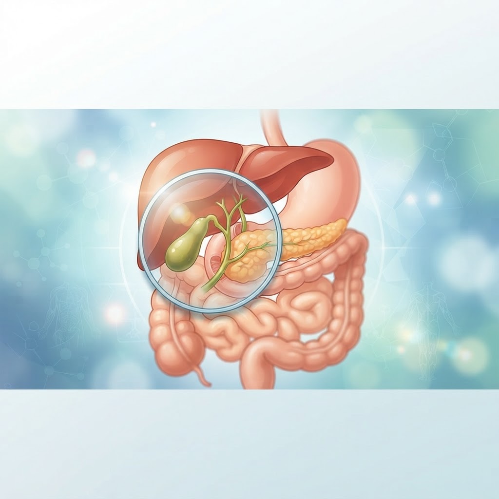
    The gallbladder is a small, pear-shaped organ that often goes unnoticed until it begins to cause trouble. For many, the onset of sharp abdominal pain serves as a jarring introduction to the world of gallstone health. Whether you are experiencing mild discomfort or debilitating attacks, understanding the underlying mechanics of your digestive system is essential for long-term wellness. From the dietary triggers that tax your gallbladder to the subtle lifestyle adjustments that promote healthy bile flow, taking a proactive approach can make a significant difference. In this comprehensive guide, the healthauthoritylife Editorial Team explores the intricacies of gallbladder function, the common signs of trouble, and evidence-based natural strategies to help you manage your health effectively.

    What Are Gallstones and How Do They Form?

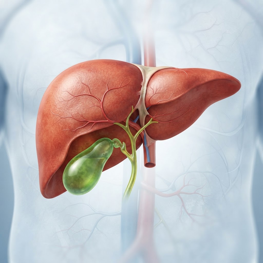
    Gallstones are small, hardened deposits that develop within the gallbladder, a digestive organ situated just beneath the liver. To understand what they are, one must first understand their primary job: storing bile. Bile is a greenish-yellow digestive fluid produced by the liver that helps the body break down fats during digestion. When you consume a meal containing fats, your gallbladder contracts, pushing this stored bile into the small intestine through a series of ducts.

    Gallstones essentially represent a chemical imbalance within this fluid. When bile becomes over-saturated with certain components—most commonly cholesterol or bilirubin—it can no longer maintain its liquid state. Over time, these excess substances crystallize and harden into pebble-like structures. These stones can range in size from a grain of sand to a large golf ball, and their composition dictates their appearance and behavior.

    The most common type of stone is the cholesterol stone. These typically appear yellow or green and are composed primarily of undissolved cholesterol. They form when the liver excretes more cholesterol than the bile can keep in solution. The second type, pigment stones, are generally darker and smaller, formed from an excess of bilirubin. Bilirubin is a byproduct created when the body breaks down old red blood cells. While less common, these pigment stones are often associated with underlying liver conditions or blood disorders.

    The formation process is rarely instantaneous. It is a progressive journey that begins with “biliary sludge.” This is a thicker, more viscous version of bile that contains microscopic crystals. If the gallbladder does not empty effectively or if the bile chemistry remains imbalanced for a prolonged period, these crystals eventually clump together. Once they aggregate, they become stones. Because these stones are located in a confined space, they can migrate into the bile ducts, causing blockages that trigger severe pain and complications. Understanding that these stones are essentially a localized “clog” in your digestive plumbing helps clarify why dietary interventions and movement are so frequently recommended as a foundational aspect of gallbladder health.

    Common Symptoms of Gallstones to Watch For

    Recognizing gallstone symptoms early can prevent significant distress. It is important to note that many individuals carry gallstones without ever experiencing symptoms—a state often referred to as “silent stones.” However, when a stone moves or blocks a duct, the body responds with distinct warning signals that are difficult to ignore.

    The hallmark symptom is sudden, rapidly intensifying pain in the upper right quadrant of the abdomen. This sensation is often colloquially described as a gallbladder attack. The pain typically radiates to the back, specifically between the shoulder blades or directly toward the right shoulder. Because this pain can be sharp and stabbing or a dull, constant ache, it is frequently mistaken for heart-related issues or severe indigestion.

    Another classic indicator is the timing of the discomfort. Gallbladder pain often flares up shortly after eating, particularly after consuming meals high in fat or processed oils. Because the gallbladder is responsible for releasing bile to emulsify these fats, a meal high in grease demands a forceful contraction. If a stone is obstructing the outlet, that contraction causes pressure to build up rapidly, resulting in the characteristic pain.

    Beyond acute pain, there are several systemic signs that suggest your gallbladder is struggling. Nausea and vomiting are common during an active episode. In more persistent cases, individuals may notice changes in bowel movements, such as clay-colored or pale stools, which indicate that bile is not reaching the intestines correctly. Furthermore, persistent jaundice—a yellowing of the skin or the whites of the eyes—is a significant red flag. Jaundice suggests that a stone has potentially lodged in the common bile duct, preventing the normal drainage of bilirubin and forcing it back into the bloodstream.

    Finally, some people experience “silent” symptoms that manifest as chronic, low-level digestive issues. This includes frequent bloating, excessive gas, or an intolerance to fatty foods that causes an overall feeling of heaviness in the stomach. While these symptoms can also be attributed to other digestive conditions like GERD or IBS, they should be investigated if they occur consistently after fatty meals. If you find yourself frequently reaching for antacids or avoiding entire food groups to prevent discomfort, it is highly advisable to consult with a healthcare professional to rule out gallbladder involvement.

    Approach Primary Focus Best For
    Low-Fat Diet Reducing gallbladder demand Immediate pain management
    Hydration Strategy Thinning bile consistency Long-term sludge prevention
    Fiber Supplementation Managing cholesterol levels Regulating bile chemistry
    Regular Movement Physical gallbladder stimulation Maintaining consistent flow

    Root Causes and Risk Factors for Gallstone Development

    What causes gallstones is rarely a single, isolated event. Instead, it is typically the result of a combination of genetic predispositions, dietary patterns, and metabolic health. While some risk factors remain outside of our control—such as age, gender, and family history—many others are deeply tied to lifestyle choices that can be modified to reduce overall risk.

    One of the most prominent risk factors is diet. High consumption of refined carbohydrates and added sugars has been linked to increased stone formation. These types of foods trigger spikes in insulin, which can alter cholesterol metabolism in the liver. When the liver is overloaded with metabolic work, it often excretes excess cholesterol into the bile. Additionally, rapid weight loss is a well-documented contributor. When a person sheds weight too quickly through extreme calorie restriction, the body releases large amounts of cholesterol into the bile while simultaneously causing the gallbladder to contract less frequently. This “stagnation” creates the perfect environment for crystals to settle and grow.

    Gender and hormonal factors play a significant role as well. Women are significantly more likely to develop gallstones than men. This is largely attributed to the hormone estrogen. High levels of estrogen, whether natural, from pregnancy, or through hormone replacement therapy, are known to increase the concentration of cholesterol in the bile while slowing gallbladder motility. This hormonal influence explains why many women experience their first symptoms during or shortly after pregnancy.

    Metabolic conditions like insulin resistance and obesity also create a high-risk environment. Obesity often involves a diet high in calories and fat, which keeps the gallbladder in a constant state of overwork. Meanwhile, insulin resistance interferes with the healthy processing of fats in the liver. Over time, these systemic issues lead to bile that is “lithogenic,” meaning it is prone to stone formation. Age is another factor, as the risk of gallstone development increases as we grow older; the gallbladder’s ability to empty completely can diminish with time, leading to the accumulation of older, thicker bile.

    Finally, it is important to consider the role of chronic inflammation in the body. If the digestive system is under constant stress from food sensitivities or an unbalanced microbiome, the resulting inflammation can affect liver function. Since the liver is the “gatekeeper” of bile quality, anything that compromises its efficiency—such as alcohol consumption, certain medications, or environmental toxins—will inevitably impact the composition of your bile. By understanding these root causes, it becomes clear that preventing gallstones is less about “avoiding fat” and more about optimizing metabolic and liver health through a balanced, nutrient-dense lifestyle.

    How Gallbladder Sludge Transitions Into Hard Stones

    The progression from healthy, free-flowing bile to a solid gallstone is a fascinating yet problematic biological process. This transition is not always linear; it often happens in stages, characterized by what clinicians call “biliary sludge.” When we discuss gallbladder health, we often focus on the hard stones that show up on imaging, but the sludge that precedes them is arguably more important for those interested in prevention and natural relief.

    Biliary sludge is a suspension of tiny crystals, mucus, and calcium salts. It is essentially the “sediment” at the bottom of the bile reservoir. Under normal, healthy conditions, the gallbladder empties completely during digestion, flushing out any microscopic debris. However, if the gallbladder lacks the proper signal to contract, or if the bile is too thick to move through the narrow cystic duct, this debris stays behind. Over time, the watery portion of the bile is reabsorbed by the gallbladder wall, leaving behind a concentrated, pasty substance that is even more difficult to eject.

    As this sludge sits, it creates a hospitable environment for chemical binding. Cholesterol molecules, which are hydrophobic, begin to nestle together to avoid the aqueous environment of the bile. As they cluster, they provide a scaffold for other substances like bilirubin and calcium. This is the nucleation phase. Once the microscopic crystals reach a certain size, they are no longer just floating in the sludge; they become solid entities. Once these entities are formed, they act as magnets, attracting more cholesterol and other components to build layer upon layer.

    The speed at which this transition happens varies from person to person. For some, the environment remains in the sludge phase for years, causing only intermittent digestive symptoms. For others, a single trigger—perhaps a period of extreme stress, a change in diet, or a medication-induced slowdown of gallbladder motility—can catalyze the hardening process into mature stones. This is why many people report that their symptoms seemed to appear “out of nowhere.” In reality, the physical conditions for stone formation had been slowly building for a significant amount of time.

    Addressing this transition is the core focus of many natural gallstone treatment strategies. The goal is to improve the “lithogenicity index” of your bile—essentially, making it more soluble and less likely to crystallize. By incorporating foods that encourage gallbladder contraction and staying adequately hydrated, you can discourage the accumulation of sludge. Preventing that first layer of crystal formation is much easier than trying to break down a hard, mature stone. Consequently, viewing your gallbladder as a dynamic organ that requires regular “flushing” through consistent, healthy eating patterns is a powerful mental shift for anyone looking to support their digestive health.

    The Role of Bile Flow in Preventing Gallstone Formation

    If there is one “secret” to maintaining gallbladder health, it is the importance of consistent, robust bile flow. Bile is designed to be a liquid, but in the modern sedentary, processed-food environment, it often becomes stagnant. Ensuring that bile moves regularly from the liver, through the gallbladder, and into the small intestine is the single most effective way to prevent the formation of gallstones and the accumulation of sludge.

    Bile flow is stimulated primarily by the presence of fats in the diet. When you eat a meal with a healthy portion of fats—such as avocados, olive oil, or nuts—the small intestine releases a hormone called cholecystokinin (CCK). CCK travels through the bloodstream and signals the gallbladder to contract. This contraction acts like a reset button, squeezing out the stored bile and preventing it from sitting long enough to become concentrated and crystalline. Problems arise when people go on ultra-low-fat diets, thinking they are helping their gallbladder. By eliminating fat, they effectively stop the signal for the gallbladder to empty. The bile sits in the gallbladder, loses water, becomes sludgy, and the likelihood of stone formation increases drastically.

    Beyond dietary fat, physical movement is a secondary, yet powerful, pump for the gallbladder. The physical pressure of abdominal muscles during movement, particularly exercises like walking, swimming, or yoga, can physically assist in the contraction of the gallbladder. When we remain stationary for long periods, the lymphatic and digestive systems slow down, leading to a sluggishness that extends to the bile ducts. Integrating regular, moderate activity is a simple but highly effective gallbladder health tip.

    Fiber intake also plays a critical, often overlooked, role. Soluble fiber, found in foods like oats, legumes, and certain fruits, binds to bile acids in the intestines and encourages their excretion. This forces the liver to produce fresh, high-quality bile using available cholesterol from the bloodstream. By constantly cycling through this process, your body maintains a healthier cholesterol profile within the bile itself. It’s a self-regulating loop: healthy fiber intake leads to healthier bile, which prevents the stagnation that causes stones.

    Finally, we must consider hydration. Bile is mostly water. If you are chronically dehydrated, the liver may struggle to produce bile with the appropriate viscosity. Think of it like a river: when the water levels are high, the current is strong and carries debris downstream. When the water level is low, the current becomes weak, and mud and debris settle on the riverbed. Drinking plenty of water throughout the day ensures that your bile remains fluid and easy to transport. By combining these three elements—healthy fats to signal contraction, fiber to manage chemistry, and water for flow—you create an environment that is naturally resistant to the conditions that allow gallstones to form. Maintaining this flow is the hallmark of a gallbladder-friendly lifestyle.

    Dietary Changes to Support Healthy Gallbladder Function

    The gallbladder acts as a reservoir for bile, a digestive fluid produced by the liver that is essential for breaking down dietary fats. When the gallbladder becomes sluggish or congested, the bile can crystallize, leading to the formation of gallstones. Consequently, the cornerstone of gallbladder health lies in your daily plate. A gallstone diet focuses on reducing the workload on your gallbladder while ensuring bile flow remains consistent and thin.

    One of the primary dietary shifts recommended for those seeking gallbladder pain relief is the transition to a high-fiber, plant-forward eating pattern. Fiber, particularly soluble fiber found in oats, beans, lentils, and apples, helps to bind to bile acids in the digestive tract, encouraging the liver to pull more cholesterol from the blood to produce new bile. This process can help maintain a healthier balance within the gallbladder.

    Equally important is the quality of fats consumed. While severe fat restriction can actually cause the gallbladder to stop contracting—which ironically leads to stone formation—choosing the right fats is critical. Focus on healthy, unsaturated fats such as those found in avocados, raw walnuts, and extra virgin olive oil. Avoid trans fats and heavily processed vegetable oils like soybean or corn oil, which can trigger inflammatory responses in the digestive system.

    Incorporating bitter foods into your meals can serve as a natural gallbladder stimulant. Bitter greens such as arugula, radicchio, dandelion greens, and kale signal the digestive system to ramp up bile production. Many traditional wellness practices advocate for consuming these bitters 15 to 20 minutes before a main meal to prime the gallbladder for the digestive process.

    Staying adequately hydrated is another non-negotiable dietary habit. Bile is largely composed of water, and when the body is dehydrated, bile becomes more viscous and prone to sludge formation. Aiming for consistent water intake throughout the day, rather than large amounts at once, ensures that your liver has the necessary fluid volume to maintain healthy bile consistency.

    Food Category Examples Impact on Gallbladder Best For
    Soluble Fiber Oats, Apples, Flaxseeds Reduces cholesterol levels in bile Long-term prevention
    Bitter Greens Dandelion, Arugula Stimulates natural bile flow Improving digestion
    Healthy Fats Avocado, Olive Oil Promotes healthy gallbladder contraction Preventing sludge
    Anti-Inflammatory Herbs Turmeric, Ginger Reduces biliary tract inflammation Pain management

    Natural Supplements That May Help Improve Bile Quality

    While food is the foundation, specific nutrients can offer targeted support for individuals looking into natural gallstone treatment options. These supplements are often used to thin the bile, improve liver detoxification pathways, or reduce the localized inflammation that causes discomfort during a gallbladder attack.

    Magnesium is frequently cited for its role in muscle relaxation, including the smooth muscles of the bile ducts. When the bile ducts are relaxed, bile flow is less obstructed, which may reduce the intensity of biliary colic. Many people find that a magnesium glycinate supplement taken before bed helps support general digestive comfort and calm the autonomic nervous system.

    Taurine is an amino acid that plays a vital role in the conjugation of bile acids. Essentially, taurine makes bile more water-soluble, which prevents it from becoming thick or “sludgy.” By ensuring the bile remains fluid, taurine may help prevent the stagnation that allows crystals to grow into larger, more problematic stones.

    Lecithin is another supplement often discussed in the context of gallbladder health. Sunflower-derived lecithin is a rich source of phospholipids, which are natural emulsifiers. By emulsifying fats in the bile, lecithin helps keep cholesterol dissolved, theoretically making it more difficult for cholesterol-based stones to form or grow. It is often used by those looking to support bile composition.

    Herbal extracts such as milk thistle and artichoke leaf extract are also highly regarded. Milk thistle contains silymarin, which supports overall liver function, the factory where bile is produced. If the liver is functioning optimally and producing high-quality bile, the gallbladder is under significantly less stress. Artichoke leaf is a classic choleretic, meaning it promotes the flow of bile from the gallbladder into the small intestine, acting as a gentle physical cleaner for the biliary tree.

    Lifestyle Habits to Reduce Gallbladder Inflammation

    Addressing gallstone symptoms requires more than just dietary changes; it requires a holistic look at how your lifestyle affects systemic inflammation. High levels of chronic inflammation can exacerbate gallbladder pain and slow down the body’s natural healing processes.

    Weight management is perhaps the most significant lifestyle factor. Rapid weight loss, often triggered by “crash diets” or extreme caloric restriction, is a known trigger for gallstone formation. When you lose weight too quickly, the liver secretes extra cholesterol into the bile, while the gallbladder empties less frequently. A slow, steady approach to weight loss is much safer for gallbladder health. Aim for lifestyle changes that support gradual progress rather than aggressive, short-term fixes.

    Stress management is often overlooked in digestive health. The gallbladder is intimately connected to the parasympathetic nervous system—the “rest and digest” state. When we are chronically stressed, our bodies operate in “fight or flight” mode, which inhibits digestive processes, including gallbladder contraction. Incorporating mindfulness practices, such as deep diaphragmatic breathing or gentle yoga, can help transition the body into a digestive state where bile can be released effectively.

    Regular, low-to-moderate intensity physical activity also plays a part. Exercise helps regulate metabolic health and improves insulin sensitivity. Since insulin resistance is often associated with higher concentrations of cholesterol in the bile, keeping blood sugar stable through consistent movement is an effective way to protect the gallbladder over the long term.

    Finally, avoid the common habit of late-night snacking. Giving your digestive system a long break overnight—often called intermittent fasting or time-restricted eating—allows the gallbladder to rest and fully reset its bile storage levels. A 12-hour overnight fast is a common recommendation to allow the digestive organs the downtime they need to prevent congestion.

    When to Seek Medical Attention for Gallbladder Pain

    While natural strategies are powerful tools for maintenance and support, it is critical to recognize when gallbladder issues have moved beyond the realm of self-care. Gallbladder attacks can be severe, and in some cases, can lead to complications such as infection, pancreatitis, or ductal blockage that require emergency intervention.

    You should seek immediate medical evaluation if you experience severe, persistent abdominal pain in the upper right quadrant that lasts for more than a few hours. Pain that radiates to the back or the right shoulder blade is a hallmark of gallbladder distress. If this pain is accompanied by a fever or chills, it may indicate an infection, which is a medical emergency.

    Jaundice—the yellowing of the skin or the whites of the eyes—is a red flag that a stone may have slipped into the common bile duct and is causing an obstruction. This is a serious condition that requires prompt diagnosis. Additionally, if your stool becomes unusually pale or clay-colored, or if your urine becomes dark, these are physical signals that bile flow is being blocked, necessitating a trip to the physician.

    Even if pain is intermittent, it is wise to consult a healthcare provider for an ultrasound to confirm the presence of stones. Understanding the size, location, and composition of your stones can help you determine whether your condition can be managed through lifestyle changes or if you are at high risk for complications that require more direct medical intervention.

    Frequently Asked Questions

    Can gallstones be completely dissolved through natural methods?

    While some small, cholesterol-based gallstones may occasionally dissolve through specific dietary changes and bile-supporting supplements, there is no guarantee. Most large or calcified stones are unlikely to dissolve completely. Natural strategies are generally most effective for preventing new stones from forming and managing discomfort rather than removing existing large stones.

    What does a typical gallbladder attack feel like?

    A gallbladder attack typically presents as intense, steady pain in the upper right abdomen, just below the ribs. This pain often comes on suddenly, particularly after eating a high-fat meal, and may last from 30 minutes to several hours. Many people describe it as a feeling of pressure or cramping that can radiate to the back or shoulder.

    Is coffee good or bad for the gallbladder?

    Some research suggests that moderate coffee consumption may be protective for the gallbladder. Caffeine can stimulate gallbladder contraction and improve bile flow. However, for some individuals, coffee can be an irritant. It is best to monitor your own symptoms; if coffee triggers pain, it is best to avoid it regardless of potential benefits.

    Should I avoid all fats if I have gallstones?

    No, avoiding all fats is actually counterproductive. The gallbladder needs the signal of dietary fat to contract and empty its contents. If you stop eating fat entirely, the gallbladder becomes stagnant, which can increase the risk of more stones forming. Instead, focus on small, consistent portions of healthy, plant-based fats.

    Can stress really cause a gallbladder attack?

    Stress impacts the autonomic nervous system, which controls digestion. When the body is in a state of high stress, digestive motility slows down. This can lead to bile stasis, where bile sits in the gallbladder too long, becoming thick and sludge-like, which may trigger or worsen an existing gallbladder issue.

    How long does it take for dietary changes to show results?

    Digestive health is a gradual process. While some people may notice a reduction in mild bloating or discomfort within a few weeks of improving their diet, significant changes to bile quality and gallbladder function typically take several months of consistent habit changes to manifest. Patience and consistency are key.

    Conclusion

    Navigating gallbladder health requires a balance of informed lifestyle choices, a deep understanding of your body’s signals, and a commitment to long-term dietary wellness. By focusing on bile-thinning foods, stress reduction, and maintaining a healthy pace of life, you can significantly support your gallbladder function and minimize the impact of gallstone symptoms. Remember that your digestive system thrives on consistency and quality nourishment.

    If you have found these strategies helpful, start by making one small change this week—such as incorporating a bitter green into your daily meal or increasing your water intake. For those currently managing discomfort, keep a food journal to identify your personal triggers and work toward a sustainable, pain-free lifestyle. Take control of your digestive health today by prioritizing the organ that does so much work behind the scenes to keep you feeling your best.

    By healthauthoritylife Editorial Team

    This article is for informational purposes only and is not a substitute for professional medical advice. Always consult your doctor before making changes to your diet, exercise routine, or treatment plan.

  • Gallbladder Sludge: Symptoms, Causes, and Natural Relief Tips

    Gallbladder Sludge: Symptoms, Causes, and Natural Relief Tips

    Key Takeaways

    • Gallbladder sludge is a precursor to gallstones, consisting of a mixture of bile pigments, cholesterol crystals, and calcium salts.
    • Common biliary sludge symptoms often mimic indigestion, including upper abdominal pain, nausea, and bloating after fatty meals.
    • Sludge accumulation is frequently driven by stasis in the gallbladder, often caused by rapid weight loss, pregnancy, or dietary imbalances.
    • Improving bile flow support is essential for preventing the hardening of sludge into solid, painful gallstones.
    • Natural treatment for sludge primarily focuses on hydration, dietary adjustments, and supporting healthy liver and gallbladder function.

    If you have ever experienced an unexplained, dull ache in your upper right abdomen or a recurring sense of bloating after a meal, your gallbladder might be trying to tell you something. While many people are familiar with gallstones, far fewer understand the silent precursor known as gallbladder sludge. This “pre-stone” condition acts as a warning sign that your digestive system is struggling to process fats effectively and that your internal filtration system—your bile—has become stagnant. Understanding the nuances of this condition is the first step toward reclaiming your digestive comfort and supporting long-term gallbladder health.

    What Is Gallbladder Sludge and Why Does It Form?

    At its core, gallbladder sludge is a collection of microscopic particles that accumulate within the gallbladder. Imagine your bile—the digestive fluid produced by the liver—as a complex, fluid mixture of water, cholesterol, bile salts, and bilirubin. Under healthy conditions, this mixture remains fluid, flowing easily from the liver through the gallbladder and into the small intestine to aid in the digestion of fats. However, when the concentration of these substances becomes unbalanced, the fluid begins to thicken, much like the difference between water and a dense smoothie.

    The sludge itself is primarily composed of cholesterol crystals, calcium salts, and other bile pigments that have settled out of the liquid suspension. When this thick, sediment-like material lingers in the gallbladder instead of being emptied into the digestive tract, it is referred to as biliary sludge. Think of it as a form of “stasis.” Because the gallbladder is a muscular sac, it relies on timely contractions to push bile out during digestion. If the gallbladder does not contract fully or if the bile becomes too viscous, this sludge begins to layer at the bottom of the organ.

    Why does this form in the first place? It is largely a matter of chemistry and kinetics. If the gallbladder stays inactive for too long—perhaps due to a very low-fat diet or long periods of fasting—the bile remains inside the sac, losing its water content and becoming hyper-concentrated. Over time, these concentrated solutes begin to precipitate. Once the process of sludge formation begins, it often creates a vicious cycle; the sludge itself can irritate the gallbladder lining, leading to inflammation, which in turn makes it even more difficult for the gallbladder to contract and empty properly. Many experts generally agree that if this sludge is ignored, it can eventually provide a foundation for the development of solid, calcified gallstones, which are significantly more difficult to manage.

    From an anatomical perspective, the gallbladder serves as a storage tank. When you consume fat, the body releases a hormone called cholecystokinin (CCK), which signals the gallbladder to squeeze. If your diet lacks healthy fats, that signal may be weak, causing the gallbladder to remain dormant. This dormancy is the primary culprit in why so many individuals begin to develop sediment. By maintaining a consistent, healthy flow of bile through a balanced diet, you effectively keep your gallbladder “flushed,” preventing the tiny crystals from ever finding a place to settle and grow into a larger, more problematic mass.

    Common Symptoms of Biliary Sludge to Watch For

    Recognizing the biliary sludge symptoms can be challenging because they often masquerade as garden-variety indigestion, heartburn, or general GI distress. Because the symptoms are non-specific, many individuals live with them for months or even years before ever realizing their gallbladder is the source of the trouble. The most classic presentation is a sensation of discomfort in the upper right quadrant of the abdomen, specifically situated just below the rib cage. This pain may radiate toward the back or even up toward the right shoulder blade, which is a common pattern for gallbladder-related issues.

    This discomfort often intensifies significantly after consuming a meal high in fat. When you eat a fatty meal, your gallbladder is commanded to empty its contents into the small intestine. If the gallbladder is filled with thick, viscous sludge rather than healthy, free-flowing bile, the act of contracting becomes strained and painful. This is frequently accompanied by feelings of extreme bloating or a sensation of “fullness” that seems disproportionate to the amount of food you actually consumed. Some report a “tight” feeling in the abdomen that persists long after the meal is over, creating a heavy, uncomfortable physical state.

    Nausea is another hallmark symptom. It may not always manifest as full-blown vomiting, but rather a persistent, low-level queasiness, particularly when you first wake up in the morning or immediately following lunch. You might also notice shifts in your bowel habits. Because bile is essential for breaking down fats, insufficient bile flow—or bile that is sluggish—can lead to light-colored or “clay-colored” stools. Furthermore, because fats are not being properly emulsified, they may pass through the digestive tract undigested, potentially leading to greasy or floating stools, a condition known as steatorrhea.

    It is important to note that these symptoms can be intermittent. You might have days where you feel completely fine, followed by a flare-up after a particularly rich meal. This fluctuating nature often leads people to dismiss the warning signs as simple “food sensitivity.” However, if you find that you are regularly reaching for antacids, digestive enzymes, or peppermint tea to manage your post-meal comfort, it is likely that your body is signaling that its bile quality is compromised. Paying attention to these subtle cues is a critical part of supporting long-term gallbladder health before the sludge develops into something more acute, such as biliary colic or cholecystitis.

    Primary Causes and Risk Factors for Sludge Accumulation

    The causes of gallbladder sludge are diverse, though most paths lead back to the same result: impaired bile concentration and reduced gallbladder motility. One of the most significant risk factors is rapid weight loss. While shedding excess weight is generally beneficial for health, the speed at which it occurs matters. When you lose weight too quickly, especially through crash dieting or very-low-calorie intake, the body mobilizes large amounts of cholesterol from fat stores. This cholesterol is then processed by the liver and secreted into the bile, creating a “supersaturated” environment that is a perfect breeding ground for crystals and sludge.

    Pregnancy is another major factor often cited in health literature. Hormonal changes—specifically elevated levels of estrogen—can decrease the contractility of the gallbladder and increase the concentration of cholesterol in the bile. During pregnancy, the gallbladder often does not empty as effectively as it does under non-pregnant conditions, which contributes to the formation of sediment. Fortunately, in many cases, this sludge may resolve on its own after delivery, but it remains a notable risk factor for developing more permanent issues later in life.

    Factor Mechanism of Action Best For
    Rapid Weight Loss Increases cholesterol secretion into bile Understanding weight management risks
    Low-Fat Diets Reduces gallbladder stimulation/emptying Dietary planning and adjustments
    Hormonal Imbalance Decreases gallbladder muscle tone Addressing root systemic causes
    Medication Side Effects Interferes with normal biliary function Medical review and safety

    Dietary habits play perhaps the most controllable role. In our modern food environment, there is a tendency to oscillate between high-processed, high-sugar diets and overly restrictive “clean” diets that are nearly devoid of healthy fats. Both extremes are problematic. A diet high in refined carbohydrates and sugar can contribute to liver fat accumulation, which alters bile composition. Conversely, the “low-fat” craze of past decades has caused many people to neglect the very nutrients required to trigger gallbladder contraction. Without adequate healthy fats (like those from avocados, olive oil, or nuts), the gallbladder remains dormant, allowing bile to thicken and sludge to accumulate.

    Certain medical conditions and medications also contribute to the risk. Diabetes, which affects lipid metabolism, is frequently associated with gallbladder dysfunction. Furthermore, certain medications, particularly those that affect the liver or are used for cholesterol management, can alter the chemical makeup of bile. Genetics also play an undeniable role; some individuals naturally produce bile that is more prone to crystallization. While you cannot change your genetic predisposition, understanding these risk factors allows for a proactive, rather than reactive, approach to your daily wellness routine.

    The Critical Link Between Bile Quality and Digestion

    Many people view digestion as starting in the stomach, but true, efficient digestion is a team effort that relies heavily on the liver-gallbladder-intestine axis. Bile is the unsung hero of this process. It functions essentially as a biological detergent. When you consume fat, your body cannot simply dissolve it in the water-based environment of the small intestine. It needs to be emulsified—broken down into tiny droplets—so that enzymes like lipase can get to work and extract the nutrients your body needs, including essential fat-soluble vitamins like A, D, E, and K.

    When your bile is thin and healthy, it performs this emulsification with ease. However, when you have gallbladder sludge, the bile becomes thick and sluggish. This prevents it from effectively breaking down fats. As a result, those fats sit in the digestive tract, where they can be fermented by gut bacteria, leading to gas, bloating, and discomfort. But the problem goes beyond just digestive discomfort; it affects your entire nutritional profile. If your bile quality is compromised, you may find yourself suffering from vitamin deficiencies even if you are eating a nutrient-dense diet, simply because your body is unable to absorb the fat-soluble components of your food.

    Supporting healthy bile flow is a form of foundational health. When bile moves freely, it also acts as a natural “sweep” for the intestines. Bile contains waste products and toxins that the liver has processed and sent out of the body; if this bile flows correctly, it carries those toxins out via your bowel movements. When flow is stagnant, these waste products can be reabsorbed or cause irritation within the biliary tree. This is why biliary sludge is not just a localized gallbladder issue—it is a systemic concern that impacts your energy, your skin health, and your metabolic efficiency.

    To improve bile quality, one must look at both hydration and nutrient intake. Bile is composed largely of water; dehydration causes the bile to become concentrated and viscous. Staying adequately hydrated is one of the simplest yet most overlooked steps in ensuring the liver can produce high-quality, fluid bile. Furthermore, supporting the liver—the “factory” that produces bile—is vital. Foods that support phase two detoxification in the liver, such as cruciferous vegetables (broccoli, cauliflower, kale), provide the sulfur-containing compounds necessary to keep bile flowing optimally. By focusing on bile quality, you aren’t just treating the sludge; you are optimizing the entire metabolic engine that keeps your digestive system humming.

    Diagnostic Methods for Identifying Gallbladder Sludge

    Because biliary sludge is often asymptomatic or mild in its initial stages, diagnosing it requires specific imaging techniques. You cannot typically identify sludge through blood tests alone, although certain liver function panels might hint at an issue if the bile ducts are obstructed. When a health professional suspects sludge, the gold standard for identification is usually a transabdominal ultrasound. This non-invasive, painless procedure uses sound waves to create a real-time image of the gallbladder. During the exam, a technician will look for “echoes” within the gallbladder that do not cast a shadow (which is a defining feature of hard, solid gallstones).

    In many cases, the sludge may appear as a shifting, gravity-dependent layer of sediment that moves as the patient changes positions. This is a very clear marker of the condition. However, there are times when ultrasound may miss very small amounts of sludge. If a person continues to experience classic symptoms despite a negative initial ultrasound, a doctor might consider more advanced diagnostic tools, such as an endoscopic ultrasound. This procedure involves passing a small camera on the end of a flexible tube down the throat into the stomach and duodenum, providing a much higher-resolution view of the gallbladder and the surrounding bile ducts.

    Another, more specialized test involves the examination of bile under a microscope. This is known as bile microscopy or “bile aspiration.” In this procedure, a sample of bile is extracted—usually during an endoscopic procedure—and tested for the presence of microscopic cholesterol crystals. While this is rarely done as a first-line diagnostic due to its invasive nature, it is considered the most accurate method for confirming the presence of microscopic sludge that other imaging might miss. It is essentially the “final word” in diagnosis when clinical symptoms are severe but imaging remains inconclusive.

    Finally, health professionals will often correlate imaging results with a detailed clinical history. If an ultrasound shows a sludge-filled gallbladder, but the patient is entirely asymptomatic, the approach may simply be “watchful waiting” or preventative lifestyle shifts. Conversely, if the ultrasound shows sludge and the patient is suffering from chronic pain and digestive distress, it creates a much stronger mandate for intervention. Regardless of the diagnostic path taken, the ultimate goal is to define the extent of the accumulation so that a personalized strategy for gallbladder detox and bile flow support can be implemented. Understanding what is happening inside the gallbladder empowers you to take actionable steps toward better health, moving away from confusion and toward a clear, symptom-free future.

    Lifestyle Changes to Improve Your Gallbladder Function

    Supporting your gallbladder health requires a holistic approach that moves beyond temporary fixes. Since the gallbladder acts as a reservoir for bile, the goal of lifestyle modification is to ensure that bile remains thin, fluid, and capable of being released efficiently during digestion. When bile becomes stagnant, it thickens into sludge. By addressing sedentary habits and stress levels, you can directly influence the motility of your biliary system.

    Regular physical activity is one of the most effective ways to encourage bile flow. The mechanical action of moving your body helps stimulate peristalsis, the wave-like muscle contractions that move food and bile through the digestive tract. Even moderate, low-impact exercise like brisk walking, yoga, or swimming can help prevent the “sluggish” feeling associated with biliary stasis. Furthermore, maintaining a stable and healthy weight is vital. Rapid weight loss or extreme fluctuations can ironically trigger the formation of gallbladder sludge, so slow, steady, and sustainable lifestyle habits are always preferred over crash dieting.

    Stress management also plays an often-overlooked role in digestive health. Chronic stress activates the sympathetic nervous system—the “fight or flight” mode—which effectively puts the brakes on the parasympathetic nervous system, the “rest and digest” state. When your body is under stress, digestive processes, including gallbladder contraction, are deprioritized. Integrating techniques like deep diaphragmatic breathing, meditation, or consistent sleep hygiene can shift your body into a restorative state, allowing your gallbladder to function without the interference of stress-related chemical signaling.

    Lastly, hydration is a foundational requirement for bile consistency. Bile is composed primarily of water, cholesterol, bile salts, and bilirubin. When the body is dehydrated, the concentration of these substances shifts, making bile more prone to becoming viscous. Aiming for consistent water intake throughout the day, rather than large amounts at once, ensures that your liver has the necessary fluid volume to produce healthy, free-flowing bile.

    Dietary Adjustments for Reducing Biliary Stasis

    Dietary choices are perhaps the most direct lever you can pull to manage gallbladder sludge. Because the gallbladder is stimulated to contract when you consume dietary fats, your intake must be balanced—neither too low nor too high. If you consume too little fat, the gallbladder may not receive the signal to empty, leading to accumulation. If you consume highly processed or inflammatory fats, you may put excessive strain on your liver and gallbladder.

    The “Goldilocks” approach involves consuming moderate amounts of healthy, easily digestible fats throughout the day. Focus on fats that support healthy cell membranes and do not require excessive bile for breakdown, such as those found in avocados, raw nuts, and cold-pressed extra virgin olive oil. High-fiber foods are equally essential. Soluble fiber, found in oats, chia seeds, flaxseeds, and legumes, helps bind to excess cholesterol in the digestive tract. By preventing this cholesterol from being reabsorbed back into the liver, you reduce the overall burden on the bile and decrease the likelihood of sludge formation.

    Additionally, incorporating bitter-tasting foods into your meals can act as a natural stimulant for bile production. Traditional diets frequently included bitter herbs and vegetables like arugula, dandelion greens, radicchio, and artichokes. These foods are known as “bitters,” and they work by stimulating taste receptors that signal the digestive system to ramp up enzyme and bile secretion. Reducing the intake of refined carbohydrates and high-glycemic sugars is also critical, as high insulin levels have been linked to an increased risk of cholesterol crystallization in the bile.

    Dietary Category Examples Best for
    Bitter Vegetables Arugula, Radicchio, Dandelion Greens Stimulating natural bile secretion
    Soluble Fiber Chia seeds, Flaxseeds, Oats Binding to cholesterol in bile
    Healthy Fats Extra virgin olive oil, Avocado Gentle gallbladder contraction
    Hydration Boosters Cucumber, Celery, Lemon water Maintaining thin, liquid bile

    Herbs and Supplements That Support Bile Production

    When dietary adjustments are not enough, certain botanical interventions and supplements can provide targeted support for bile flow. These agents are often referred to as “cholagogues,” which are substances that promote the flow of bile from the gallbladder into the intestines, and “choleretics,” which increase the production of bile by the liver.

    Milk thistle is perhaps the most well-known herb for liver support. Its active compound, silymarin, helps protect liver cells from oxidative stress and may assist in the overall efficiency of liver detoxification pathways, which in turn improves the quality of bile produced. Another excellent option is artichoke leaf extract. Clinical interest in artichoke extract often centers on its ability to increase bile output, making it a staple in many digestive support protocols. Turmeric, specifically the compound curcumin, also offers anti-inflammatory benefits and has been shown to support the solubility of bile, potentially helping to prevent the hardening of sludge into stones.

    For those looking to improve bile flow directly, taurine is an amino acid that acts as a primary building block of bile salts. Taurine deficiency can lead to poor bile acid conjugation, resulting in thicker, more stagnant bile. Supplementing with taurine, under appropriate guidance, may help “thin out” the sludge by ensuring the chemical components of bile are balanced. Similarly, digestive enzymes containing lipase can help take the workload off the gallbladder by ensuring fats are broken down efficiently even when bile flow is suboptimal. Always ensure that any supplement chosen is sourced from a reputable manufacturer to ensure purity and potency.

    When to Consult a Specialist About Your Gallbladder Health

    While lifestyle and natural remedies are powerful tools for managing minor sludge, there is a clear threshold where professional medical intervention becomes necessary. The gallbladder is a critical organ, and complications such as cholecystitis (inflammation), choledocholithiasis (stone in the common bile duct), or pancreatitis can arise if sludge transitions into full-blown gallstones or leads to a blockage.

    You should consult a gastroenterologist or a surgeon if you experience persistent or intensifying upper-right abdominal pain, especially after high-fat meals. This pain may radiate to the right shoulder blade or back. Other warning signs include jaundice (a yellowing of the skin or the whites of the eyes), which indicates that bile is backing up into the bloodstream; unexplained clay-colored stools; or dark-colored urine. These are red flags that the biliary tree may be obstructed.

    Furthermore, if you experience sudden, sharp, and unrelenting pain accompanied by a fever or chills, this could indicate an infection. In these scenarios, natural remedies are not appropriate, and emergency care may be required. A doctor can use imaging tools like abdominal ultrasounds to visualize the gallbladder and determine the exact amount of sludge present and whether there is any evidence of inflammation or tissue damage. Never ignore persistent digestive discomfort; getting a clear diagnosis is the safest way to determine if you can manage the situation with lifestyle changes or if you require medical intervention to prevent long-term damage.

    Frequently Asked Questions

    Can gallbladder sludge eventually turn into gallstones?

    Yes, gallbladder sludge is often considered a precursor to gallstones. If the sludge remains in the gallbladder for an extended period, the cholesterol and other substances within it can begin to crystallize and aggregate, eventually forming solid stones. Regular management of bile flow is key to preventing this transition.

    Is it possible to completely clear sludge from the gallbladder naturally?

    Many individuals find success in reducing or eliminating gallbladder sludge through a combination of a low-inflammatory diet, regular movement, and targeted bile-supporting supplements. However, results vary significantly based on the severity of the condition and individual health history. It is essential to monitor your progress with your healthcare provider.

    What does “biliary colic” feel like in relation to sludge?

    Biliary colic is a sharp, cramping pain in the upper right quadrant of the abdomen that occurs when the gallbladder contracts against a temporary blockage, such as sludge or a stone. While it often subsides once the contraction stops, it is a significant signal that the gallbladder is struggling to empty efficiently.

    Should I avoid all fats if I have gallbladder sludge?

    No, avoiding all fats is generally counterproductive. Fats are the primary stimulus for gallbladder contraction. If you stop eating fat entirely, your gallbladder may stop contracting, which actually promotes more stagnation and sludge buildup. The goal is to consume small, moderate amounts of healthy fats to keep the system moving.

    Are there certain foods that definitely trigger sludge accumulation?

    Foods high in refined sugars, processed carbohydrates, and trans fats are frequently associated with increased risk. These foods can cause blood sugar spikes and liver stress, both of which affect the composition of bile and the liver’s ability to process cholesterol effectively, leading to a higher concentration of sludge.

    How long does it take to improve bile flow through diet?

    There is no fixed timeline, as everyone responds differently. Generally, people notice improvements in digestive comfort within a few weeks of consistent dietary changes. However, clearing long-standing sludge is a gradual process that may take several months of dedicated lifestyle support.

    Conclusion

    Gallbladder sludge is more than just a minor digestive annoyance; it is a clear signal from your body that your biliary system is in need of support. By understanding the causes—such as poor diet, lack of movement, and internal imbalances—you can take actionable steps to restore proper bile flow. Integrating bitter greens, supporting your liver with the right herbs, and managing your stress levels are not just “wellness trends”; they are foundational practices that protect one of your body’s most vital detoxification organs.

    If you have been feeling the symptoms of biliary stasis, start small. Begin by hydrating more effectively, incorporating one bitter vegetable into your daily routine, and speaking with a medical professional to ensure your digestive health is on the right track. Remember, the journey to a healthier gallbladder is built on consistency and informed choices. Start prioritizing your digestive flow today to experience the relief and vitality your body deserves.

    By healthauthoritylife Editorial Team

    This article is for informational purposes only and is not a substitute for professional medical advice. Always consult your doctor before making changes to your diet, exercise routine, or treatment plan.
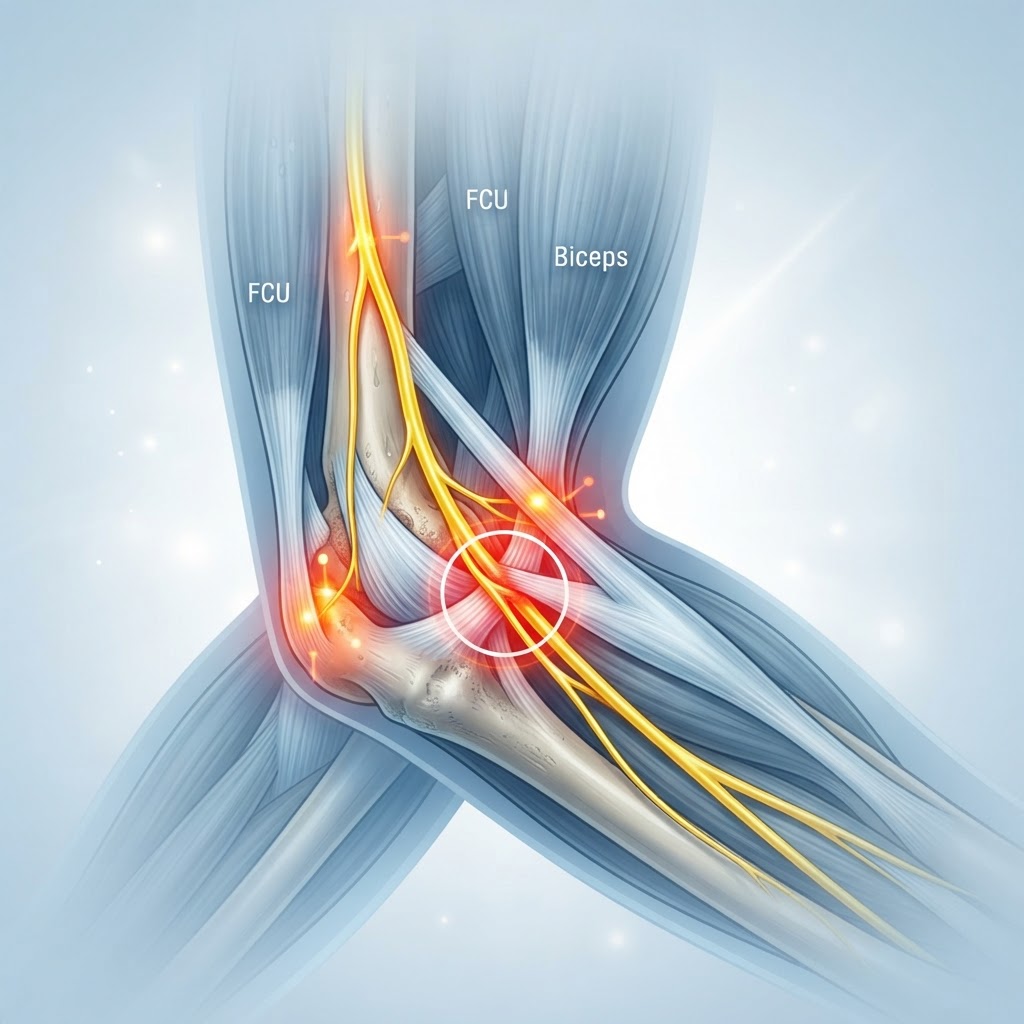
  • Restless Leg Syndrome: Causes, Triggers, and Natural Relief

    Restless Leg Syndrome: Causes, Triggers, and Natural Relief

    Key Takeaways

    • Restless leg syndrome is a neurological sensory disorder characterized by an uncontrollable urge to move the legs, especially during periods of inactivity.
    • Common underlying factors include iron deficiency, magnesium imbalances, and disruptions in dopamine signaling within the brain.
    • Symptoms are often most severe in the evening and nighttime, leading to significant sleep deprivation and decreased quality of life.
    • Natural remedies for RLS often focus on optimizing nutritional status, improving sleep hygiene, and incorporating gentle movement or compression.
    • Addressing chronic RLS frequently requires a multi-faceted approach that balances medical supervision with lifestyle modifications.

    For those who struggle to find peace at the end of a long day, the sensation often begins as a subtle, creeping irritation. It is not quite pain, yet it is impossible to ignore—a deep, rhythmic ache or a “crawling” feeling that demands movement. This is the reality for millions living with restless leg syndrome, a condition that transforms the simple act of resting into a nightly battle. While the medical community continues to explore the complexities of this neurological disorder, the path to reclaiming restful sleep often begins with understanding the intersection of internal physiology and external triggers. As we navigate the nuances of this condition, it becomes clear that relief is rarely found in a single solution, but rather through a comprehensive understanding of the body’s internal signals and the evidence-based strategies that provide sustainable comfort.

    Understanding Restless Leg Syndrome (RLS)

    Restless leg syndrome, or Willis-Ekbom disease, is classified as a neurological sensory disorder that significantly impacts the ability to rest. At its core, the condition is defined by an irresistible urge to move the limbs, which typically happens when an individual is sedentary, such as when sitting for a long commute, attending a movie, or attempting to fall asleep. The sensation is often described as deep, internal, and uniquely bothersome—patients frequently report feelings of pulling, itching, burning, or even a sensation described as “ants crawling under the skin.”

    What makes this condition particularly challenging is its cyclical nature. The sensations almost universally subside or vanish entirely when the individual engages in physical movement, such as walking or stretching. However, the relief is usually temporary. Once the movement ceases, the symptoms return, often with increased intensity if the person has become overtired. This phenomenon creates a vicious cycle where the inability to find comfort leads to profound sleep fragmentation.

    Experts generally agree that RLS is not merely a “behavioral” issue or a simple case of nerves. It is deeply rooted in the central nervous system. While the exact neurological mechanisms are still being mapped, current research points toward a complex interplay involving the brain’s basal ganglia—the area responsible for controlling movement—and the neurotransmitter dopamine. Dopamine acts as the body’s primary messenger for smooth, controlled muscle activity. When dopamine pathways are disrupted, the body’s ability to “turn off” the urge to move during rest is compromised, resulting in the characteristic restlessness.

    Furthermore, RLS is often categorized into two types: primary and secondary. Primary RLS is considered idiopathic, meaning it appears to have a genetic component and often manifests earlier in life. Many people with primary RLS report a strong family history, suggesting that certain genetic predispositions make some individuals more susceptible to these neural signals. Conversely, secondary RLS typically develops later in life and is often linked to underlying health conditions, medication side effects, or nutritional deficiencies. Understanding which category one falls into is the first step toward effective management. Because the symptoms are subjective, diagnosis often relies on a clinical interview rather than a single blood test or scan, highlighting the importance of clear communication with healthcare providers about the specific nature, timing, and frequency of these symptoms. By observing when the sensations occur and what triggers them, those suffering can begin to differentiate between occasional discomfort and the chronic nature of RLS.

    Common Symptoms and Daily Impact

    The symptoms of restless leg syndrome extend far beyond simple “twitching.” While the urge to move is the hallmark, the daily impact of this disorder ripples through every facet of a person’s existence. Symptoms typically peak during the evening and night hours, creating a direct conflict with the body’s natural need for recovery. The physical sensations are frequently accompanied by periodic limb movements in sleep (PLMS), which are involuntary jerks or kicks that occur throughout the night. Even if the person remains asleep, these movements can prevent the body from entering the restorative, deep stages of sleep, leaving them exhausted upon waking.

    The emotional and cognitive toll of chronic sleep deprivation is substantial. Individuals often describe a “brain fog” that persists throughout the day, manifesting as difficulty concentrating, irritability, and a diminished capacity to handle stress. Because the symptoms are invisible to others, friends, family, and coworkers may struggle to grasp the severity of the condition. This can lead to feelings of isolation, anxiety about upcoming travel, or dread as the evening hours approach. The anticipation of the “nightly crawl” often creates a hyper-vigilance that makes falling asleep even more difficult, establishing a psychological component to the physiological problem.

    Living with chronic RLS often necessitates significant lifestyle adjustments. Social activities that require long periods of sitting—such as long-haul flights, theater performances, or extended dinners—can become sources of extreme stress. Many people find they must pace the aisles of an airplane or stand during events to mitigate the symptoms, which can be both physically exhausting and socially draining. The lack of control over one’s own body during these vulnerable times can erode confidence and lead to a retreat from the activities that once brought joy.

    Approach Mechanism Best For
    Compression Therapy Increases circulation and provides proprioceptive feedback Reducing nocturnal leg sensations
    Magnesium Supplementation Supports muscle relaxation and neurotransmitter balance Relieving muscle twitching and tension
    Warm/Cold Therapy Modulates nerve signaling through thermal stimulation Immediate, short-term symptom relief
    Sleep Hygiene Audit Stabilizes circadian rhythms and lowers cortisol Preventing RLS flare-ups at bedtime

    Ultimately, the daily impact of RLS is characterized by a persistent feeling of being “on edge.” The body is constantly signaling that something is wrong, even when one is trying to relax. Recognizing these symptoms early—and acknowledging that they are not just “part of getting older” or a temporary phase—is crucial. When symptoms are managed through a combination of consistent routines and targeted natural remedies for RLS, many individuals find that they can reclaim their nights and restore their daytime energy, effectively breaking the cycle of fatigue and frustration.

    Underlying Causes and Risk Factors

    While the exact origins of restless leg syndrome remain a subject of ongoing investigation, researchers have identified a variety of potential drivers that contribute to the manifestation of symptoms. It is rarely the case that a single cause is responsible for the entirety of an individual’s struggle; rather, RLS often emerges from a “perfect storm” of biological, chemical, and environmental factors. By examining these risks, individuals can better navigate their health path, moving from a position of confusion to one of informed management.

    One of the most frequently discussed factors is the role of genetics. As mentioned, primary RLS often displays a clear familial pattern, with symptoms surfacing in early adulthood or even childhood. In these cases, researchers suspect that variations in specific genes related to the regulation of dopamine might be involved. When these genetic markers are present, the nervous system may be inherently more sensitive to subtle fluctuations in neurotransmitters, leading to the sensory irritation that characterizes the condition. Knowing that there is a genetic component can be both validating and helpful, as it emphasizes that the condition is a physiological reality rather than a product of poor lifestyle choices.

    Beyond genetics, secondary RLS introduces a different layer of causality. Several chronic health conditions are strongly correlated with the onset of symptoms. Renal failure, for example, is a well-documented risk factor. The metabolic changes that occur in the kidneys, particularly regarding waste product accumulation, can irritate nerves and exacerbate RLS sensations. Similarly, individuals with peripheral neuropathy—a condition involving damage to the nerves in the feet and hands—often report an overlap with RLS symptoms. In these instances, treating the underlying condition is often the primary strategy for managing the restlessness.

    Environmental and lifestyle factors also play a substantial role. Substance use is a common trigger; alcohol, nicotine, and caffeine are known to disrupt sleep architecture and, in many people, significantly worsen RLS symptoms. Medications, particularly certain antidepressants, antihistamines, and anti-nausea drugs, can also antagonize the dopamine receptors in the brain, thereby acting as triggers for those who are already susceptible. It is for this reason that a medication audit is often a recommended step for those seeking RLS relief.

    Finally, pregnancy is a notable, albeit temporary, risk factor for RLS. The physiological demands placed on the body during pregnancy, including changes in blood volume, potential iron depletion, and hormonal shifts, often trigger transient RLS symptoms in women who have no prior history of the condition. While these symptoms typically resolve shortly after delivery, they highlight how sensitive the neurological system is to systemic metabolic changes. By evaluating these various causes—genetic, systemic, and environmental—one can begin to isolate the specific “triggers” that are unique to their own physiology, which is essential for creating an effective, personalized plan for natural relief.

    The Connection Between Iron Levels and RLS

    If there is one physiological factor that dominates the conversation regarding RLS, it is the status of iron in the body. The link between iron deficiency and RLS is so robust that many medical professionals view it as the most critical point of investigation for anyone presenting with these symptoms. Iron is not merely essential for hemoglobin and oxygen transport; it is a vital cofactor in the synthesis of dopamine. Without sufficient iron, the brain’s ability to produce and regulate this neurotransmitter is compromised, which directly impacts the neural pathways that signal the body to stop moving.

    Crucially, one can have systemic iron levels that appear “normal” on standard blood tests—such as hemoglobin levels—yet still suffer from a brain-based iron deficiency. This is why testing for serum ferritin, which measures the body’s stored iron, is considered the gold standard for RLS assessment. Experts generally suggest that when ferritin levels drop below a certain threshold, the brain’s dopamine receptors become less effective, and the hallmark restlessness begins. Even for individuals who do not meet the clinical definition of anemia, “low-normal” ferritin stores can be enough to trigger or sustain RLS symptoms.

    The relationship is further complicated by the body’s inability to easily transport iron across the blood-brain barrier. Even when a person takes iron supplements, it can take significant time for those levels to reach the brain in the concentrations required to restore normal dopamine signaling. This is why immediate, overnight relief is rarely achieved through iron supplementation alone. It requires patience and consistent monitoring, usually under the guidance of a professional, to ensure that iron levels are brought into an optimal range without causing the issues associated with iron overload.

    Beyond simply checking levels, it is important to consider how the body absorbs iron. Dietary choices, gut health, and even the timing of supplement intake can dictate how much iron actually makes it into the bloodstream. For example, consuming caffeine or calcium-rich foods alongside iron sources can inhibit absorption, while Vitamin C acts as a catalyst for better uptake. For those attempting to improve their iron status as part of their strategy for natural remedies for RLS, it is essential to focus on high-bioavailability sources—such as lean meats or specifically formulated iron supplements if recommended by a practitioner—while avoiding the inhibitors that can render those efforts ineffective. When managed correctly, addressing iron deficiency is often the single most impactful step in reducing the frequency and intensity of RLS symptoms.

    How Magnesium Deficiency Triggers Leg Twitching

    While iron is the engine of dopamine production, magnesium acts as the system’s “governor” or regulator. Magnesium is an essential mineral involved in over three hundred biochemical reactions in the human body, but its role in neuromuscular function is particularly relevant for those dealing with restless leg syndrome. Magnesium is known as the body’s natural muscle relaxant; it helps to regulate muscle contractions and nerve signaling, ensuring that the body moves fluidly rather than sporadically.

    When the body is deficient in magnesium, the nervous system becomes hyper-excitable. Without enough magnesium to antagonize calcium—which promotes muscle contraction—the muscles and the nerves governing them may begin to fire spontaneously. This manifests as the twitching, cramping, and “creepy-crawly” sensations that define RLS. Magnesium for RLS is a popular area of exploration because the mineral is relatively safe and plays a foundational role in maintaining the balance between the sympathetic and parasympathetic nervous systems—the “fight or flight” and “rest and digest” branches, respectively.

    The modern diet, which is often high in processed foods and low in nutrient-dense greens, beans, and seeds, frequently fails to provide the recommended intake of magnesium. Furthermore, stress is a notorious magnesium-depleting factor. When the body is under chronic stress, it burns through its magnesium stores at an accelerated rate, leaving it vulnerable to the very muscle tension and neurological instability that contribute to RLS. For this reason, many people find that symptoms are significantly worse during periods of high life stress or poor dietary adherence.

    Incorporating magnesium for RLS can take several forms, each with different absorption profiles. Oral supplements, such as magnesium glycinate or magnesium citrate, are often used to increase systemic levels. However, many people find topical applications—such as magnesium oil or soaking in Epsom salt baths before bed—to be especially effective for localized symptom relief. The transdermal route allows the mineral to be absorbed directly into the tissues, potentially providing a more immediate calming effect on the lower extremities before the nighttime “symptoms window” opens. While it is not a standalone “cure” for everyone, improving magnesium status is a cornerstone of natural remedies for RLS. By stabilizing the nerve signaling in the legs and promoting overall systemic relaxation, magnesium can provide the physical support necessary to move past the threshold of irritation and into a state of genuine, restful comfort.

    Identifying Common Dietary and Lifestyle Triggers

    Managing chronic restless leg syndrome often begins with a detective-like approach to your daily routine. Because RLS symptoms are notoriously reactive, what you consume and how you spend your waking hours can have a profound impact on the severity of your nighttime sensations. While iron deficiency and genetics remain primary RLS causes, environmental triggers frequently exacerbate the frequency and intensity of the “creepy-crawly” sensations associated with the condition.

    Caffeine is perhaps the most well-documented dietary trigger. Because it acts as a stimulant, caffeine can overexcite the nervous system, making the involuntary motor movements associated with RLS more pronounced. Many individuals find that even a morning cup of coffee can impact their nighttime comfort, as the stimulant properties may linger throughout the day. Alcohol consumption is another significant factor; while it may initially act as a sedative to help you fall asleep, it often causes “rebound” RLS symptoms as the body metabolizes it, leading to fragmented sleep and increased leg restlessness in the middle of the night.

    Beyond stimulants, certain medications can inadvertently worsen symptoms. Commonly used antihistamines, anti-nausea medications, and certain types of antidepressants are known to interfere with dopamine pathways, which can provoke RLS flare-ups. If you are experiencing worsening symptoms, reviewing your medicine cabinet with a healthcare provider to check for these triggers is a crucial step toward finding genuine RLS relief.

    Lifestyle habits also play a role. Prolonged periods of inactivity, such as sitting for long hours during a commute or at a desk, can lead to a buildup of tension in the lower extremities. While physical activity is encouraged, intense exercise performed too close to bedtime can have the opposite effect, overstimulating the muscles and leading to increased restlessness when you finally attempt to settle into bed.

    The Role of Dopamine in Restless Leg Syndrome

    To understand the physiological mechanisms behind restless leg syndrome, one must look at the role of dopamine in the brain. Dopamine is a vital neurotransmitter, acting as a chemical messenger that facilitates communication between nerve cells. It is particularly essential for the regulation of muscle movement and the coordination of motor control. In the context of RLS, experts generally agree that a dysfunction in the dopaminergic system—the network of neurons that produce and transport dopamine—is a central factor.

    In a healthy nervous system, dopamine helps to inhibit excessive motor activity, allowing the limbs to remain still and relaxed during rest. However, in individuals with chronic RLS, the dopaminergic pathways appear to function inefficiently, particularly during the evening and nighttime hours. This cyclical dip in dopamine activity coincides precisely with the timeframe when most individuals report their symptoms are at their worst.

    This biological “clock” is why symptoms are rarely a problem in the morning but become nearly impossible to ignore by the time the sun goes down. Because iron is a necessary cofactor for the brain to produce dopamine, the connection between iron deficiency and RLS becomes clear; without adequate iron stores, the brain’s ability to synthesize this critical neurotransmitter is compromised, directly impacting the regulation of limb movement. Understanding this link is fundamental to managing the condition, as many medical strategies for RLS aim to either support iron levels or modulate dopamine activity to prevent the “urge to move” that defines the condition.

    Natural Supplements to Support Nervous System Calm

    For those seeking natural remedies for RLS, the goal is often to provide the nervous system with the essential minerals and nutrients required to maintain stable function. Magnesium for RLS is one of the most frequently recommended natural interventions, and for good reason. Magnesium acts as a natural muscle relaxant and plays a vital role in nerve signal transmission. Many people with chronic RLS report that magnesium deficiencies contribute to muscle cramping and twitching, and replenishing these levels can help induce a state of physiological calm.

    In addition to magnesium, certain herbal approaches may offer support for the nervous system. Valerian root and chamomile have long been utilized for their mild sedative properties, helping to lower the “arousal” level of the brain as you prepare for sleep. Furthermore, maintaining adequate levels of Vitamin D and B12 is essential, as deficiencies in these vitamins are frequently observed in individuals struggling with nerve-related symptoms. Because supplements can interact with other health conditions or medications, it is important to choose high-quality sources and introduce them one at a time to monitor your body’s response.

    Supplement Mechanism of Action Best For
    Magnesium Glycinate Supports muscle relaxation and nerve signal health. Chronic muscle tension and twitching.
    Iron Bisglycinate Replenishes low iron stores to support dopamine production. Individuals with low ferritin or documented iron deficiency.
    Valerian Root Promotes relaxation via GABA receptor pathways. Falling asleep despite the urge to move.
    Vitamin B12 Supports myelin health and nervous system efficiency. Nerve-related tingling or sensory disturbances.

    Lifestyle Habits to Improve Sleep Quality with RLS

    Achieving restful sleep with RLS requires a dedicated “sleep hygiene” protocol that minimizes sensory triggers. Establishing a consistent schedule is the first priority; going to bed and waking up at the same time every day helps regulate the body’s circadian rhythm, which can have a stabilizing effect on the dopaminergic system.

    Temperature control is also surprisingly effective. Many individuals find that a warm bath before bed, combined with a cool bedroom environment, helps soothe the nerves and signals to the body that it is time to transition into sleep. Conversely, keeping the legs warm—perhaps by wearing soft socks—can prevent the cold-triggered sensory discomfort that often leads to RLS flare-ups.

    Incorporating mindful movement, such as gentle stretching, yoga, or a brief walk, can help discharge the restless energy in the legs before they hit the mattress. It is equally important to manage the “mental” side of sleep. When the urge to move strikes, instead of ruminating on the frustration, try engaging in a distracting cognitive task like reading, solving a puzzle, or listening to calming audio. This helps prevent the anxiety of not sleeping from becoming an additional stressor that worsens the physical sensation of the RLS itself.

    When to See a Professional for Persistent Symptoms

    While many individuals successfully manage their symptoms through lifestyle adjustments and natural support, there are instances where professional medical guidance is necessary. If your symptoms are frequent enough to cause significant sleep deprivation, or if the “urge to move” is impacting your work, mood, or relationships, it is time to schedule a consultation with a healthcare provider.

    A doctor can conduct blood panels to specifically measure your ferritin (iron) levels, which is the most definitive way to determine if iron deficiency and RLS are connected in your specific case. Furthermore, if you are experiencing symptoms that are atypical, such as severe pain, weakness, or localized numbness, professional evaluation is required to rule out other neurological conditions or peripheral neuropathy. A specialist may discuss pharmacological options if natural methods are insufficient, helping you weigh the risks and benefits of various treatments aimed at regulating dopamine activity in the brain.

    Frequently Asked Questions

    Can dehydration cause restless leg syndrome symptoms?

    While dehydration itself is not listed as a primary cause of RLS, it can certainly exacerbate muscle irritability and cramping. When the body lacks sufficient fluids and electrolytes, the nerves may become more sensitive, potentially worsening the sensations of discomfort in the legs at night.

    Is restless leg syndrome hereditary?

    Evidence suggests that RLS often has a genetic component. Many people who suffer from chronic RLS report that one or more close family members also deal with the condition, particularly when the symptoms begin at a younger age. However, genetics are only one piece of the puzzle, as environmental and nutritional factors also heavily influence the severity of the condition.

    What is the fastest way to stop an RLS flare-up at night?

    There is no universal “off switch,” but many people find immediate, temporary relief through movement. Getting out of bed to pace, performing deep calf stretches, or using a heating pad or ice pack on the affected area can often disrupt the sensation cycle long enough to allow you to return to sleep.

    Does magnesium for RLS really work for everyone?

    Magnesium is highly effective for some because it helps relax muscles and supports the nervous system, but it is not a “cure-all.” Its efficacy depends largely on whether the individual has an underlying magnesium deficiency. It is most effective as part of a comprehensive approach to sleep health rather than as a standalone solution.

    Could my medications be causing my RLS?

    Yes, several common classes of medications can trigger or intensify RLS. These include certain antidepressants, anti-nausea drugs, and over-the-counter antihistamines. If you suspect your medication is a trigger, it is essential to consult your doctor before discontinuing any prescribed treatment to find safer alternatives.

    Is it possible to have RLS symptoms in the arms as well?

    While RLS is primarily defined by symptoms in the legs, it is possible for the “urge to move” to manifest in the arms and, in rare cases, even the torso. As the condition progresses, the sensory disturbances may spread, though the primary focus remains the lower extremities in the vast majority of cases.

    Conclusion

    Managing chronic restless leg syndrome is rarely about finding a single “magic bullet.” Instead, it is about cultivating a holistic understanding of your body’s unique needs. By addressing potential iron deficiencies, moderating stimulants like caffeine and alcohol, and supporting your nervous system with targeted nutrients like magnesium, you can significantly lower the intensity and frequency of your symptoms. The journey to RLS relief requires patience, consistency, and a commitment to observing how your daily choices influence your nighttime comfort. If you find your symptoms are persistent or limiting your quality of life, reach out to a healthcare professional to explore a customized management plan. Take the first step today by auditing your evening routine and assessing your nutritional status; your path to more restful nights begins with these small, intentional changes.

    By healthauthoritylife Editorial Team

    This article is for informational purposes only and is not a substitute for professional medical advice. Always consult your doctor before making changes to your diet, exercise routine, or treatment plan.
  • Ulnar Nerve Entrapment: Symptoms, Causes, and Natural Relief

    Ulnar Nerve Entrapment: Symptoms, Causes, and Natural Relief

    Key Takeaways

    • Ulnar nerve entrapment is most frequently caused by prolonged pressure on the elbow, often leading to cubital tunnel syndrome symptoms.
    • Chronic elbow nerve pain is frequently linked to repetitive occupational activities and poor static posture.
    • Early intervention through ergonomic adjustments and posture correction can significantly improve nerve compression relief outcomes.
    • Tingling in fingers, particularly the ring and small fingers, serves as a primary indicator of ulnar nerve involvement.
    • Natural nerve healing begins with reducing mechanical stress on the nerve pathway and restoring healthy circulation to the arm.

    If you have ever experienced a sudden, sharp shock radiating through your arm after bumping your elbow, you are familiar with the “funny bone” sensation. While that jolt is usually fleeting, for millions of people, that same area becomes the site of chronic, persistent discomfort. Ulnar nerve entrapment is a condition that, when left unaddressed, can evolve from an occasional annoyance into a debilitating obstacle that hinders sleep, work, and simple daily tasks. Because the ulnar nerve travels through a narrow path known as the cubital tunnel, it is particularly susceptible to mechanical stress and inflammation. Understanding the root causes of this nerve compression—and learning how to fix ulnar nerve pain through conservative, natural, and ergonomic strategies—is the first step toward regaining comfort in your extremities. In this comprehensive guide, the healthauthoritylife Editorial Team explores the mechanics of this condition and actionable paths toward recovery.

    Understanding Ulnar Nerve Entrapment and Cubital Tunnel Syndrome

    To truly grasp the nature of ulnar nerve entrapment, one must first visualize the journey of the ulnar nerve itself. Originating from the neck, this major nerve bundle travels down the arm, passing behind the medial epicondyle—the bony bump on the inner side of your elbow. This specific anatomical corridor is known as the cubital tunnel. When the nerve passes through this tunnel, it is relatively superficial, meaning it sits close to the skin and is protected by little more than a thin layer of connective tissue. When this tunnel becomes narrowed or the nerve is subjected to repeated friction, the result is cubital tunnel syndrome.

    The syndrome occurs when the nerve is compressed, stretched, or restricted. Think of the ulnar nerve as a power cable transmitting signals between your brain and your hand. If that cable is pinched or bent at a sharp angle for extended periods, the signal transmission becomes compromised. Unlike other nerves in the arm, the ulnar nerve does not have a high degree of “slack” to move around, making it highly sensitive to the position of the elbow. When the elbow is flexed (bent) for long durations, the ulnar nerve is physically stretched around the elbow bone, which naturally increases the internal pressure within the cubital tunnel. Over time, this repetitive stretching or compression leads to inflammation and the onset of discomfort.

    Many people mistake these sensations for general muscle soreness or localized injury. However, the distinction is vital: muscle pain usually resides in the fleshy parts of the limb, whereas nerve pain follows a specific sensory map. In the case of the ulnar nerve, that map covers the ring finger and the pinky finger. When the nerve is consistently agitated, the body reacts with protective mechanisms that can actually exacerbate the problem, such as localized swelling or muscle tightening around the elbow. Recognizing that this is a neurological issue rather than a muscular one is the critical turning point for anyone seeking nerve compression relief.

    As the condition progresses, the nerve may become scarred or thickened due to persistent irritation. This is often described as chronic entrapment. While the body possesses an innate capacity for natural nerve healing, it cannot achieve this if the offending pressure remains constant. The goal, therefore, is to identify the mechanical triggers—whether they are sleeping habits, work desk setups, or specific recreational activities—that keep the ulnar nerve in a state of high tension. By lowering this mechanical load, we provide the environment necessary for the nerve to decompress and for the associated tingling and pain to subside naturally.

    Common Symptoms of Ulnar Nerve Compression

    The clinical presentation of cubital tunnel syndrome is often subtle at first. Many individuals report noticing a faint, intermittent “buzzing” or tingling in fingers, specifically the ring and little fingers. It is common for these cubital tunnel syndrome symptoms to fluctuate based on activity levels. For instance, you might notice the sensations flare up while you are driving with your arms draped over the steering wheel or while speaking on a cell phone for long periods with your elbow bent.

    As the compression persists, the sensations tend to move from intermittent to more constant. You may experience a pins-and-needles sensation that becomes more pronounced during the night. Because many of us naturally sleep with our elbows flexed, the nerve remains under tension for hours at a time during rest. This explains why many patients report waking up with a numb or “asleep” hand. If you find yourself frequently waking up to shake your hand out in an attempt to restore sensation, this is a classic hallmark of ulnar nerve entrapment.

    Beyond the tingling and numbness, the physical sensation of elbow nerve pain can manifest as a deep, aching discomfort on the inner side of the elbow. This ache can sometimes radiate upward toward the shoulder or downward into the forearm. It is important to note that if you are experiencing severe weakness in your grip or a tendency to drop objects, these are signs that the nerve is significantly affected and the motor signals are being interrupted. At this stage, the fine motor skills of the hand—such as typing, buttoning a shirt, or playing an instrument—may become difficult.

    Another often-overlooked symptom is the gradual thinning of the small muscles of the hand. Because the ulnar nerve provides the electrical stimulation for these delicate muscles, chronic entrapment can lead to atrophy. You might notice the area between your thumb and index finger appears slightly sunken, or that the palm of your hand looks different compared to your unaffected side. These symptoms should be treated as a signal that the nerve requires immediate attention. Monitoring the frequency, duration, and intensity of these symptoms is a valuable practice, as it provides a baseline to see if your efforts to fix ulnar nerve pain are moving you in the right direction.

    Strategy Mechanism Best For
    Nighttime Splinting Keeps elbow in a neutral, extended position. Reducing nocturnal numbness.
    Ergonomic Mouse Pads Supports the wrist and forearm alignment. Desktop computer users.
    Gentle Nerve Gliding Promotes movement of the nerve through the tunnel. Early-stage stiffness/discomfort.
    Hot/Cold Compresses Reduces local inflammation and increases blood flow. Acute pain flare-ups.

    Primary Causes and Risk Factors for Nerve Entrapment

    The development of ulnar nerve entrapment is rarely the result of a single event; instead, it is typically the culmination of chronic mechanical stressors. The most common cause is prolonged pressure, often resulting from habits that seem innocuous. For example, the habit of leaning on your elbows while working at a desk or while reading creates direct external pressure on the cubital tunnel. Over time, this pressure forces the nerve against the bone, causing localized irritation. This is particularly problematic for those who work in environments where hard surfaces (like glass or metal desks) are the primary contact point for their elbows.

    Beyond external pressure, internal anatomy plays a significant role. Some individuals have a naturally shallow cubital tunnel, or they may possess an extra muscle or band of tissue that crosses the tunnel, leaving the nerve with less “room to breathe.” When these anatomical predispositions meet repetitive flexion, the nerve becomes trapped. Occupations that require repetitive bending of the elbows—such as assembly line work, carpentry, or even excessive smartphone usage—are high-risk activities. In these scenarios, the constant bending and straightening of the arm acts like a seesaw, grinding the nerve against the bony prominences of the elbow.

    Lifestyle factors also contribute to the risk profile. Health experts often point to the link between metabolic health and nerve vulnerability. Conditions that cause systemic inflammation, such as diabetes or chronic thyroid issues, can make nerves more susceptible to compression. When the body is in a state of high systemic inflammation, nerve tissues are less resilient to mechanical stress. This is why natural nerve healing often involves a holistic approach that includes anti-inflammatory nutrition and hydration, in addition to physical changes. If your body is already struggling to manage systemic inflammation, a minor nerve pinch can quickly turn into a chronic, symptomatic issue.

    Additionally, athletes and those who engage in high-impact physical hobbies are not immune. Sports that involve throwing, such as baseball or tennis, involve powerful, repetitive elbow movements that can put significant strain on the ulnar nerve. Even strength training, if done with improper form, can lead to muscle imbalances that tighten the tissues around the elbow, further narrowing the space for the nerve. Understanding your specific risk factors is paramount. By identifying which activities, habits, or systemic conditions are contributing to your nerve compression, you can create a targeted plan to mitigate those triggers and give your body the best chance to recover.

    The Connection Between Posture and Nerve Health

    Posture is not merely about having a straight back; it is the fundamental framework upon which your entire nervous system hangs. When we discuss nerve health, particularly in the upper extremities, we must look at the “nerve chain” that starts in the cervical spine (the neck) and extends all the way to the fingertips. Poor posture often leads to a phenomenon known as “forward head carriage” or “rounded shoulders.” When the shoulders slump forward, the space in the collarbone area and the chest can become compressed, which affects the flow of signals and blood supply down the entire length of the arm.

    Consider the “chain reaction” of poor posture: when you slump at your desk, your shoulders rotate inward. This changes the angle of your arms and places more of your body weight on your elbows when you finally do rest them on the desk. Furthermore, if the muscles in your chest (the pectorals) become tight from this slumped position, they can pull on the nerves that originate in your neck, which can heighten your sensitivity to compression at the elbow. In essence, the ulnar nerve is not working in isolation. If it is being stretched or pinched at the neck, the shoulder, and the elbow, the aggregate tension becomes significantly higher than if it were only being pinched in one location.

    Improving your posture is one of the most effective ways to facilitate natural nerve healing. By keeping the chest open and the head aligned with the spine, you reduce the structural strain on the nervous system. When you sit with an upright spine, your shoulders naturally retract to a neutral position. This, in turn, takes the tension off the nerve pathway in the arm. It is important to emphasize that this change does not need to be forced or rigid. Instead, it should be a mindful adjustment of your baseline position throughout the day.

    Many people find success by implementing “posture resets” every thirty minutes. These are simple micro-movements: rolling the shoulders back, gently tucking the chin, and straightening the spine. These resets serve two purposes: they physically break the cycle of static, slumped posture, and they increase circulation to the tissues surrounding the nerves. When you combine these postural corrections with an awareness of how you hold your elbows, you are actively addressing the “tension architecture” of your body. This is a foundational step in learning how to fix ulnar nerve pain, as it removes the postural stressors that might be perpetuating the compression even while you are resting.

    Ergonomic Adjustments to Reduce Elbow Nerve Pressure

    The workspace is often the primary battleground for individuals suffering from cubital tunnel syndrome. Because we spend so many hours of our day interacting with computers, keyboards, and mobile devices, optimizing our environment is not a luxury—it is a medical necessity. The main goal of ergonomic adjustments is to keep the elbow in a state of gentle extension, avoiding prolonged flexion beyond 90 degrees. When the elbow is bent at a sharp angle for long periods, the ulnar nerve is stretched to its limit. By adjusting your desk height, chair position, and hardware placement, you can dramatically lower this tension.

    Start with your chair. Your elbow should be approximately at the same height as your keyboard, allowing your forearms to rest comfortably without having to reach up or down. If your chair is too low, you are forced to flex your wrists and elbows awkwardly. If it is too high, you might end up leaning heavily on your elbows to compensate. Invest in a chair with adjustable armrests that are padded. If your current armrests are hard plastic, consider adding a soft foam or gel pad to distribute the pressure more evenly across the forearm rather than focusing it on the bony elbow point.

    Next, evaluate your peripheral devices. If you are using a standard mouse, consider switching to an ergonomic vertical mouse. A vertical mouse allows your hand to rest in a “handshake” position, which naturally aligns the forearm and reduces the twisting stress that travels through the elbow. Furthermore, the placement of your keyboard and mouse matters. They should be close enough to your body that you do not have to stretch your arms forward, but far enough away that your elbows remain at an open angle. Avoid the “desk lean,” where you plant your elbows on the desk and support the weight of your head with your hands.

    If you find that your work requires long hours of typing, try to integrate frequent breaks where you let your arms hang loosely at your sides. This simple action allows the nerve to return to its resting length. Additionally, consider the use of an ergonomic forearm support—a contoured pad that runs the length of the desk edge. This spreads the weight of your arms across a larger surface area, preventing the localized pressure on the cubital tunnel. By transforming your workspace into a nerve-friendly environment, you are effectively removing the mechanical irritant that keeps the condition chronic, allowing your body the necessary room to pursue its own natural recovery process.

    Natural Anti-Inflammatory Strategies for Nerve Healing

    When dealing with chronic ulnar nerve entrapment, the body often exists in a state of localized systemic inflammation. Because the nerve is housed within the tight confines of the cubital tunnel, even minor swelling can significantly increase pressure, leading to the hallmark tingling in fingers and elbow nerve pain that patients report. To foster natural nerve healing, one must adopt a multi-faceted approach to reducing inflammation without relying solely on pharmaceuticals.

    Topical applications are among the most immediate ways to soothe inflammation around the medial epicondyle. Many practitioners recommend the use of high-quality arnica montana or capsaicin-based creams. While capsaicin provides a sensation of heat, it is believed to deplete substance P, a neurotransmitter involved in transmitting pain signals, which can be highly effective for chronic nerve discomfort. Furthermore, cold and heat therapy—often referred to as contrast therapy—can encourage blood flow while simultaneously dampening the inflammatory response. Applying a cold pack for 15 minutes to reduce acute swelling, followed by a gentle warm compress to stimulate circulation, can help loosen the tissues surrounding the compressed nerve.

    Lifestyle modifications, such as managing stress, are equally critical. Chronic stress elevates cortisol levels, which, over time, can exacerbate inflammatory pathways throughout the body. Techniques such as mindfulness meditation, deep breathing exercises, and restorative sleep play a massive role in how the nervous system recovers from compression. When the body is in a state of “fight or flight,” nerve sensitivity is often heightened; by engaging the parasympathetic nervous system, you create an environment where the nerve can heal more efficiently.

    Ergonomics also serve as a foundational anti-inflammatory tool. If your daily environment includes repetitive tasks that force the elbow into prolonged flexion—such as typing or holding a smartphone—you are essentially keeping the ulnar nerve under constant mechanical tension. Utilizing desk chair adjustments to keep the elbow at an open angle (greater than 90 degrees) or using a headset to avoid holding a phone against the ear can prevent the repetitive irritation that keeps inflammation trapped in the cubital tunnel.

    Effective Nerve Gliding Exercises for Relief

    Nerve gliding, also known as nerve flossing, is a specialized physical therapy technique designed to help the ulnar nerve move freely through the cubital tunnel. Unlike traditional stretching, which pulls on the nerve, gliding encourages the nerve to slide through the surrounding tissues, breaking up adhesions and minimizing the mechanical tension that causes cubital tunnel syndrome symptoms.

    A classic ulnar nerve glide, often called the “waiter’s tip” maneuver, is a staple for nerve compression relief. To perform this, start by extending your arm out to the side with your elbow straight and palm facing the ceiling. Gradually bring your hand toward your head, bending at the elbow and turning your palm toward your face, as if you are trying to frame your ear with your hand. It is vital to move slowly; if you feel an sharp increase in tingling, you have likely pushed the nerve too far. The goal is to feel a gentle, sustained tension, not pain.

    Consistency is more important than intensity when it comes to nerve health. Rather than doing one long session, experts generally suggest performing these glides in shorter, more frequent intervals throughout the day. This prevents the nerve from becoming stagnant and stuck in a compressed position for long hours. Below is a comparison table outlining different approaches to nerve mobility to help you identify which might be most suitable for your current level of mobility.

    Technique Primary Focus Best For
    Ulnar Nerve Flossing Longitudinal mobility Breaking adhesions in the cubital tunnel
    Soft Tissue Mobilization Reducing myofascial trigger points Relieving tension in the forearm flexors
    Postural Retraining Scapular stabilization Addressing upstream nerve compression (Thoracic Outlet)
    Myofascial Release (Rolling) Fascial fluidity Reducing general arm stiffness

    It is important to remember that these exercises should not cause numbness. If you find that your symptoms worsen after performing these movements, you may be over-mobilizing the nerve. In such cases, pause the exercises and focus on static rest and icing until the nerve sensitivity settles down. Always ensure you are not clenching your jaw or shrugging your shoulders during these glides, as muscular tension elsewhere can manifest as radiating discomfort down the arm.

    Nutritional Support for Nerve Regeneration

    Nerve health is deeply tied to the intake of specific micronutrients that support myelin sheath integrity and axonal repair. When you are looking for how to fix ulnar nerve pain through diet, you are primarily looking to provide the building blocks necessary for the body to repair damaged neural pathways. Chronic compression often leads to a localized deficiency in key vitamins required for nerve conduction.

    B-vitamins, particularly B12, B6, and B1 (thiamine), are the gold standard for nerve health. Vitamin B12 is essential for the production of myelin, the protective coating that insulates your nerves and ensures clear signal transmission. A deficiency here can mimic or exacerbate symptoms of nerve entrapment, such as tingling and numbness. Integrating foods like leafy greens, wild-caught fish, eggs, and fortified nutritional yeast can provide these vital nutrients in a bioavailable form.

    Omega-3 fatty acids are another cornerstone of neurological recovery. These healthy fats are potent anti-inflammatories that help reduce the edema (swelling) around the ulnar nerve. Sources such as chia seeds, flaxseeds, walnuts, and fatty fish provide the EPA and DHA required to lower systemic inflammation. Many people also find benefit in adding turmeric (specifically with black pepper for absorption) to their diet, as its active compound, curcumin, has been shown in various studies to support nerve recovery and reduce oxidative stress.

    Finally, hydration must not be overlooked. The nervous system is highly sensitive to the electrolyte balance within the body. Proper hydration ensures that the tissues surrounding the ulnar nerve remain supple rather than dehydrated and tight. If you are consuming significant amounts of caffeine or processed sugar, you may be leaching minerals like magnesium, which is crucial for muscle relaxation and preventing the forearm cramping that often accompanies chronic elbow nerve pain.

    When to See a Specialist for Ulnar Nerve Issues

    While natural interventions are excellent for managing symptoms and promoting healing, there are specific “red flags” that indicate your ulnar nerve entrapment has progressed beyond the stage where home management is sufficient. Ignoring these signs can lead to permanent nerve damage, characterized by muscle wasting (atrophy) in the hand.

    The most pressing reason to see a specialist is the onset of motor weakness. If you notice that you are dropping objects frequently, have trouble buttoning a shirt, or feel as though your hand is becoming clumsy, this suggests that the nerve compression is severe enough to affect your motor signals, not just sensory feedback. Atrophy, or the visible thinning of the muscles between the thumb and index finger or on the palm side of the hand, is a clear sign that the nerve signal is severely interrupted and requires medical evaluation.

    If your tingling in fingers has become constant rather than intermittent, this is another signal that the nerve is under sustained, high-pressure compression. When the sensation never goes away, the nerve may be developing scar tissue (fibrosis) within the tunnel. A specialist, such as a neurologist or an orthopedic surgeon, can perform tests like an Electromyography (EMG) or Nerve Conduction Velocity (NCV) test to objectively measure how well your nerve is conducting electrical impulses. These tests are the only way to know the exact severity of the entrapment.

    Lastly, if you have diligently applied natural strategies—such as ergonomic changes, nerve gliding, and anti-inflammatory support—for six to eight weeks without any measurable improvement, it is time to consult a professional. They may suggest bracing (specifically for nighttime to prevent full elbow flexion), corticosteroid injections, or in persistent cases, a surgical decompression procedure to create more room for the nerve. Prompt medical intervention is often the difference between a full recovery and long-term, irreversible nerve dysfunction.

    Frequently Asked Questions

    Is it normal for ulnar nerve pain to be worse at night?

    Yes, it is very common for symptoms to worsen at night. Many people sleep with their elbows bent, which puts the ulnar nerve under constant stretch and increases pressure within the cubital tunnel. Additionally, the lack of movement during sleep can cause tissues to stiffen, making the morning period particularly uncomfortable.

    Can ulnar nerve entrapment lead to permanent damage?

    If left untreated for an extended period, chronic compression can lead to permanent damage. This typically manifests as muscle wasting, known as atrophy, in the hand and loss of grip strength. Early intervention is key to preventing these irreversible changes to the nerve and the muscles it controls.

    How long does it take for the ulnar nerve to heal naturally?

    Healing timelines vary significantly based on the severity of the compression and the individual’s dedication to lifestyle changes. Generally, mild cases may show improvement within four to eight weeks of consistent ergonomic adjustments and nerve gliding. More chronic cases require patience and a sustained approach to avoid relapse.

    Should I avoid all physical activity if I have elbow nerve pain?

    You do not need to avoid all physical activity, but you must avoid movements that aggravate the nerve. High-impact activities or those requiring heavy, repetitive lifting with bent elbows should be modified. Low-impact movement is actually encouraged, as it keeps the surrounding muscles healthy and promotes blood flow, provided it does not trigger sharp pain.

    Are there specific pillows or braces that help with cubital tunnel syndrome?

    Many individuals find success with nighttime elbow splints or braces that prevent the elbow from bending past a certain angle while sleeping. Keeping the elbow in a more neutral, extended position at night can significantly reduce morning symptoms. There are no specific “pillows” designed to heal the nerve, but using pillows to prop up the arm or ensure neutral alignment can be very beneficial.

    Can stress really affect nerve compression in the elbow?

    Yes, stress plays a major role in nerve sensitivity. Chronic stress keeps the body in a state of inflammation and can increase muscle tension in the shoulders and neck. If your neck and shoulders are tight, it can alter your posture, which in turn shifts the load on your arms and can worsen the compression of the ulnar nerve at the elbow.

    Conclusion

    Managing chronic ulnar nerve entrapment is an ongoing process that requires patience, consistency, and a proactive stance toward your own health. By addressing the root causes of the compression—whether through ergonomic desk setups, intentional nerve gliding exercises, or targeted nutritional support—you can significantly alleviate the painful tingling in fingers and elbow nerve pain that often disrupt daily life. Remember that the body has an incredible capacity for healing when provided with the right environment, but it is equally vital to recognize when the complexity of your symptoms requires the expertise of a medical professional.

    Take the first step today by auditing your workspace and starting a gentle, consistent nerve-gliding routine. Listen to your body, prioritize anti-inflammatory habits, and do not hesitate to seek help if your condition does not improve. You have the power to reclaim your comfort and restore the health of your nervous system.

    By healthauthoritylife Editorial Team

    This article is for informational purposes only and is not a substitute for professional medical advice. Always consult your doctor before making changes to your diet, exercise routine, or treatment plan.

  • Mold Exposure and Leaky Gut: Symptoms, Connection, and Recovery

    Mold Exposure and Leaky Gut: Symptoms, Connection, and Recovery

    Key Takeaways

    • Mold exposure can directly degrade the integrity of the intestinal lining, leading to systemic inflammation.
    • Mycotoxins often create a dysbiotic environment in the gut, favoring opportunistic pathogens over beneficial bacteria.
    • Traditional gut protocols may fail if the root cause—environmental mold toxicity—is not removed from the patient’s living or working space.
    • Symptoms of mold-induced gut issues often overlap with autoimmune and neurological conditions, requiring a nuanced diagnostic approach.
    • Successful recovery requires a two-pronged strategy: sequestering environmental toxins while simultaneously repairing the intestinal mucosa.

    When most people think of environmental illness, they picture respiratory issues, wheezing, or sinus congestion. However, functional health practitioners are increasingly uncovering a quieter, more pervasive consequence of mold exposure: the internal breakdown of the digestive system. For many individuals struggling with persistent bloating, food sensitivities, and unexplained fatigue, the root cause may not be found in their pantry, but rather in their environment. The link between mold toxicity symptoms and intestinal hyperpermeability—commonly known as leaky gut—is a critical piece of the puzzle for those navigating chronic health challenges. As we explore the mechanisms by which mycotoxins compromise gut barrier function, it becomes clear that true recovery requires addressing the toxicity within your walls before you can effectively heal the toxicity within your gut.

    How Mold Exposure Triggers Intestinal Hyperpermeability

    The human gastrointestinal tract is lined with a sophisticated, single layer of epithelial cells joined by tight junctions. These junctions act as a biological gatekeeper, allowing essential nutrients to pass into the bloodstream while blocking undigested food particles, toxins, and pathogens. When an individual suffers from chronic mold exposure, this barrier is often the first line of defense to be compromised. Mycotoxins—the secondary metabolites produced by various fungi—are potent biological toxins that can interact directly with the intestinal epithelium.

    Research suggests that once mycotoxins are ingested or enter the systemic circulation after respiratory inhalation, they can induce oxidative stress within the epithelial cells. This oxidative stress weakens the structural proteins that make up tight junctions, such as occludin and zonulin. When these structural proteins lose their integrity, the “gate” swings open, allowing large molecules—which would otherwise be expelled—to leak into the bloodstream. This process is the clinical hallmark of leaky gut. Once these foreign substances cross into the systemic circulation, the immune system launches an inflammatory response, which is why mold toxicity symptoms often manifest as systemic fatigue or skin issues rather than localized digestive pain.

    Furthermore, mold exposure often triggers the release of inflammatory cytokines, which further degrade the gut lining. It creates a feedback loop: the mold damages the gut, the damaged gut allows more toxins and food proteins into the bloodstream, and the resulting inflammation further weakens the intestinal barrier. This is why individuals often report that their food sensitivities seem to multiply overnight. Foods they once tolerated perfectly well suddenly cause bloating or brain fog, not because the food has changed, but because the intestinal landscape has been fundamentally altered by the pervasive presence of mycotoxins.

    Addressing this requires more than just high-quality probiotics or bone broth. It requires a fundamental shift in how the body handles environmental stressors. If the source of mold exposure remains active—whether in your home, office, or vehicle—the epithelial cells will continue to face a constant barrage of oxidative damage, making the repair process an uphill battle. Understanding this mechanism is the first step in moving beyond symptom management and toward a protocol that actually supports the restoration of gut barrier function.

    The Role of Mycotoxins in Gut Microbiome Imbalance

    The gut microbiome is a complex, diverse ecosystem that relies on a delicate balance of bacteria, fungi, and viruses. When you encounter mold, you are not just breathing in spores; you are often introducing potent antimicrobial substances into your digestive tract. Many mycotoxins, such as trichothecenes and ochratoxins, possess inherent antibiotic properties. While this sounds positive in theory—after all, antibiotics kill bacteria—the reality in the human gut is detrimental. These toxins do not discriminate between harmful pathogens and beneficial, commensal bacteria.

    When mycotoxins disrupt the microbiome, they often kill off the sensitive, beneficial bacteria that are responsible for producing short-chain fatty acids like butyrate. Butyrate is essential for the health of the colonocytes, the cells that line the large intestine. Without adequate butyrate, the intestinal lining becomes energy-starved and prone to atrophy. Concurrently, the reduction in beneficial bacterial populations creates a “niche” or open space in the ecosystem. This vacuum is frequently filled by opportunistic pathogens, such as Candida albicans or harmful strains of Clostridium, which are often more resistant to the toxic effects of mold than our beneficial flora.

    This dysbiosis—an imbalance in the microbial population—exacerbates the effects of environmental illness. As the microbiome shifts toward a more inflammatory state, the digestive system loses its ability to properly break down and assimilate nutrients. This can lead to nutritional deficiencies, such as low B12, magnesium, or zinc, which are common findings in those suffering from mold-related illness. These deficiencies then impair the body’s detoxification pathways, including the liver’s ability to process and eliminate the very mycotoxins that caused the problem in the first place.

    To restore balance, one must look at the gut not just as a digestive organ, but as a biological sanctuary that is currently under siege. Simply adding more bacteria via a probiotic may not be enough if the environmental pressure remains. Many experts suggest a phased approach: first, remove the source of mold; second, use gentle binders to sequester toxins in the digestive tract before they are reabsorbed; and finally, focus on prebiotic fiber and specific strains of probiotics that can withstand an inflammatory environment to slowly rebuild the diverse microbial community. Healing the microbiome is a slow, methodical process that requires patience and a deep understanding of how environmental factors dictate the microbial hierarchy within our own bodies.

    Common Symptoms of Mold-Induced Gut Inflammation

    Because the gut is the largest immune organ in the body, its inflammation rarely stays localized. When mold exposure compromises gut barrier function, the symptoms can mimic a wide array of other conditions, leading to frequent misdiagnoses. Patients often describe a constellation of symptoms that seem unrelated, yet they all share a common thread of inflammation and malabsorption. It is helpful to categorize these symptoms to better understand how they arise from a gut-originating issue.

    First, there are the “classic” digestive complaints: chronic bloating, particularly after meals; erratic bowel movements, ranging from diarrhea to constipation; and persistent abdominal discomfort. These are often the result of both the physical structural breakdown of the gut and the resulting microbial dysbiosis mentioned earlier. When the gut barrier is compromised, the body struggles to break down complex carbohydrates, leading to fermentation by bacteria and the subsequent production of gas.

    However, the more concerning symptoms are often systemic. Brain fog, often described as a “cloudy” or detached feeling, is a frequent complaint. This is likely due to the “gut-brain axis,” where the inflammatory response in the gut triggers neuro-inflammation. Similarly, many patients experience worsening skin conditions, such as unexplained rashes, hives, or recalcitrant acne. The skin is often referred to as the “third kidney,” and when the gut cannot properly filter and process substances, the body may attempt to expel toxins through the skin.

    Fatigue is another major component. When the gut is inflamed, the body diverts a significant amount of metabolic energy to the immune system. This constant, low-grade immune activation leaves the individual feeling perpetually drained, regardless of how much sleep they obtain. Additionally, the development of new food sensitivities—especially to gluten, dairy, or histamine-rich foods—is a hallmark sign that the intestinal barrier is no longer functioning as an effective filter. If you find yourself needing to eliminate more and more foods to keep your symptoms at bay, it is a strong indicator that the underlying issue of gut permeability has not yet been resolved.

    It is important to track these symptoms in a journal to observe patterns. Are your digestive symptoms worse when you are in a specific room in your home? Do your cognitive symptoms flare up after eating specific foods that usually cause bloating? By connecting these dots, you move from feeling like your body is failing you to understanding that your body is responding to an environmental insult. Recognizing these patterns is the first step toward reclaiming your health.

    Approach Mechanism Best For
    Binding Agents (Charcoal/Clay) Adsorption of mycotoxins in the gut lumen Acute exposure & reducing reabsorption
    Digestive Enzymes Enhancing nutrient breakdown under stress Bloating & malabsorption symptoms
    Gut Lining Support (L-Glutamine/Zinc) Providing building blocks for tight junctions Long-term mucosal integrity repair
    Environmental Remediation Removing the source of persistent toxin intake Foundational recovery success

    Why Traditional Gut Healing Protocols Often Fail for Mold Patients

    There is a standard “gut healing” playbook used by many practitioners: remove inflammatory foods, kill off overgrowths with herbal antimicrobials, and add in probiotics and gut-healing supplements like L-glutamine. While this approach works wonders for those dealing with simple IBS or post-antibiotic dysbiosis, it frequently falls flat for patients dealing with mold toxicity. The primary reason is that this protocol assumes the gut is an isolated system. In the case of mold exposure, the gut is merely a victim of a much larger, systemic problem.

    When you use standard antimicrobials—even herbal ones—to kill off gut bugs in a mold-toxic patient, you may inadvertently cause a “die-off” reaction that mimics the severity of the original toxicity symptoms. The body, already burdened with the processing of inhaled mycotoxins, is often unable to handle the sudden influx of debris from dying gut bacteria. Furthermore, if the patient is still being exposed to mold in their living environment, they are essentially trying to patch a hole in a boat while the boat is still being actively punctured by a drill.

    Another common failure point is the misuse of supplements. L-glutamine, for instance, is the gold standard for healing the gut lining. However, in an environment saturated with inflammatory cytokines caused by mold, the body’s priority is not necessarily building new tissue; it is managing acute immune threats. If the systemic inflammatory load is too high, the body may not utilize these building blocks efficiently. In some cases, the inflammation is so severe that it prevents the proper absorption of these supplements, leading the patient to believe the protocol is “not working” when, in reality, the dosage or the timing is simply mismatched with their body’s current physiological state.

    Finally, traditional protocols often overlook the importance of sequestration. In environmental illness, mycotoxins often cycle through the enterohepatic circulation—the liver dumps toxins into the bile, the bile enters the gut, and the gut reabsorbs the toxins. If you are not actively using binders to “catch” these toxins as they enter the digestive tract, you will continue to re-poison your own system with every meal. Without incorporating binders, traditional gut-healing supplements are like trying to fill a bucket that has a hole in the bottom. You must address the cycle of reabsorption to see meaningful progress.

    Diagnostic Signs: Linking Environmental Illness to Digestive Distress

    Identifying that your gut issues are linked to environmental illness requires looking for clues that extend beyond the digestive tract. Conventional medicine often uses the “Rome Criteria” or standard endoscopy to diagnose digestive disorders, but these tools are often inadequate for detecting the subtle, systemic inflammation characteristic of mold exposure. To effectively link environmental illness to your symptoms, you must look for the “fingerprints” of mold toxicity in your overall health presentation.

    One diagnostic indicator is the phenomenon of “the moving target.” Many mold patients report that their gut issues migrate or change based on their location. If you notice your bloating and pain subside when you spend a week away from home or your office, and return shortly after you come back, this is a massive red flag. This environmental correlation is one of the most reliable diagnostic signs you can observe. Additionally, the presence of specific co-symptoms can be highly telling. Do you also suffer from unexplained sensitivity to light and sound? Is your cognitive performance inconsistent, with “good days” and “bad days” appearing for no clear reason? These neuro-inflammatory symptoms are frequently comorbid with mold-induced gut distress.

    Laboratory markers, while not always definitive, can provide valuable data. While there is no single test for “leaky gut,” markers of systemic inflammation—such as elevated C-reactive protein or abnormal white blood cell counts—can hint at an immune system under fire. Some practitioners also use tests to measure the presence of specific mycotoxins in the urine or look for inflammatory markers related to mucosal damage. However, these tests should be viewed as part of a larger clinical picture rather than the sole diagnostic factor.

    It is also worth considering the timing of your symptom onset. Did your gut issues begin after a move to a new home, a water leak in your apartment, or a change in your workplace? Sometimes, we dismiss these events as unrelated, but they are often the inciting incidents that our bodies were unable to recover from. When you sit down with a healthcare provider, be sure to provide a thorough timeline. The more accurately you can map your digestive decline to your environmental history, the easier it will be to build a case for mold exposure as the primary driver of your health struggles. Moving from a reactive state—simply treating symptoms—to a proactive state of environmental investigation is the hallmark of the successful recovery path.

    The Connection Between Mold Toxicity and Chronic Systemic Inflammation

    When mycotoxins—the volatile organic compounds released by toxic molds—enter the body, they do not simply sit in the gut. While intestinal permeability is often the “front line” of damage, these toxins are systemic actors. Once they breach the compromised gut barrier, they enter the bloodstream, triggering a widespread immune response that characterizes chronic mold toxicity symptoms. The relationship between mold exposure and systemic inflammation is cyclical and self-perpetuating, creating a state of biological distress that can be difficult to break without a multi-pronged approach.

    At the heart of this inflammation is the immune system’s reaction to mycotoxins as foreign invaders. The body’s mast cells, which are frontline defenders located throughout the gut lining and connective tissues, often become hyper-reactive. In individuals with chronic mold exposure, these mast cells can enter a state of chronic degranulation, releasing massive amounts of histamine and inflammatory cytokines into the bloodstream. This surge doesn’t just cause localized bloating or digestive discomfort; it manifests as “brain fog,” persistent fatigue, joint pain, and skin rashes.

    Furthermore, mycotoxins are known to inhibit the body’s natural antioxidant defenses, particularly the glutathione pathways. Glutathione is the body’s master antioxidant, responsible for neutralizing reactive oxygen species. When mycotoxin exposure depletes these stores, the body enters a state of oxidative stress. This oxidative damage targets the mitochondrial membranes, which are crucial for cellular energy production. When mitochondria are damaged by systemic inflammation, the entire metabolic engine slows down, leading to the crushing fatigue often described by those suffering from environmental illness.

    The neurological component of this inflammation is equally significant. The gut-brain axis becomes disrupted as inflammatory markers cross the blood-brain barrier, triggering neuroinflammation. This is why many people dealing with mold-induced leaky gut report anxiety, depression, and cognitive deficits. The inflammation is not isolated to the intestines; it is a whole-body manifestation of the immune system attempting to fight an invisible, persistent enemy. Managing this requires more than just gut healing; it requires a systemic reduction of the inflammatory load, beginning with the removal of the toxic source and the support of the body’s own biochemical cleanup mechanisms.

    Natural Strategies to Support Detoxification Pathways

    To recover from mold toxicity, one must effectively escort mycotoxins out of the body. Because mycotoxins are fat-soluble, they are often processed by the liver and excreted through bile into the intestines. However, if the gut is unhealthy or if there is underlying constipation, these toxins can be reabsorbed back into the bloodstream through the damaged gut lining—a process known as enterohepatic recirculation. Supporting detoxification is therefore a matter of both optimizing liver phase I and II pathways and ensuring proper elimination.

    Natural binders are essential tools in this process. Binders are non-absorbable substances that “grab” mycotoxins in the gastrointestinal tract, preventing them from being reabsorbed and ensuring they are excreted via stool. Common natural agents include activated charcoal, bentonite clay, and modified citrus pectin. While these are potent, they should be used strategically—often away from food and medications—to avoid interfering with nutrient absorption.

    Supporting the liver through targeted phytonutrients can also enhance the processing of toxins. Ingredients like milk thistle, N-acetylcysteine (NAC), and sulforaphane (found in broccoli sprouts) help bolster glutathione production and support the liver’s ability to conjugate toxins for safe excretion. Additionally, sweating is a vital, non-invasive method for releasing toxins. Infrared saunas are frequently recommended in environmental medicine because they reach deeper into tissues than traditional saunas, promoting the excretion of fat-soluble toxins through the skin.

    Detox Strategy Mechanism of Action Best For
    Modified Citrus Pectin Binds to toxins and lectins in the gut Reducing overall toxic load and gut repair
    Activated Charcoal High-surface area adsorption of mycotoxins Acute toxin clearing and gas/bloating relief
    NAC (N-Acetylcysteine) Precursor to the body’s master antioxidant Replenishing glutathione and liver support
    Infrared Sauna Promotes deep-tissue mobilization of toxins Systemic detoxification and inflammatory relief

    Nutritional Support for Repairing the Intestinal Barrier

    Healing a “leaky” gut compromised by mold exposure requires a shift from an inflammatory diet to a restorative, nutrient-dense protocol. When the gut barrier is damaged, the goal is to provide the building blocks necessary for enterocyte (intestinal cell) repair while simultaneously reducing the presence of gut irritants that further tax the immune system.

    The foundation of this dietary approach is the removal of pro-inflammatory foods. Sugar, processed oils, and refined grains often feed the opportunistic pathogens—such as Candida or imbalanced bacteria—that thrive in a mold-weakened gut. Many experts suggest a transition to a whole-foods-based diet that emphasizes easily digestible proteins, such as bone broth, which contains collagen, gelatin, and glutamine. These amino acids are foundational for sealing the tight junctions between intestinal cells.

    L-glutamine is often cited as a critical supplement for gut barrier function. It acts as the primary fuel source for the cells lining the small intestine. When the body is under stress from mycotoxins, these cells turn over rapidly, and supplemental glutamine can provide the energy needed to facilitate repair. Furthermore, the inclusion of anti-inflammatory fats, such as those found in avocados, wild-caught fish (rich in Omega-3 fatty acids), and coconut oil, can help soothe the intestinal mucosa and reduce the systemic inflammatory response.

    Probiotics and fermented foods may also be beneficial, but caution is advised. In a severely compromised gut, some individuals may react poorly to high-dose probiotics, particularly those that produce d-lactate. Introducing soil-based organisms or spore-forming probiotics can often be a gentler approach to rebalancing the microbiome without triggering histamine responses. Furthermore, zinc carnosine is a specialized form of zinc that has been studied for its ability to stabilize the gut mucosal lining and promote the healing of ulcers and barrier gaps, making it an excellent addition to a gut-recovery toolkit.

    Lifestyle Adjustments to Minimize Ongoing Mold Exposure

    Even the most rigorous nutritional protocol will fall short if the individual remains in an environment contaminated by mold. Minimizing ongoing exposure is not just an aesthetic or housekeeping goal; it is a fundamental pillar of medical recovery. If the source of the mycotoxin remains, the body’s detoxification and healing systems will remain overwhelmed.

    The first step in lifestyle adjustment is a professional assessment of the living and working environment. This often involves more than just a visual inspection. Professional mold remediation experts typically utilize specialized testing methods, including ERMI (Environmental Relative Moldiness Index) testing or air quality sampling, to detect spores that are not visible to the naked eye. If mold is identified, professional remediation is usually required; attempting to scrub high-toxicity mold areas personally can often lead to further spore dispersion and symptom flares.

    Improving indoor air quality is the next logical step. The use of high-efficiency particulate air (HEPA) filters is mandatory. A true medical-grade HEPA filter captures particles as small as 0.3 microns, which includes most mold spores and many airborne mycotoxin carriers. Ensuring low indoor humidity—typically keeping levels below 50 percent—is the best preventative measure, as mold requires moisture to grow. Utilizing dehumidifiers in basement areas, bathrooms, and laundry rooms can create an inhospitable environment for fungal proliferation.

    Finally, lifestyle adjustments must extend to personal items. Mycotoxins are sticky; they adhere to clothing, books, and soft furniture. In severe cases of mold toxicity, it may be necessary to discard porous items that have been contaminated. Developing a “clean home” routine—which includes daily vacuuming with a HEPA vacuum, regular dusting with wet cloths (rather than dry dusting, which spreads spores), and the use of natural, non-toxic cleaners—will help maintain an environment that supports, rather than hinders, the body’s ongoing repair.

    Frequently Asked Questions

    Can leaky gut caused by mold exposure be completely reversed?

    While healing is often a gradual process, many people find significant relief by removing the mold source and following a structured gut-healing protocol. Recovery speed depends on the duration of exposure, the severity of the intestinal damage, and individual genetic factors. With consistent lifestyle changes, nutrition, and environmental control, the gut barrier can be effectively supported and restored.

    What are the first signs of mold exposure I should look for?

    Early signs often include unexplained fatigue, headaches, nasal congestion, and sudden digestive issues like bloating or diarrhea. Because these symptoms mirror many other health conditions, individuals often overlook the environment as a cause. If you notice your symptoms improve when you leave your home or office for an extended period, it is a strong indicator that the environment may be a factor.

    Do I need to see a doctor to treat mold-induced gut issues?

    Yes, it is highly recommended to work with a healthcare professional, particularly one familiar with environmental medicine or functional medicine. Because mold toxicity can lead to systemic complications like hormonal imbalances or nutrient deficiencies, professional oversight ensures that you are safely managing your detoxification and not inadvertently causing more stress to your body.

    How do mycotoxins specifically break down the gut lining?

    Mycotoxins cause damage primarily by inducing oxidative stress on the enterocytes, which are the cells that make up your intestinal wall. This oxidative damage weakens the proteins responsible for keeping the tight junctions of the gut closed. Once these junctions loosen, toxins, undigested food particles, and bacteria can “leak” into the bloodstream, prompting an inflammatory cascade.

    Are there specific foods I should avoid while recovering from mold exposure?

    Most experts recommend avoiding high-sugar foods, processed carbohydrates, and common allergens like gluten and dairy during the initial recovery phase. These foods can feed opportunistic fungal growth in the gut and exacerbate inflammation. Additionally, some people find it helpful to limit high-histamine foods if they are experiencing significant mast cell activation.

    How long does it take for the gut to heal from mold toxicity?

    There is no fixed timeline, as it varies from person to person. Some individuals feel initial relief within a few weeks of environmental remediation and dietary adjustments, while deep tissue healing and gut restoration can take several months. The key is to remain patient and consistent with the protocol, prioritizing the removal of the toxic stressor above all else.

    Conclusion

    Understanding the link between mold exposure and leaky gut is the first vital step toward reclaiming your health. Environmental illness is complex, but it is not insurmountable. By recognizing the symptoms of systemic inflammation, supporting your body’s natural detoxification pathways, focusing on nutrient-dense gut repair, and strictly controlling your indoor air quality, you can halt the cycle of damage and foster a terrain where healing is possible.

    If you suspect that your home or workplace environment is compromising your wellness, do not wait for symptoms to worsen. Take action by testing your space, consulting with a qualified health professional, and beginning a supportive dietary protocol today. Your health is a long-term investment, and by taking these proactive steps, you are choosing to prioritize your vitality and long-term resilience.

    By healthauthoritylife Editorial Team

    This article is for informational purposes only and is not a substitute for professional medical advice. Always consult your doctor before making changes to your diet, exercise routine, or treatment plan.

  • Chronic Bloating: Causes, Triggers, and Natural Relief Tips

    Chronic Bloating: Causes, Triggers, and Natural Relief Tips

    Key Takeaways

    • Chronic bloating is often a signal from the body that the digestive system is struggling to process specific foods or manage internal pressure.
    • Microbiome imbalances and dysbiosis are frequent drivers of persistent gas production in the gut.
    • Dietary triggers are highly individual, making the elimination diet approach a gold standard for identifying specific culprits.
    • The gut-brain axis explains why psychological stress can manifest physically as stomach distention and slowed motility.
    • Effective natural bloating relief requires a multifaceted strategy involving lifestyle adjustments, dietary awareness, and stress management techniques.

    Waking up with a flat stomach only to end the day feeling as though you are carrying a balloon in your abdomen is a frustrating experience shared by millions. While occasional puffiness is a normal byproduct of digestion, chronic bloating—the kind that lingers, repeats, and significantly impacts your daily comfort—is a message your body is sending about your internal environment. Understanding the nuances of why your stomach distends requires looking beyond just “what you ate” and diving into the intricate harmony of your digestive system. From the delicate bacterial balance in your microbiome to the way your nervous system communicates with your gut, achieving lasting relief is about restoring balance rather than finding a quick-fix solution. In this guide, the healthauthoritylife Editorial Team explores the root causes of persistent digestive distress and provides actionable steps to help you reclaim your digestive health.

    1. Understanding Chronic Bloating and Abdominal Distention

    To address the challenge of chronic bloating, one must first distinguish between the sensation of “fullness” and the physical phenomenon of abdominal distention. Bloating is often defined as the subjective feeling of a tight, gassy, or swollen abdomen, while distention refers to a measurable, visible increase in the girth of the waistline. When these conditions become chronic, they disrupt daily life, affect self-esteem, and often point toward underlying irregularities in digestive function.

    At its core, bloating occurs when gas accumulates in the gastrointestinal tract, or when the digestive tract is hypersensitive to the normal processes of digestion. The human gut is a complex ecosystem that relies on a precise sequence of events: ingestion, enzymatic breakdown, microbial fermentation, and elimination. When any part of this chain is interrupted, the resulting gas—a byproduct of microbial activity—can become trapped. In healthy individuals, gas is typically moved along through muscle contractions known as peristalsis or absorbed through the lining of the colon. However, in those dealing with chronic issues, this transit process often slows down, allowing gas to pool and create pressure.

    Another factor in the causes of abdominal bloating is the structural and functional integrity of the gut. Anatomical issues, such as pelvic floor dysfunction or slow colonic transit, can prevent gas from escaping efficiently. Furthermore, some individuals experience visceral hypersensitivity, meaning their internal pain receptors are more reactive to normal amounts of gas than the average person. This means that even a “normal” amount of digestive gas can cause significant discomfort and the sensation of being bloated.

    It is important to note that digestive health is not an isolated system. Chronic bloating is frequently linked to broader metabolic and hormonal environments. For instance, many people notice increased distention during specific phases of their hormonal cycle, suggesting that progesterone or estrogen fluctuations can influence gut motility. Because the reasons for persistent bloating are so varied, the first step in how to stop bloating is to observe patterns rather than jumping to broad, restrictive conclusions. Keeping a food and symptom journal can be an invaluable tool. By documenting exactly when the distention occurs—whether it is immediately after a meal, hours later, or consistently at night—you can begin to categorize the potential triggers. This analytical approach moves you from a state of reactive discomfort to one of informed management, allowing you to identify whether your issues are linked to food choices, stress, or perhaps deeper functional imbalances.

    2. Common Dietary Triggers for Persistent Bloating

    What we ingest is perhaps the most significant modifiable factor in our gut health. While some foods are universally recognized as gas-producing, the “trigger” list is highly personal. Identifying your unique sensitivities is the cornerstone of effective gut health tips, as removing a healthy but reactive food can often provide immediate relief.

    Among the most common culprits are high-FODMAP foods. This acronym refers to a group of fermentable carbohydrates: Fermentable Oligosaccharides, Disaccharides, Monosaccharides, and Polyols. These carbohydrates are poorly absorbed in the small intestine, meaning they travel to the large intestine where they act as fuel for gut bacteria. When bacteria “feed” on these sugars, they produce hydrogen, methane, or carbon dioxide as a byproduct. In individuals with sensitive digestive tracts, this rapid fermentation leads to significant gas accumulation and abdominal pain. Common high-FODMAP foods include onions, garlic, certain beans, legumes, wheat, and some stone fruits.

    Beyond FODMAPs, carbonated beverages are a frequent but often overlooked source of distention. The act of drinking carbonated water, soda, or seltzer introduces direct air into the digestive tract. While some of this is released through belching, much of it travels downward, contributing to stomach swelling. Similarly, artificial sweeteners like sorbitol, xylitol, and mannitol—often found in “sugar-free” gums and candies—are notoriously difficult for the human body to digest. Because they reach the colon largely intact, they are fermented by bacteria, frequently resulting in bloating and, in some cases, digestive upset.

    High-fiber foods are a double-edged sword. While essential for overall wellness and bowel regularity, a sudden increase in fiber intake can overwhelm a system that is not accustomed to it. When bacteria are suddenly presented with large amounts of fiber, they work in overdrive, creating gas as they break it down. To avoid this, it is recommended to increase fiber intake slowly, allowing your microbiome time to adapt. For those seeking natural bloating relief, the table below highlights common dietary approaches to managing these triggers.

    Approach Methodology Best For
    Low-FODMAP Diet Systematic reduction of fermentable carbohydrates. IBS-related chronic bloating.
    Food Elimination Removing common allergens like gluten or dairy. Suspected food sensitivities.
    Slow Fiber Increase Gradual addition of soluble fiber sources. General digestive irregularity.
    Mindful Eating Chewing thoroughly and slowing meal pace. Aerophagia (swallowing air).

    Ultimately, the goal is not to eliminate entire food groups long-term, but to identify the specific categories that cause your system the most distress. Through a period of careful observation, you can refine your diet to one that provides nutrition without the painful cost of distention.

    3. The Role of Microbiome Imbalances in Digestive Gas

    The human gut microbiome is an intricate, dense community of trillions of bacteria, fungi, and viruses that play a vital role in everything from immunity to mood. When this internal ecosystem is in balance—a state known as eubiosis—digestion typically proceeds smoothly. However, when the diversity or population density of these microbes is shifted, it can lead to chronic bloating. This state, known as dysbiosis, is often at the heart of persistent digestive gas production.

    One of the primary ways dysbiosis manifests is through Small Intestinal Bacterial Overgrowth (SIBO). In a healthy digestive system, the majority of bacteria reside in the large intestine. If these bacteria migrate into the small intestine, they begin to interact with food particles before they have been properly absorbed. This premature fermentation leads to intense gas production, often occurring shortly after eating. Because the small intestine is not equipped to handle the high levels of gas and bacterial activity found in the colon, the result is often severe distention and discomfort.

    Dysbiosis can be triggered by a variety of factors, including the overuse of antibiotics, chronic stress, poor dietary habits, or even an infection that alters the gut environment. For example, a “gut bug” or food poisoning incident can sometimes leave the microbiome in a state of flux, where non-beneficial bacteria or yeast populations take advantage of the disrupted terrain. When these populations grow unchecked, they can produce excessive amounts of methane or hydrogen gas. Research suggests that methane-producing microbes in particular can slow down gut motility, creating a feedback loop where slow transit times provide more time for gas to build up, further aggravating the bloating.

    Restoring balance to the microbiome is a key pillar of gut health tips. This does not always mean reaching for a probiotic supplement, which can sometimes worsen symptoms if the individual already has an overgrowth issue. Instead, it often involves a combination of dietary adjustments—such as emphasizing prebiotic foods that selectively feed beneficial bacteria—and supporting the body’s natural cleansing processes. Prebiotics, such as those found in artichokes, chicory root, and flaxseeds, act as “fertilizer” for healthy bacteria. However, it is important to introduce these slowly, as they can also be fermentable. Additionally, some experts advocate for herbal support or specific lifestyle interventions that promote the migrating motor complex (MMC). The MMC is a wave of muscle contractions that clears the small intestine of debris and excess bacteria between meals. Supporting the MMC through spacing out meals—avoiding constant snacking—can be a surprisingly effective way to curb the bacterial overgrowth that leads to chronic distention.

    4. How Food Intolerances Contribute to Stomach Swelling

    While many people conflate food allergies with food intolerances, the two are fundamentally different in how they impact the digestive system. A true food allergy involves an immune system response that can be dangerous or even life-threatening. A food intolerance, by contrast, is a chemical or metabolic reaction that occurs in the digestive tract. It is often the result of the body’s inability to properly break down or absorb certain components of food, and it is a leading cause of localized abdominal swelling.

    Lactose intolerance is perhaps the most well-known example. It occurs when an individual lacks sufficient lactase, the enzyme required to break down lactose, which is the primary sugar in dairy products. When undigested lactose reaches the large intestine, it draws water into the gut and serves as a feast for bacteria. The combination of water retention and gas production leads to the classic symptoms of bloating, cramping, and distention. Similarly, fructose malabsorption occurs when the body struggles to process fructose, a sugar found in many fruits, honey, and high-fructose corn syrup. When fructose is not absorbed correctly, it exerts osmotic pressure and, like lactose, becomes a fermentable substrate for gut bacteria.

    Beyond sugars, gluten sensitivity has become an increasingly recognized trigger for digestive distress. Even in individuals who do not have Celiac disease—an autoimmune condition—a non-celiac gluten sensitivity can cause significant inflammation in the gut lining. This inflammation can impede the proper digestion and absorption of nutrients, leading to increased pressure and visible distention. It is theorized that proteins like gluten may affect the permeability of the gut wall, sometimes referred to as “leaky gut,” which triggers a low-grade inflammatory response that manifests as bloating.

    Identifying which intolerances are impacting your system can feel like a game of trial and error. Because the reaction to an intolerance may not be immediate, it can be difficult to link a specific meal to the sensation of bloating hours later. This is why many clinicians recommend an elimination protocol. During this process, common irritants—most frequently dairy, gluten, soy, corn, and eggs—are removed from the diet for a period of time to allow the gut to “reset” and reduce any current inflammation. After a set period, these foods are systematically reintroduced one by one. By monitoring how your body responds to each specific food, you can create a personalized roadmap of what you can tolerate and what causes your digestive symptoms to flare. This empowers you to move away from a generic, restrictive diet and toward one that is nutritionally dense while specifically excluding your unique triggers.

    5. The Connection Between Stress and Digestive Function

    The gut is often referred to as the “second brain” because of the enteric nervous system, a vast network of neurons that lines the gastrointestinal tract and communicates directly with the central nervous system. This bidirectional relationship, known as the gut-brain axis, explains why periods of high stress so often manifest as physical symptoms in the stomach. When you are feeling anxious, overwhelmed, or “stressed out,” your body activates the sympathetic nervous system—the “fight or flight” response—which effectively shuts down non-essential processes, including digestion.

    Under chronic stress, the blood flow to the digestive tract is diverted toward the muscles and heart. This inhibits the production of necessary digestive enzymes and slows down the rhythmic contractions of the gut. When digestion slows, food sits in the stomach and intestines for longer than it should. This extended transit time creates an ideal environment for fermentation and gas buildup. Consequently, many people find that even when they are eating a “perfect” diet, they still experience chronic bloating because their nervous system is keeping their digestive system in a state of partial paralysis.

    Furthermore, stress can directly alter the composition of the gut microbiome. High levels of cortisol, the primary stress hormone, can change the environment in the gut to favor the growth of opportunistic bacteria while reducing the presence of beneficial species. This shift in the microbial landscape can increase sensitivity to gas, leading to a vicious cycle where digestive distress creates more anxiety, which in turn leads to more bloating. This is a common pattern for those living with conditions like Irritable Bowel Syndrome (IBS), where psychological triggers are often just as impactful as dietary ones.

    How to stop bloating caused by stress involves incorporating practices that shift the body back into a parasympathetic state—the “rest and digest” mode. Techniques such as diaphragmatic breathing, also known as deep belly breathing, can be remarkably effective. By stimulating the vagus nerve, which runs from the brain to the gut, deep breathing helps downregulate the stress response and encourages the digestive tract to resume normal motility. Similarly, practices like mindfulness-based eating—where you remove distractions, chew slowly, and eat in a calm environment—can significantly reduce the amount of air swallowed and improve the initial stages of digestion. For those who find themselves perpetually bloated, prioritizing nervous system regulation is not a luxury; it is a fundamental requirement for digestive health. By calming the mind, you provide your gut with the stability it needs to function efficiently and comfortably.

    Identifying Hidden Causes Like SIBO and Motility Issues

    When dietary adjustments and common lifestyle changes fail to provide relief, the root cause of chronic bloating may lie in more complex physiological imbalances. Many individuals struggling with persistent stomach distention find that their issues are not merely related to “what” they are eating, but rather “how” their digestive system is processing that intake.

    One of the most frequently overlooked contributors to chronic bloating is Small Intestinal Bacterial Overgrowth (SIBO). Under normal conditions, the majority of your gut bacteria reside in the large intestine. SIBO occurs when an excessive amount of bacteria migrates into the small intestine, where they do not belong. When these bacteria interact with carbohydrates, they ferment them prematurely, producing excess gas that becomes trapped, leading to severe distention, pain, and discomfort shortly after meals.

    Closely related to this is the concept of motility. The digestive tract relies on a rhythmic series of muscular contractions known as the Migrating Motor Complex (MMC) to move food and bacteria through the system. If this “housekeeping” mechanism is sluggish—often due to past infections, stress, or nervous system dysregulation—undigested food and bacteria can linger too long in the small intestine, creating the perfect environment for fermentation and bacterial overgrowth. This is often referred to as a motility issue, and it can significantly exacerbate the sensation of fullness even when one has eaten very little.

    Furthermore, structural or functional issues such as visceral hypersensitivity can play a role. In this state, the nerves in the gut wall become over-responsive to normal amounts of gas. Even if a person does not have more gas than the average individual, their brain perceives the normal expansion of the bowel as painful or uncomfortable. Recognizing these hidden triggers is essential; if you have tried broad-spectrum dietary changes without success, these underlying clinical presentations should be part of your investigation into your digestive health.

    Natural Lifestyle Strategies to Reduce Bloating Fast

    Reducing bloating requires a multi-pronged approach that targets the nervous system and physical movement patterns simultaneously. Because the gut is often called the “second brain,” the state of your central nervous system dictates how well you digest your meals.

    One of the most effective immediate strategies is the practice of mindful eating. Many people eat while distracted, standing, or under high stress. When you eat in a “fight or flight” state, your body diverts blood flow away from the digestive tract and toward your extremities. To counteract this, experts suggest taking five deep, diaphragmatic breaths before your first bite. This simple act shifts your system into “rest and digest” mode (parasympathetic dominance), which optimizes stomach acid production and enzyme secretion.

    Physical movement is also paramount. Gentle movement, such as a ten-minute walk after a meal, aids mechanical digestion by stimulating peristalsis. However, it is important to distinguish between helpful movement and excessive strain. High-intensity exercise immediately after a meal can pull energy away from digestion, potentially worsening bloating. Instead, consider restorative yoga poses like “wind-relieving pose” (Apanasana) or gentle, sustained walking to assist the body in moving gas through the digestive tract.

    Hydration timing is another often-overlooked factor. While water is vital for gut health, gulping large quantities of fluid during a meal can dilute stomach acid and enzymes, potentially slowing digestion. Aim to drink the majority of your water between meals rather than with them. Additionally, managing your posture after eating is crucial. Avoid reclining or lying down immediately after consuming food, as this can increase the risk of reflux and bloating. Remaining upright for at least 30 to 60 minutes allows gravity to assist in the movement of food through the stomach.

    The Best Herbal Remedies for Soothing Digestive Distress

    Nature provides a wealth of botanical allies that have been used for centuries to manage gas and ease abdominal pressure. These remedies often work by relaxing the smooth muscles of the digestive tract or by accelerating the transit time of gas through the bowels.

    Herbal Remedy Primary Mechanism Best For
    Peppermint Oil Antispasmodic / Muscle relaxant Cramping and pressure relief
    Ginger Root Prokinetic / Enzyme support Sluggish digestion and nausea
    Fennel Seed Carminative / Gas-reducing Immediate post-meal distention
    Chamomile Tea Anti-inflammatory / Nervine Stress-induced bloating
    Dandelion Root Mild diuretic / Bile stimulant Water retention and liver support

    Peppermint oil is widely regarded as one of the most effective tools for soothing the gut. It contains menthol, which acts as a natural antispasmodic, helping to calm the involuntary contractions of the intestinal walls. This can be particularly helpful for those who feel significant cramping alongside their bloating.

    Ginger is perhaps the gold standard for motility. Known as a “prokinetic,” it encourages the stomach to empty more efficiently. For those who feel like food “sits like a brick” in their stomach, ginger tea or fresh ginger slices can provide significant relief. It not only reduces bloating but also helps to calm the gastric lining.

    Fennel seeds are traditional favorites for a reason. Chewing on a teaspoon of fennel seeds after a meal or brewing them as a concentrated tea can help to dissipate gas bubbles trapped in the intestines. Their carminative properties help move gas downward and outward, providing a sense of lightness that many find beneficial.

    Finally, consider the role of bitter herbs. Herbs like gentian or dandelion stimulate the body’s natural production of digestive enzymes and bile. By taking a small amount of “digestive bitters” before a meal, you prepare your gut for the work of breaking down proteins and fats, thereby reducing the likelihood that undigested particles will reach the large intestine and cause fermentation.

    When to See a Professional for Chronic Digestive Symptoms

    While natural remedies and lifestyle adjustments are excellent for managing occasional or mild bloating, there are specific signs that indicate your digestive health issues require the attention of a healthcare professional. Chronic bloating is sometimes a symptom of an underlying condition that cannot be managed with diet or herbs alone.

    You should prioritize scheduling an appointment if your bloating is accompanied by what are known as “red flag” symptoms. These include unintended weight loss, persistent blood in the stool, unexplained iron-deficiency anemia, or night sweats. These symptoms may suggest more serious conditions like inflammatory bowel disease (IBD), celiac disease, or even occult structural issues that need to be ruled out by a physician.

    Additionally, if your bloating is accompanied by severe, localized pain that does not resolve after passing gas or having a bowel movement, professional evaluation is warranted. Changes in bowel habits—such as a sudden and persistent switch between constipation and diarrhea—also warrant a conversation with a doctor. These changes can sometimes point to conditions like irritable bowel syndrome (IBS) or food intolerances that require specific diagnostic testing, such as breath tests for SIBO or blood panels for nutritional deficiencies.

    Even if you do not have “red flag” symptoms, if your digestive health is significantly impacting your quality of life, your ability to sleep, or your social wellbeing, you deserve a more thorough investigation. A physician can help coordinate advanced testing or provide a personalized approach to managing chronic conditions, ensuring that your path to wellness is both safe and evidence-based.

    Frequently Asked Questions

    Why does my stomach get flatter in the morning and bloated by the end of the day?

    This is a common phenomenon often linked to the accumulation of food and gas throughout the day. As you consume meals, your digestive system works to break them down, and the resulting gas can gradually build up in the intestines. Additionally, postural fatigue and the natural slowing of digestion toward the evening hours can make the abdomen appear more distended by nighttime.

    Is it possible that my bloating is caused by drinking too much water?

    While water is essential for health, drinking large volumes of fluid during a meal can lead to a sensation of fullness and distention. This is often because the liquid can dilute the stomach’s digestive enzymes and acid, potentially slowing down the breakdown of your food. Try sipping water between meals rather than gulping it during your food intake to see if symptoms improve.

    Can stress really cause my stomach to bloat?

    Absolutely. The gut-brain axis is a direct communication highway between your central nervous system and your digestive system. When you are stressed, your body enters a “fight or flight” mode, which diverts energy away from the digestive organs. This slows down gut motility and can lead to trapped gas and a bloated, heavy sensation, even if you have eaten a very healthy meal.

    What is the difference between food intolerance and food allergy when it comes to bloating?

    A food allergy typically involves an immune system reaction, often causing hives, itching, or, in severe cases, anaphylaxis. A food intolerance, by contrast, is a digestive issue. It occurs when your body lacks the necessary enzymes to break down a specific food (like lactose) or has difficulty processing certain compounds (like FODMAPs). This results in fermentation, gas, and significant abdominal bloating rather than an immediate immune response.

    Are probiotics always helpful for chronic bloating?

    Not necessarily. While probiotics can be very beneficial for gut flora balance, they are not a “one size fits all” solution. In cases of Small Intestinal Bacterial Overgrowth (SIBO), introducing more bacteria—even “good” ones—into an already overcrowded small intestine can actually make bloating worse. It is often wise to consult with a professional to determine if your gut microbiome requires balancing or if your symptoms stem from an overgrowth issue.

    Does chewing gum contribute to a bloated stomach?

    Yes, for many people, chewing gum is a silent contributor to bloating. The act of chewing gum forces you to swallow excess air, a condition known as aerophagia. This swallowed air accumulates in the digestive tract and must be passed as gas, which can cause significant bloating and abdominal discomfort throughout the day.

    Conclusion

    Chronic bloating is more than just a passing discomfort; it is a signal from your body that your digestive system requires support and attention. By understanding the root causes—ranging from SIBO and motility issues to the impact of stress and diet—you can move away from merely managing symptoms and toward addressing the core health of your gut. Whether you are incorporating gentle movement, experimenting with soothing herbal teas, or choosing to work with a healthcare professional to rule out underlying conditions, you have the agency to reclaim your comfort.

    Consistency is the cornerstone of digestive healing. Start by implementing one or two of the strategies mentioned here, such as mindful eating or evening herbal tea, and observe how your body responds. You do not have to live with the persistent sensation of distention; with the right approach, you can restore your digestive health and feel lighter, more energized, and more at home in your body.

    By healthauthoritylife Editorial Team

    This article is for informational purposes only and is not a substitute for professional medical advice. Always consult your doctor before making changes to your diet, exercise routine, or treatment plan.

  • Chronic Cholecystitis: Symptoms, Causes, and Natural Relief

    Chronic Cholecystitis: Symptoms, Causes, and Natural Relief

    Key Takeaways

    • Chronic cholecystitis is a long-term condition characterized by repeated bouts of gallbladder inflammation, often linked to gallstones.
    • Common gallbladder inflammation symptoms include recurring abdominal pain, digestive discomfort, and systemic fatigue.
    • Identifying specific dietary triggers and understanding the role of bile flow are essential components of natural gallbladder support.
    • Lifestyle modifications, such as maintaining a healthy weight and staying hydrated, are primary strategies for managing chronic gallbladder pain relief.
    • Chronic inflammation can lead to structural changes in the gallbladder wall, necessitating a proactive approach to digestive health.

    The gallbladder is often an overlooked organ until it begins to signal its distress through persistent, aching pain or digestive disruption. When this small, pear-shaped pouch beneath the liver undergoes repeated, low-grade irritation rather than a single acute event, it is classified as chronic cholecystitis. For many individuals, this condition creates a cycle of discomfort that influences every meal and impacts overall quality of life. Understanding the nuances of this condition—from the root triggers to the potential for natural gallbladder support—is the first step toward reclaiming digestive comfort. In this comprehensive guide, the healthauthoritylife Editorial Team explores the mechanics of this chronic inflammation and provides evidence-based insights on how to manage and potentially soothe your gallbladder naturally.

    What Is Chronic Cholecystitis and Why It Develops

    Chronic cholecystitis represents a persistent, long-term state of gallbladder wall inflammation. Unlike acute cholecystitis, which often manifests as a sudden, medical emergency characterized by intense, unbearable pain, fever, and nausea, the chronic iteration tends to present in a more subtle, recurring fashion. It is frequently the result of repeated episodes of mild inflammation, which are almost always associated with the presence of gallstones, or cholelithiasis. Over time, these stones can cause mechanical irritation to the gallbladder mucosa, leading to thickening and scarring of the gallbladder wall.

    The development of chronic cholecystitis is a gradual process. When gallstones are present, they may periodically obstruct the cystic duct—the narrow tube that allows bile to flow from the gallbladder into the small intestine. Even if the blockage is transient and not severe enough to cause the medical crisis of an acute attack, it creates a “backup” of bile. This trapped bile, combined with the physical rubbing of stones against the gallbladder lining, causes the tissues to become irritated. As this cycle repeats over months or even years, the gallbladder wall loses its elasticity and becomes fibrous. This fibrotic change impairs the organ’s ability to contract effectively and store or release bile, which is essential for the proper digestion of dietary fats.

    Experts often describe this as a self-perpetuating loop. The scarred gallbladder wall cannot empty as effectively as a healthy one, which means bile stays in the organ for longer periods. Stagnant bile is more prone to forming further sludge or stones, which in turn leads to more irritation and inflammation. This is why many people who suffer from chronic cholecystitis find that their symptoms fluctuate; there may be weeks of relative stability followed by days of increased sensitivity. Furthermore, the body’s inflammatory response to this irritation can sometimes trigger systemic symptoms, making the individual feel generally unwell, fatigued, or bloated long after the physical pain of a specific episode has subsided.

    Understanding why this develops is crucial for long-term management. While the presence of stones is the primary mechanical culprit, the biochemical environment—your overall metabolic health, dietary intake, and hydration levels—plays a massive role. When the composition of bile becomes “lithogenic,” or stone-forming, due to high cholesterol levels or a lack of certain bile salts, the gallbladder is put under unnecessary stress. By addressing these root causes, rather than just waiting for the next “flare,” individuals can work toward a more sustainable, stable digestive environment. Chronic cholecystitis is not merely a localized issue; it is a signal from the body that the delicate balance of your biliary system has been disrupted, and it requires consistent, long-term attention to stabilize.

    Common Symptoms of Chronic Gallbladder Inflammation

    Identifying the symptoms of chronic cholecystitis can be challenging because they often mimic other digestive complaints, such as irritable bowel syndrome (IBS), acid reflux, or gastritis. The “signature” pain of the gallbladder is often referred to as biliary colic, but in a chronic setting, this pain may be less about acute, sharp spikes and more about a dull, persistent ache in the upper right quadrant of the abdomen, just below the ribs. This discomfort may radiate toward the right shoulder or the area between the shoulder blades, a phenomenon known as referred pain.

    One of the hallmark gallbladder inflammation symptoms is a clear relationship between the intake of high-fat meals and the onset of distress. Because the gallbladder is stimulated to contract when you consume fat, a compromised gallbladder often struggles to keep up with a heavy, greasy, or fried meal. Within 30 to 90 minutes after eating, you might experience a sense of fullness, sharp localized pain, or a feeling of “heaviness” in the upper abdomen. This is the gallbladder attempting to force bile through a partially blocked or sluggish system. Chronic sufferers often learn to avoid specific foods, but even with dietary adjustments, the underlying inflammation can cause symptoms to surface unexpectedly.

    Beyond the typical pain profile, digestive disturbances are incredibly common. Chronic gallbladder issues frequently manifest as chronic bloating, excessive gas, and changes in bowel habits. Because bile is essential for breaking down fats and absorbing fat-soluble vitamins, an inadequate flow of bile can result in stools that are light in color, greasy, or float (steatorrhea). Furthermore, many individuals report an persistent feeling of nausea or a sour, bitter taste in the mouth, particularly in the mornings. This is often linked to poor biliary output and the subsequent backup of digestive juices.

    Systemic symptoms should not be ignored either. Persistent inflammation, even at a low level, demands energy from the immune system. Many people living with chronic cholecystitis report a vague sense of fatigue or brain fog. They may feel “run down” without a clear explanation. While it is easy to attribute these feelings to stress or lack of sleep, when they occur in tandem with digestive complaints, they are often part of the broader symptom cluster associated with chronic biliary inflammation. Paying close attention to the timing of these symptoms—specifically how they correlate with food intake and physical movement—is a vital exercise in self-awareness for anyone seeking chronic gallbladder pain relief.

    Below is a comparison of common management approaches for those dealing with persistent gallbladder sensitivity:

    Approach Focus Best For
    Low-Fat Diet Reducing biliary demand Immediate symptom management
    Gentle Hydration Thinning bile consistency Prevention of sludge formation
    Digestive Enzymes Supporting fat breakdown Improving nutrient absorption
    Stress Reduction Calming nervous system Reducing spasm-related pain

    Primary Causes and Risk Factors for Gallbladder Issues

    To understand chronic cholecystitis, one must look at the multifactorial causes that lead to gallbladder inflammation. At the center of these causes is the formation of gallstones. Gallstones are hardened deposits of digestive fluid that form within the gallbladder. They range in size from as small as a grain of sand to as large as a golf ball. While not everyone with gallstones experiences chronic inflammation, the presence of these stones is the most significant risk factor for developing the condition. When these stones move or irritate the lining of the gallbladder, the resulting inflammatory response is the primary mechanism of injury.

    Biological and genetic factors play a significant role. Statistically, individuals with a family history of gallbladder disease are at a higher risk. Certain metabolic conditions also exacerbate the problem. For instance, high cholesterol levels significantly alter the composition of bile, making it more saturated and, consequently, more likely to crystallize into stones. This is why maintaining metabolic health—specifically managing healthy blood sugar and lipid levels—is an integral part of long-term gallbladder health. The interplay between blood sugar fluctuations and gallbladder contraction is well-documented; when insulin levels are poorly managed, it can affect the overall motility of the gallbladder, leading to bile stasis.

    Dietary habits are another major variable. A diet chronically high in refined carbohydrates, processed vegetable oils, and low in fiber is often linked to the development of gallbladder issues. Fiber, in particular, is essential because it binds to bile acids in the digestive tract and helps eliminate them, prompting the liver to produce fresh, healthy bile. When fiber intake is low, bile acids are recycled excessively, which can lead to a more “sludgy” and stagnant bile. Additionally, rapid weight loss or extreme “yo-yo” dieting can trigger the formation of gallstones. When the body loses weight too quickly, the liver secretes extra cholesterol into the bile, and the gallbladder doesn’t empty as often, creating a perfect environment for stone growth.

    Hormonal influences, particularly regarding estrogen, also appear to be a major factor for many. Elevated estrogen levels, whether endogenous or through hormone replacement therapies, are known to increase cholesterol secretion into bile and decrease gallbladder motility. This explains why gallbladder issues are statistically more common in women, particularly those who are pregnant or have reached middle age. By understanding these risk factors, individuals can move beyond the “bad luck” narrative and identify the specific lifestyle levers they can adjust. Whether it is stabilizing hormones, improving fiber intake, or managing blood sugar, identifying the root causes is the first step toward effective, long-term natural gallbladder support.

    How Chronic Inflammation Affects Your Digestive System

    The digestive system is a sophisticated, interconnected machine, and when one component—like the gallbladder—is chronically inflamed, the entire process of nutrient assimilation is disrupted. Chronic cholecystitis fundamentally alters the efficiency of fat digestion. Bile is the primary emulsifier in the body; it breaks down large fat globules into smaller, manageable particles so that lipase, an enzyme from the pancreas, can effectively process them. When the gallbladder is inflamed, the timing, concentration, and volume of bile release are often compromised. This leads to a situation where fats are poorly digested and pass through the small intestine largely intact.

    When fat is not properly emulsified, it acts as an irritant to the lining of the intestines. This leads to common complaints such as loose stools, diarrhea, or urgent bowel movements after meals. Furthermore, the malabsorption of fats means that the body is also losing out on essential fat-soluble vitamins, specifically vitamins A, D, E, and K. Over time, a chronic deficiency in these micronutrients can lead to a host of other health issues, including compromised skin integrity, weakened immunity, and bone health concerns. The body struggles to derive the full nutritional value from the food consumed, leading to a “starvation in the midst of plenty” scenario where you might be eating enough calories but remain nutritionally deficient.

    The inflammation also impacts the gut microbiome. Bile acids have natural antimicrobial properties, which help keep the bacterial population in the small intestine balanced. If the gallbladder is not releasing bile effectively or at the right times, the delicate balance of the small intestine’s flora can shift. This often leads to Small Intestinal Bacterial Overgrowth (SIBO). Bacteria that would normally be kept in check by the acidity and bile flow can colonize the small intestine, leading to excessive fermentation of food, which manifests as severe bloating, gas, and abdominal pain shortly after eating. This secondary issue often confuses the diagnosis, as the symptoms of SIBO and chronic gallbladder inflammation are strikingly similar.

    Finally, the chronic stress on the digestive system impacts the nervous system. The “gut-brain axis” is a bidirectional highway, and constant inflammatory signals from the gallbladder can keep the nervous system in a state of “fight or flight.” This can lead to increased muscle tension in the abdomen and a lowered threshold for pain. People with chronic cholecystitis often find that their digestion becomes even more sensitive during periods of high emotional stress, illustrating that the health of the gallbladder is deeply woven into the overall state of the body’s internal environment. Managing the physical inflammation is therefore not just about soothing the gallbladder—it is about calming the entire digestive tract and resetting the gut-brain connection.

    The Role of Bile Flow in Managing Gallbladder Pain

    Bile flow is the cornerstone of biliary health. Think of the gallbladder as a reservoir and the bile ducts as the plumbing; for the system to remain healthy, the fluid must move efficiently, remain free-flowing, and avoid stagnation. In the context of chronic cholecystitis, the primary goal for many natural approaches is to ensure that bile remains thin and moves consistently. When bile is thick, sluggish, or “sludgy,” it is far more likely to cause blockages and initiate the pain signals associated with gallbladder inflammation. Optimizing this flow is a fundamental strategy for achieving chronic gallbladder pain relief.

    Hydration is the most overlooked aspect of bile viscosity. Bile is composed primarily of water, cholesterol, bile salts, and bilirubin. When the body is dehydrated, the liver produces more concentrated, viscous bile. Staying adequately hydrated throughout the day helps maintain the proper fluidity of bile, making it easier for the gallbladder to eject it during meals. Many wellness experts recommend starting the day with warm water and perhaps a splash of lemon, which may help stimulate the liver and prepare the digestive system for the intake of fats later in the day. While this seems like a simple step, consistent hydration is the foundation upon which all other natural gallbladder support strategies are built.

    The timing and composition of meals also dictate bile flow. The gallbladder is designed to contract in response to a meal containing fat. If you eat a very low-fat diet for an extended period, the gallbladder may become dormant and “atrophy,” leading to an accumulation of bile that becomes increasingly sludgy. However, for those with chronic cholecystitis, the key is not “zero fat” but “managed fat.” Incorporating small, consistent amounts of high-quality, easily digestible fats (like those found in avocado or small portions of olive oil) can act as a gentle signal to the gallbladder to contract and empty regularly. This prevents the “storage and stagnation” cycle that characterizes chronic inflammation.

    Bitter herbs and foods play an ancient, time-honored role in supporting biliary function. Many traditional healing systems utilize “bitters”—such as dandelion root, burdock, artichoke, and milk thistle—to support the liver and bile flow. These substances stimulate the production of bile in the liver (acting as choleretics) and encourage the gallbladder to contract (acting as cholagogues). By incorporating these bitter flavors into your routine—perhaps through a tea or as a salad base—you can encourage a more robust and efficient flow of bile. This natural stimulation helps keep the “plumbing” clear, reducing the likelihood that stones or sludge will cause the obstruction and pain associated with chronic cholecystitis.

    Essential Dietary Changes to Calm a Chronic Gallbladder

    The management of chronic cholecystitis is deeply rooted in how you nourish your body. Because the gallbladder’s primary job is to store and concentrate bile—a substance required to break down fats—chronic inflammation essentially creates a digestive bottleneck. When the organ is inflamed, the standard high-fat Western diet becomes a significant stressor, often triggering those familiar waves of discomfort. Adapting your dietary approach is not merely about restriction; it is about gallbladder management through strategic nutrient timing and composition.

    The cornerstone of a gallbladder-friendly diet is the reduction of saturated and trans fats. When you ingest heavy fats, your gallbladder is commanded to contract forcefully to release bile. If the organ is chronically inflamed or struggling with sluggish flow, these forceful contractions lead to intense pressure and pain. Instead of heavy animal fats, prioritize small, frequent meals. Large meals act as a major trigger for gallbladder inflammation symptoms, as they require a larger “bile dump” all at once. By eating four to five smaller meals throughout the day, you provide a steady, manageable trickle of bile rather than a flood.

    Focusing on anti-inflammatory whole foods is critical for long-term chronic gallbladder pain relief. This means increasing your intake of soluble fiber, which is found in abundance in oats, apples, carrots, and legumes. Soluble fiber binds to bile acids in the digestive tract and helps move them through the system, which can take the pressure off a congested gallbladder. Additionally, integrating lean proteins like lentils, chickpeas, or white fish ensures you meet your protein requirements without taxing the gallbladder with the heavy fats often found in red meat or processed dairy.

    Hydration acts as the silent partner in bile flow. Bile is primarily composed of water. If you are chronically dehydrated, your bile becomes thick, viscous, and “sludge-like,” which increases the likelihood of stone formation and further inflammation. Aim for consistent water intake throughout the day, perhaps enhanced with a squeeze of fresh lemon, which many practitioners associate with stimulating digestive enzymes. Finally, prioritize bitter greens. Arugula, radicchio, dandelion greens, and kale are traditional foods used to stimulate bile production naturally and support the liver-gallbladder axis.

    Food Category Examples to Prioritize Best for
    Soluble Fiber Oats, Pears, Lentils, Chia Seeds Regulating bile cholesterol levels
    Anti-Inflammatory Fats Flaxseed oil, Hemp seeds, Avocado (small portions) Providing energy without bile overload
    Bitter Vegetables Dandelion greens, Arugula, Radicchio Naturally stimulating bile flow
    Lean Proteins White fish, Tofu, Turkey breast Minimizing gallbladder contraction stress

    Natural Herbs and Supplements for Gallbladder Health

    Beyond dietary shifts, natural gallbladder support can be found in a variety of herbs and nutrients that have been used traditionally for centuries to address sluggish digestion. It is vital to understand that supplements are meant to complement, not replace, a balanced lifestyle. When dealing with chronic gallbladder inflammation, the goal is twofold: thinning the bile for better flow and reducing the systemic inflammatory response in the gallbladder wall.

    Milk thistle is perhaps the most well-regarded herb for liver and gallbladder health. Its active component, silymarin, is recognized for its ability to protect hepatic cells and potentially improve the composition of bile. By supporting the liver’s detoxification processes, milk thistle indirectly eases the burden on the gallbladder. Similarly, dandelion root tea is a classic “bitter” tonic. Bitters work on the tongue and the digestive tract to trigger a physiological reflex that signals the gallbladder to empty rhythmically rather than spasmically.

    Turmeric, specifically its active compound curcumin, is a potent anti-inflammatory agent. Many individuals dealing with chronic gallbladder pain relief find that regular supplementation with high-quality, bioavailable curcumin helps reduce the frequency and intensity of flare-ups. Because chronic cholecystitis is inherently an inflammatory condition, calming the body’s internal immune response can have downstream benefits for the organ.

    Magnesium is another critical nutrient for those experiencing gallbladder symptoms. Many people are deficient in magnesium, and this mineral plays a significant role in muscle relaxation—including the smooth muscles of the bile ducts. Ensuring adequate magnesium levels may help prevent the biliary spasms that often characterize a gallbladder attack. Lastly, artichoke leaf extract is frequently used to support bile production. Artichoke contains cynarin, which has been shown in various observations to aid in fat digestion and increase the bile output, helping to “flush” the system and prevent the stagnation that often leads to chronic inflammation.

    Lifestyle Habits to Prevent Gallbladder Flare-ups

    Lifestyle factors are the unsung heroes of gallbladder wellness. While many people focus exclusively on what they put on their plate, how you move your body and manage your internal stress levels can be equally influential in preventing future flare-ups. The gallbladder does not operate in a vacuum; it is highly responsive to the body’s stress hormones, particularly cortisol.

    Physical activity is a primary defense against gallbladder stagnation. Sedentary lifestyles often coincide with sluggish digestive motility. Regular, low-to-moderate intensity exercise—such as brisk walking, swimming, or restorative yoga—helps stimulate digestive transit and keeps the bile flowing through the biliary tree. Avoid high-intensity, jarring movements if you are currently experiencing a flare-up, as the physical jostling can sometimes exacerbate abdominal tenderness. Instead, focus on consistent, daily movement that keeps the metabolism steady.

    Weight management is another critical component. While crash dieting is extremely dangerous for the gallbladder—as rapid weight loss often causes bile to become highly concentrated with cholesterol, leading to stone formation—gradual, sustainable weight loss is beneficial for reducing gallbladder stress. Aiming for a steady pace of one to two pounds per week ensures that the liver does not release excessive cholesterol into the bile, which is a common precursor to gallbladder inflammation causes.

    Stress management is arguably the most overlooked factor in gallbladder health. The “rest and digest” parasympathetic nervous system is responsible for optimal digestion. When you are in a chronic state of “fight or flight,” blood flow is diverted away from the digestive organs and toward your muscles. Practices like deep diaphragmatic breathing, meditation, or progressive muscle relaxation can help shift your body into a parasympathetic state, which encourages the smooth functioning of the gallbladder and bile ducts. Creating a ritual around mealtimes, where you eat slowly in a calm environment rather than “on the go,” can make a profound difference in your digestive comfort.

    When to See a Doctor for Gallbladder Complications

    While natural approaches offer excellent support for chronic management, there are clear clinical red flags that necessitate immediate medical intervention. Chronic cholecystitis can occasionally evolve into acute episodes, or lead to complications such as infection, organ perforation, or the blockage of common bile ducts. Ignoring these symptoms can lead to life-threatening conditions like sepsis or severe biliary pancreatitis.

    Seek urgent medical attention if you experience the “triple threat” of acute gallbladder symptoms: fever, chills, and intense, unremitting abdominal pain. A fever indicates that your body is fighting a potential infection within the gallbladder or bile ducts, which requires antibiotic intervention and clinical assessment. Similarly, if you notice jaundice—a yellowing of the skin or the whites of the eyes—this suggests that bile is blocked from entering the intestines and is backing up into the bloodstream. This is a medical emergency.

    Furthermore, if you find that your pain becomes constant rather than intermittent, or if you develop severe nausea and vomiting that prevents you from keeping down fluids, do not wait for the symptoms to subside on their own. Recurring pain that radiates sharply toward your right shoulder blade, or pain that is accompanied by a rigid, board-like abdomen, are signs that the gallbladder may be severely inflamed or infected. Always keep an open line of communication with your primary care provider or a gastroenterologist. If you have been diagnosed with chronic cholecystitis, having an “action plan” in place—knowing exactly which symptoms warrant an emergency room visit—can provide peace of mind and ensure your safety.

    Frequently Asked Questions

    Can chronic cholecystitis ever be reversed naturally?

    While the underlying anatomical damage of a chronically inflamed gallbladder cannot typically be “undone,” many individuals successfully manage symptoms and prevent progression through rigorous dietary changes and lifestyle modifications. By reducing the workload on the gallbladder, you can often achieve long-term comfort and avoid surgical intervention, though this requires consistent, lifelong commitment to these habits.

    Why does fat trigger gallbladder pain?

    The gallbladder’s primary function is to store bile produced by the liver and release it into the small intestine when fat is consumed. In a compromised gallbladder, the organ’s ability to contract effectively is hindered. When you consume fat, the body signals the gallbladder to empty; if the organ is inflamed or obstructed by sludge, this contraction causes significant pressure, spasms, and pain.

    Is intermittent fasting safe for someone with gallbladder issues?

    Intermittent fasting can be a “double-edged sword” for gallbladder health. While it gives the digestive system a break, very long fasting windows can lead to bile stagnation, as the gallbladder may not empty for extended periods, potentially encouraging the formation of sludge or stones. Many experts suggest sticking to smaller, balanced meals at regular intervals rather than extreme fasting.

    What are the early warning signs of a gallbladder attack?

    Early warning signs often include a feeling of fullness or bloating after fatty meals, mild indigestion, or a dull ache in the upper right quadrant of the abdomen. Some people also report increased gas, burping, or a sensation of discomfort just beneath the ribs on the right side. If these symptoms become regular, it is a signal to evaluate your diet and consult a healthcare provider.

    Are gallstones always present with chronic cholecystitis?

    No, not always. While gallstones are the most common cause of chronic cholecystitis, a condition known as “acalculous cholecystitis” can occur without stones. This is often related to issues with bile flow, sludge buildup, or systemic inflammation that prevents the gallbladder from emptying correctly. Regardless of the presence of stones, the management strategies for reducing inflammation remain similar.

    Can coffee affect my gallbladder inflammation?

    Coffee is a complex topic for gallbladder health. Some research suggests that moderate coffee consumption may stimulate gallbladder contraction and help bile flow, potentially reducing the risk of stone formation. However, for some individuals with existing inflammation, the acidity of coffee can be a digestive irritant. It is recommended to test your individual tolerance and prioritize decaffeinated or lower-acidity options if you find that coffee causes discomfort.

    Conclusion

    Living with chronic cholecystitis requires a proactive and informed approach to your health. By understanding the causes of gallbladder inflammation—from the dietary triggers that force the organ into overdrive to the lifestyle habits that impact bile flow—you can regain a sense of control over your body. While the journey toward relief involves significant dietary shifts and the potential integration of supportive herbs and nutrients, the payoff is a improved quality of life and a significant reduction in the frequency of painful flare-ups.

    Remember that your gallbladder is an essential part of your digestive system, and respecting its limitations is the first step toward healing. Focus on smaller, nutrient-dense meals, stay well-hydrated, and prioritize the stress-reducing activities that keep your digestive system in a state of rest and repair. Above all, listen to your body’s signals and maintain a close partnership with your healthcare providers to ensure you are managing your condition safely and effectively. You do not have to live in fear of your next meal—start making these foundational changes today to support your long-term digestive wellness.

    By healthauthoritylife Editorial Team

    This article is for informational purposes only and is not a substitute for professional medical advice. Always consult your doctor before making changes to your diet, exercise routine, or treatment plan.

  • Sjogren’s Syndrome: Natural Ways to Manage Dryness and Fatigue

    Sjogren’s Syndrome: Natural Ways to Manage Dryness and Fatigue

    Key Takeaways

    • Sjogren’s syndrome is a systemic autoimmune condition that primarily targets moisture-producing glands, leading to chronic dryness and profound fatigue.
    • Identifying personal triggers—such as extreme temperatures, stress, and certain environmental pollutants—is a critical step in reducing flare frequency.
    • Anti-inflammatory nutrition acts as a foundation for managing Sjogren’s symptoms by cooling the internal fire of systemic immune activation.
    • Persistent dry mouth syndrome and ocular dryness require a multi-faceted hydration approach that goes beyond simply drinking more water.
    • Targeted natural supplements, when used under guidance, can help modulate the immune system and provide systemic moisture support.

    Living with Sjogren’s syndrome can feel like navigating an internal drought. When the body’s own immune system shifts its focus toward the exocrine glands—those responsible for lubricating our eyes, mouth, skin, and internal organs—the result is a complex, systemic experience that goes far beyond simple dryness. For many, the condition manifests as a persistent, exhausting struggle that impacts daily productivity, emotional resilience, and overall vitality. While conventional medicine focuses on pharmaceutical management, a growing number of individuals are seeking holistic Sjogren’s support to regain control of their quality of life. By combining lifestyle modifications, targeted nutritional shifts, and mindful symptom management, it is often possible to find meaningful natural relief for Sjogren’s and dampen the intensity of the disease’s most difficult days. In this comprehensive guide, the healthauthoritylife Editorial Team explores the evidence-based, natural strategies that can help you reclaim your comfort and energy.

    Understanding Sjogren’s Syndrome and Its Impact on Health

    Sjogren’s syndrome is frequently misunderstood as a condition characterized only by mild dry eyes or a slightly parched throat. In reality, it is a complex systemic autoimmune disorder. In a healthy state, the immune system acts as a protective barrier against external pathogens. In those with Sjogren’s, however, the immune system becomes misdirected, often infiltrating the lacrimal (tear) and salivary glands. This is why dry mouth syndrome and ocular dryness are considered the “hallmark” symptoms. Yet, because these glands exist throughout the body, the impact is rarely confined to just the face.

    The systemic nature of the condition means that autoimmune dryness can affect the skin, the nasal passages, the throat, and the entire digestive tract. When mucous membranes are deprived of their natural moisture, they become vulnerable to irritation, infection, and chronic discomfort. Beyond the surface-level dryness, the “hidden” symptom that often proves most debilitating is Sjogren’s fatigue. This is not the typical tiredness that follows a long day; it is a profound, marrow-deep exhaustion that often persists regardless of how much sleep one obtains. Experts generally suggest that this fatigue is a byproduct of the body’s constant, low-grade inflammatory state.

    Furthermore, Sjogren’s syndrome treatment often requires a shift in perspective. Instead of viewing the symptoms as isolated inconveniences, one must view them as indicators of a systemic imbalance. When we address the root causes of inflammation and provide the body with the building blocks it needs to protect its tissues, we can begin to manage Sjogren’s symptoms more effectively. The immune system, in its hyper-vigilant state, is essentially “overreacting.” Natural approaches to this syndrome aim to soothe this reactivity, calming the immune system’s aggressive behavior and preserving the integrity of our glandular tissues. Recognizing the depth of this impact is the first step toward building a sustainable, long-term plan for wellness that addresses the whole person rather than just the surface symptoms.

    Identifying Common Triggers That Flare Sjogren’s Symptoms

    To effectively manage Sjogren’s symptoms, one must become a detective in their own life. A “flare” is essentially a period where the immune system’s activity intensifies, leading to increased dryness, joint pain, or an overwhelming surge in fatigue. While the exact cause of a flare can be elusive, many individuals find that specific environmental, emotional, and physical triggers act as catalysts for these systemic setbacks. By keeping a symptom diary, you can begin to spot patterns that might otherwise go unnoticed.

    Environmental factors are frequently the primary culprits. For those suffering from dry mouth syndrome or ocular dryness, the moisture content of the ambient air plays a vital role. High-altitude environments, dry winter air, and air-conditioned or heated rooms that lack humidity can strip the body of moisture faster than the glands can replenish it. Furthermore, chemical pollutants, including cigarette smoke, harsh cleaning chemicals, and strong fragrances, can act as irritants that trigger an inflammatory response in the eyes and sinuses.

    Emotional and physical stress also represent significant triggers. The endocrine system and the immune system are inextricably linked; when we are under chronic stress, the body releases cortisol and other stress hormones that can exacerbate systemic inflammation. This is why a period of high emotional strain at work or at home is often followed by a noticeable decline in physical health. Similarly, overexertion—pushing past one’s energy envelope—is a classic trigger for Sjogren’s fatigue. It is essential to recognize the difference between “good” exercise and “inflammatory” overexertion.

    Lastly, nutritional triggers cannot be overlooked. Some individuals find that specific food sensitivities—often linked to inflammatory proteins like gluten or excessive processed sugar—can set off a chain reaction in their immune system. By systematically removing potential irritants and reintroducing them one by one, many people are able to identify personal triggers. Taking control of your environment and your stress response is a foundational pillar of holistic Sjogren’s support, allowing you to proactively reduce the frequency of these immune flares.

    The Role of Anti-Inflammatory Nutrition in Symptom Management

    Diet is arguably the most powerful tool in the arsenal of natural relief for Sjogren’s. Because this is an inflammatory autoimmune condition, the goal of nutritional intervention is to lower systemic inflammatory markers and support the structural health of the mucous membranes. An anti-inflammatory diet is not about restriction; it is about nourishment that fuels the body while calming the immune system.

    The cornerstone of this approach is the focus on whole, unprocessed foods. Leafy greens, cruciferous vegetables, and colorful berries are packed with antioxidants, which help neutralize the oxidative stress that often accompanies autoimmunity. Omega-3 fatty acids are particularly critical for those dealing with autoimmune dryness. Sources like wild-caught salmon, sardines, flaxseeds, and chia seeds provide the essential building blocks for healthy cellular membranes and are known to support moisture production in the eyes and mouth.

    Many individuals find relief by minimizing pro-inflammatory triggers. Highly processed vegetable oils, refined sugars, and excessive intake of gluten or dairy can provoke an immune response in some people. While this is highly individual, testing a “clean” diet for a few weeks can yield surprising results. Hydration is also a nutritional strategy; incorporating water-rich foods like cucumber, watermelon, and celery can provide a form of hydration that is more easily absorbed by the body than plain water alone.

    To help you organize your dietary transition, consider the following table of common interventions and their primary benefits:

    Approach Key Components Best For
    Omega-3 Enrichment Fish oil, walnuts, flaxseed Reducing systemic inflammation and improving tear quality
    Elimination Diet Removing gluten, sugar, dairy Identifying specific reactive triggers causing fatigue
    Plant-Based Hydration Cucumber, melon, herbal teas Supporting mucosal health and reducing dry mouth syndrome
    Antioxidant Loading Blueberries, kale, turmeric Neutralizing oxidative stress that exacerbates Sjogren’s

    It is important to remember that nutritional changes are not a “quick fix.” The body needs time to down-regulate its inflammatory pathways. Consistency is key. By consistently choosing anti-inflammatory fats, high-fiber vegetables, and clean protein sources, you are essentially signaling to your body that it is safe, which can significantly dampen the intensity of your symptoms over time.

    Hydration Strategies for Persistent Dry Mouth and Eyes

    Hydration for someone with Sjogren’s involves more than just drinking eight glasses of water. When you suffer from chronic dryness, the water you drink often passes through your system before it has the chance to nourish the tissues that need it most. Holistic Sjogren’s support requires a strategy that improves both the quantity and the *retention* of hydration.

    For dry mouth syndrome, the challenge is twofold: stimulating saliva production and keeping the mouth lubricated. Sipping water throughout the day is standard advice, but adding electrolytes or a small pinch of high-quality sea salt can help your body hold onto that moisture more effectively. Many people find that herbal teas—specifically those that are caffeine-free—help maintain moisture without the diuretic effect of coffee or black tea. Avoiding alcohol and caffeine is critical, as both substances are potent dehydrators that can significantly worsen dry mouth and eye symptoms within minutes of consumption.

    For the eyes, environmental hydration is just as important as internal hydration. Utilizing a humidifier in the bedroom, especially during dry seasons, can prevent the eyes from drying out overnight. Many individuals also find relief in using specialized protective eyewear or moisture-chamber glasses that create a micro-environment around the eyes, preventing the tear film from evaporating too quickly.

    Another strategy is the use of botanical rinses and oils. For oral health, swishing with a high-quality coconut oil or using a xylitol-based mouth rinse can protect the teeth and gums, which are prone to decay when salivary flow is diminished. Xylitol, in particular, is often recommended by specialists because it not only provides a refreshing sensation but can also help stimulate saliva production naturally. By addressing hydration as a logistical challenge—optimizing how we intake, hold, and preserve moisture—we can achieve significant relief from the daily discomforts of autoimmune dryness.

    Natural Supplements to Support Immune Balance and Moisture

    When dietary changes and lifestyle modifications are in place, targeted supplementation can serve as the “finishing touch” for managing Sjogren’s symptoms. Supplements are not intended to replace medical treatment but rather to provide the body with specific compounds that help modulate the immune system and support the structural health of the glands.

    One of the most widely researched supplements for Sjogren’s is high-quality Omega-3 fatty acids, specifically those with a high ratio of EPA to DHA. These fatty acids are essential components of the cell membranes and are known to support the lacrimal glands, potentially helping to improve the quality of tears. Some individuals also look toward GLA (gamma-linolenic acid), found in evening primrose oil or borage oil, which has been studied for its potential to support skin health and reduce internal inflammation.

    Vitamin D3 is another essential supplement for those with autoimmune conditions. Many people with Sjogren’s are found to have sub-optimal levels of this critical nutrient. Vitamin D acts more like a hormone than a vitamin, playing a profound role in regulating the immune system. Ensuring that your levels are in a healthy, therapeutic range can be a vital step in reducing the severity of Sjogren’s fatigue.

    Probiotics also play a significant role. Since a large portion of the immune system resides in the gut, maintaining a healthy microbiome is essential for immune regulation. A balanced gut can mean a more balanced response to triggers. Additionally, certain herbal support, such as turmeric (curcumin) or ginger, can be used to manage the background inflammation that drives systemic discomfort. However, it is always important to consult with a professional when adding supplements to your routine, as some can interact with medications or have contraindications for specific health histories. By using these tools to support your body’s natural wisdom, you can create a more resilient foundation for your health journey.

    Managing Sjogren’s-Related Fatigue Through Lifestyle Pacing

    Fatigue in Sjogren’s syndrome is often described not merely as tiredness, but as a profound, systemic depletion that can render daily tasks overwhelming. Because autoimmune dryness drains the body’s energy reserves, managing this “Sjogren’s fog” requires a fundamental shift in how one approaches daily productivity. Lifestyle pacing, or energy envelope theory, is a vital tool for those seeking natural relief for Sjogren’s fatigue.

    The core concept of pacing is to distribute physical and mental activity across the day to prevent the “boom-bust” cycle—a pattern where individuals overexert themselves on high-energy days, only to experience days or weeks of debilitating exhaustion as a consequence. To implement this, consider the “Five-Minute Rule.” For every 25 to 30 minutes of activity, prioritize a mandatory five-minute break that involves zero stimulation. This means stepping away from screens, closing your eyes, or performing gentle, repetitive breathing. By avoiding the point of total exhaustion, you preserve the functional threshold of your immune system.

    Furthermore, it is helpful to categorize tasks by their “metabolic cost.” Activities involving overhead reaching, prolonged standing, or heavy focus should be balanced with low-impact tasks like light cleaning or seated correspondence. Experts often recommend using a “spoon theory” framework—mentally allocating a limited number of units of energy to each task before the day begins. If you have ten “spoons” of energy, you must decide if a grocery trip is worth three of them, or if that energy would be better spent on restorative tasks like meal prepping or stretching. By planning your output in advance, you reduce the inflammatory response often triggered by unpredictable physical exertion.

    Addressing Gut Health and Nutrient Malabsorption

    Autoimmune dryness is rarely confined to the eyes and mouth; it frequently impacts the gastrointestinal tract, leading to issues with digestion and nutrient absorption. When the mucosal linings of the gut are dry or inflamed, the body may struggle to extract essential vitamins and minerals from food. This malabsorption often exacerbates the systemic fatigue associated with Sjogren’s syndrome, creating a cycle where the body lacks the raw materials required to repair tissues and regulate the immune system.

    A holistic Sjogren’s support plan must focus on gut integrity. Incorporating bone broth, which is rich in collagen and amino acids like glutamine, can support the mucosal lining of the stomach and intestines. Additionally, focusing on prebiotics—fibrous foods that feed healthy gut bacteria—is essential. However, because those with Sjogren’s often have sensitive digestive tracts, it is important to introduce fiber gradually, such as through cooked vegetables rather than raw, which are generally easier to digest.

    Consider the following comparison of gut-supporting nutrients often recommended for autoimmune wellness:

    Nutrient Source Primary Benefit for Sjogren’s Best For
    Fermented Foods (Sauerkraut, Kimchi) Introduces probiotics to balance gut flora Overall digestive regularity
    Omega-3 Fatty Acids (Fish Oil/Algae) Reduces systemic inflammation Managing joint pain and dry eyes
    L-Glutamine Supports intestinal lining repair Reducing nutrient malabsorption
    Zinc Picolinate Supports mucosal health Boosting immune system resilience

    Beyond supplementation, hydration remains the cornerstone of gut health in Sjogren’s. Drinking water at room temperature or warm, rather than ice-cold, can stimulate digestive enzymes without “shocking” a sensitive system. It is also beneficial to practice mindful eating; chewing food until it is nearly liquid reduces the mechanical stress on the digestive tract and allows for better enzyme-to-food contact, improving the efficiency of nutrient uptake.

    The Importance of Stress Reduction for Autoimmune Flare-ups

    The relationship between the nervous system and autoimmune activity is intimate. High levels of chronic stress trigger the release of cortisol and adrenaline, which, in the long term, can exacerbate the inflammatory nature of Sjogren’s syndrome. Managing Sjogren’s symptoms successfully requires a deliberate effort to shift the body from a “fight or flight” sympathetic state into a “rest and digest” parasympathetic state.

    Deep diaphragmatic breathing is one of the most accessible natural remedies for Sjogren’s stress management. By consciously engaging the diaphragm, you signal to the vagus nerve that the environment is safe, which can dampen the inflammatory cascade. Aim for a 4-7-8 breathing pattern: inhale for four seconds, hold for seven, and exhale slowly for eight. This simple, free intervention can be practiced anywhere to preemptively manage symptoms before they reach a crisis point.

    In addition to breathing techniques, consider the role of “sensory regulation.” Sjogren’s can cause hyper-sensitivity to lights and sounds during a flare. Creating a “low-sensory” environment at home—using blackout curtains, white noise machines, or soft, ambient lighting—can significantly reduce the nervous system’s load. By limiting the amount of sensory input the brain needs to process, you free up energy that the immune system can use to regulate inflammation, potentially shortening the duration of a flare-up.

    Environmental Factors to Optimize at Home for Comfort

    Your living space acts as a third skin when managing dry mouth syndrome and ocular dryness. Because Sjogren’s affects the body’s ability to maintain its own hydration, environmental control is non-negotiable. Humidity is the most significant factor. Dry air, particularly during winter months or in air-conditioned environments, pulls moisture directly from your eyes, nose, and throat. Using cool-mist humidifiers in the bedroom and living area can maintain a consistent humidity level, which acts as a gentle, passive treatment for dry mucosal surfaces.

    Furthermore, minimize exposure to indoor pollutants that exacerbate dryness. Synthetic fragrances in laundry detergents, cleaning supplies, and candles can irritate the already sensitive mucosal tissues of the sinuses. Switching to fragrance-free, hypoallergenic alternatives is a simple but profound way to reduce daily inflammation. Also, consider the air quality in your home; a HEPA filter can remove microscopic dust and particles that often trigger burning sensations in the eyes and nose for those with autoimmune dryness.

    Another often overlooked factor is clothing and bedding. Natural fibers like organic cotton, silk, or bamboo are far more breathable than synthetics. These materials allow for better temperature regulation, preventing the overheating that can occasionally accompany autoimmune inflammation. Ensuring your sleep environment remains cool and adequately humidified will result in more restorative rest, which is arguably the most powerful natural tool for managing Sjogren’s fatigue.

    When to Seek Professional Guidance for Autoimmune Support

    While natural approaches offer substantial benefits, they should work in harmony with medical care. Knowing when to escalate your concerns to a healthcare professional is a critical part of managing your health. If you notice a sudden, severe change in your ability to swallow or speak, or if you experience persistent, localized pain in your parotid glands (the glands near your ears), these may indicate secondary complications that require medical investigation.

    Additionally, if you find that lifestyle modifications are not curbing the severity of your fatigue or joint pain, or if you are losing weight unintentionally, consult your primary doctor or a rheumatologist. Sometimes, Sjogren’s patients develop co-existing conditions, such as thyroid dysfunction or vitamin deficiencies, that require specific medical intervention. Being proactive does not mean abandoning natural methods; it means ensuring that your wellness plan is comprehensive, safe, and tailored to your specific physiological needs.

    Frequently Asked Questions

    Can diet truly impact Sjogren’s syndrome symptoms?

    Yes, diet plays a significant role. An anti-inflammatory diet—rich in whole foods, omega-3 fatty acids, and antioxidants—can help manage systemic inflammation. Many individuals find that reducing processed sugars and gluten helps alleviate fatigue and joint discomfort, though everyone’s trigger foods vary slightly.

    Is it common to feel exhausted all the time with Sjogren’s?

    Fatigue is one of the most frequently reported symptoms of Sjogren’s syndrome. It is often described as a deep, soul-level exhaustion that does not fully resolve with sleep. Managing this involves a combination of lifestyle pacing, prioritizing high-quality rest, and addressing potential nutrient deficiencies that could be worsening the exhaustion.

    How can I naturally manage dry mouth syndrome throughout the day?

    Staying hydrated is the baseline, but the quality of your hydration matters. Sipping water with a squeeze of lemon can stimulate saliva production. Additionally, using xylitol-based lozenges or sprays, chewing sugar-free gum, and using essential oils (like lavender or chamomile) in a diffuser to keep the air moist can help alleviate the feeling of dryness in the throat and mouth.

    Are there natural ways to improve dry eyes?

    Beyond over-the-counter lubricating drops, you can improve dry eye symptoms by using warm compresses to help open the meibomian glands in the eyelids, which release oil to keep eyes moist. Protecting eyes from wind and dry air with wraparound sunglasses and ensuring your environment is adequately humidified are also effective natural strategies.

    Can stress trigger a Sjogren’s flare-up?

    There is a strong connection between the nervous system and autoimmune activity. High-stress periods can lead to an increase in systemic inflammation, often triggering a flare-up of fatigue, joint pain, and increased dryness. Developing a toolkit for stress reduction, such as meditation, deep breathing, or yoga, is essential for managing the condition long-term.

    Should I stop all medication and only use natural treatments?

    No. Natural treatments are designed to be complementary to, not a replacement for, standard medical care. Abruptly stopping prescribed medications can lead to dangerous health complications. Always consult with your healthcare team to ensure that any natural or lifestyle additions are safe to combine with your existing treatment plan.

    Conclusion

    Navigating life with Sjogren’s syndrome is a continuous process of learning to listen to your body’s unique rhythms and requirements. By integrating natural strategies—such as pacing your daily energy, prioritizing gut health, managing environmental triggers, and cultivating a nervous system that favors calm over stress—you can significantly improve your quality of life and minimize the impact of symptoms. Remember that you do not need to make these changes all at once; wellness is built through small, consistent adjustments. As you continue to advocate for your health and fine-tune your routine, you will find that a more comfortable, energized, and balanced life is entirely achievable. Start with one change today, observe how your body responds, and build your foundation for long-term health from there.

    By healthauthoritylife Editorial Team

    This article is for informational purposes only and is not a substitute for professional medical advice. Always consult your doctor before making changes to your diet, exercise routine, or treatment plan.

  • Craniosacral Therapy: Benefits for Nervous System Regulation

    Craniosacral Therapy: Benefits for Nervous System Regulation

    Key Takeaways

    • Craniosacral therapy (CST) is a gentle, hands-on approach that aims to release tensions deep in the body to relieve pain and improve whole-body health.
    • By balancing the craniosacral system, the therapy helps shift the body from a sympathetic “fight-or-flight” state into a parasympathetic “rest-and-digest” state.
    • CST is particularly effective for autonomic nervous system stabilization, making it a powerful tool for those struggling with chronic stress and anxiety.
    • Unlike traditional deep-tissue massage, CST focuses on subtle energy and rhythm rather than muscle manipulation, making it ideal for those with nervous system sensitivity.
    • Somatic healing through CST provides a non-invasive way to address physical symptoms that are rooted in nervous system dysregulation.

    In an era defined by relentless digital connectivity and high-pressure lifestyles, the human body often remains trapped in a state of perpetual alertness. For millions, the quiet exhaustion of chronic stress is not merely a mental burden but a physiological reality manifesting as physical tension, digestive distress, and sensory overload. While traditional interventions often target the symptoms of this dysregulation, a growing number of individuals are turning toward modalities that address the body’s internal environment directly. Craniosacral therapy stands at the forefront of this movement, offering a bridge between the physical structure of the body and the intricate, often overlooked, rhythms of the nervous system. As we explore the science and somatic wisdom behind this gentle practice, it becomes clear that true healing requires more than just masking pain—it requires an intentional return to the body’s natural state of equilibrium.

    What Is Craniosacral Therapy and How Does It Work?

    Craniosacral therapy (CST) is a light-touch manual therapy that focuses on the craniosacral system, which encompasses the membranes and cerebrospinal fluid (CSF) that surround and protect the brain and spinal cord. While the concept may sound abstract to those accustomed to more forceful physical therapies, the fundamental principles are rooted in the physical anatomy of the central nervous system. A practitioner uses a touch that is typically no heavier than the weight of a nickel, applying this pressure to specific points along the cranium, the spine, and the pelvis. The objective is not to manipulate muscles or bones in the traditional sense, but to palpate and influence the subtle, rhythmic pulse of the cerebrospinal fluid.

    The practice relies on the observation that the craniosacral system exhibits a rhythmic motion, often referred to as the “cranial rhythm.” Proponents suggest that when this rhythm is balanced and unobstructed, the body’s internal environment is optimal for self-repair. When a person experiences physical trauma, emotional distress, or long-term chronic stress, these experiences can lead to restrictions in the connective tissues—or fascia—that encase the nervous system. By gently encouraging the release of these restrictions, a practitioner helps the body shed accumulated tension.

    During a typical session, the recipient remains fully clothed and lies on a massage table. The therapist begins by tuning into the client’s body, locating areas of tension or asymmetry. Because the nervous system is highly interconnected, the therapist may find that tension in the sacrum at the base of the spine corresponds to restricted movement in the bones of the skull. By providing a stable, quiet space for the body to reorganize, CST allows the nervous system to “down-regulate.” This process is often described as somatic healing; it is an invitation for the body to drop its defensive armor. In this state of profound relaxation, the body’s natural intelligence takes over, addressing the root causes of chronic tension that might be contributing to headaches, insomnia, or persistent physical fatigue. It is a therapy of subtraction rather than addition, removing the obstacles that prevent the body from operating in its most resilient state.

    The Connection Between Cranial Rhythm and Nervous System Health

    To understand why craniosacral therapy benefits the body, one must appreciate the importance of the cranial rhythm. While mainstream medical physiology primarily focuses on the cardiovascular and respiratory rhythms, many alternative and integrative health experts observe a third, more subtle rhythm associated with the fluctuation of cerebrospinal fluid. This fluid acts as a cushion for the central nervous system, and its rhythmic movement is essential for the delivery of nutrients and the removal of metabolic waste from the brain. When this fluid motion becomes stagnant or blocked, it can have downstream effects on the efficiency of the entire nervous system.

    The connection between this rhythm and overall health is profound. The central nervous system acts as the master control center for every other system in the body. When the craniosacral system is in balance, the brain and spinal cord can communicate more effectively with the peripheral nervous system. However, when the fascia surrounding these structures becomes tightened—often as a byproduct of chronic stress—this communication can become garbled. Imagine a kink in a garden hose; the flow is restricted, and the result is inefficient delivery. Similarly, chronic physical and emotional stressors create “kinks” in the soft tissue, which may contribute to a feeling of being “on edge” or physically rigid.

    For those navigating the challenges of chronic nervous system regulation, the cranial rhythm serves as a barometer for internal safety. When the rhythm is erratic or weak, the body often perceives a lack of safety, triggering further stress responses. Conversely, when a practitioner helps stabilize this rhythm, they are providing a form of sensory feedback that tells the brain it is safe to relax. This is not merely a psychological experience; it is a neurological one. As the rhythm settles into a more consistent pattern, the body often experiences a spontaneous release of stored physical energy. This creates a feedback loop: as the rhythm improves, the nervous system becomes better equipped to self-regulate, leading to long-term improvements in resilience. By focusing on the rhythm rather than the surface musculature, CST addresses the nervous system at its structural foundation, providing a level of care that reaches deep into the core of human physiology.

    How Craniosacral Therapy Impacts the Autonomic Nervous System

    The autonomic nervous system (ANS) governs the processes we don’t have to think about: heart rate, digestion, respiration, and the inflammatory response. It operates through two primary branches: the sympathetic nervous system (SNS), which drives the “fight-or-flight” response, and the parasympathetic nervous system (PNS), responsible for “rest-and-digest” functions. In a healthy organism, these two systems remain in a dynamic balance. However, in the modern world, many individuals exist in a state of sympathetic dominance, where the “fight-or-flight” response is perpetually activated. This leads to the symptoms of chronic stress, such as racing thoughts, digestive issues, and shallow breathing.

    Craniosacral therapy functions as a powerful tool for vagus nerve stimulation and parasympathetic activation. The vagus nerve is the primary component of the parasympathetic system, winding from the brainstem through the neck and down to the abdomen. Because this nerve is in close proximity to the structures manipulated during CST, the therapy encourages the vagus nerve to increase its “vagal tone.” High vagal tone is associated with better emotional regulation, improved heart rate variability, and a faster recovery from stress. When a therapist works on the cranium, they are often gently encouraging the muscles around the brainstem to relax, which reduces physical impingement on the vagus nerve and encourages the body to switch gears into a recovery state.

    Many clients report that during or shortly after a session, they experience a sudden, involuntary shift. This might manifest as a deep yawn, a sigh, or a feeling of heaviness in the limbs. These are classic physiological markers of the parasympathetic nervous system coming online. By shifting the body out of sympathetic dominance, CST helps dampen the inflammatory response that often accompanies chronic stress. For individuals with conditions like fibromyalgia, migraines, or irritable bowel syndrome—all of which are linked to nervous system dysregulation—this shift is vital. It provides the nervous system with a “reset,” allowing it to recalibrate its sensitivity. Instead of reacting to minor stressors as threats, the body learns to remain grounded, effectively lowering the baseline level of physiological agitation. This form of somatic healing is essential for long-term health, as it teaches the body that it is possible to exist in a state of stillness without being in danger.

    Modality Primary Focus Best For
    Craniosacral Therapy Cerebrospinal fluid rhythm & central nervous system Nervous system regulation & sensory sensitivity
    Swedish Massage Surface muscles & blood circulation General relaxation & muscle stiffness
    Deep Tissue Massage Deep muscle layers & trigger points Chronic pain & sports-related injury
    Myofascial Release Fascial tissue constriction Restricted range of motion

    Top Benefits for Those With Chronic Stress and Anxiety

    For individuals struggling with chronic stress and anxiety, the path to healing can feel like an uphill battle. Often, talking through issues is not enough because the trauma or the stress is physically encoded in the body. Craniosacral therapy offers a distinct advantage here because it bypasses the cognitive brain and communicates directly with the nervous system. One of the primary benefits is the reduction of “hyper-arousal.” When the nervous system is chronically stressed, it becomes sensitive to internal and external stimuli, often leading to a feeling of being perpetually overwhelmed. CST helps to soothe this over-active state by providing consistent, rhythmic, and gentle input that the brain interprets as a signal of safety.

    Another significant benefit is the improvement of sleep quality. Anxiety often manifests as the inability to “switch off” at night. Because CST promotes parasympathetic dominance, many clients find that the therapy helps them transition into deeper stages of sleep more easily. By calming the nervous system during the day, the body is less prone to the late-night spikes in cortisol that cause insomnia. Furthermore, the therapy can assist in releasing the physical “armor” that accompanies chronic anxiety. When we are stressed, we often unconsciously hold tension in our jaw, neck, shoulders, and pelvic floor. These areas are heavily impacted by the nervous system’s stress response. Through the gentle manipulation of the cranial bones and the sacrum, the body releases this physical gripping, which in turn signals to the brain that the threat has passed.

    Finally, CST aids in building interoceptive awareness—the ability to perceive the internal state of the body. Many people with chronic anxiety are disconnected from their bodies, viewing them as sources of discomfort or unreliability. Through the non-judgmental, slow-paced nature of craniosacral therapy, individuals learn to re-establish a positive relationship with their physical selves. They begin to notice the subtle signals of their own nervous system—the slight tightening of the chest or the shift in breath—before those signals escalate into full-blown panic or fatigue. This heightened awareness empowers individuals to implement their own self-regulation tools, creating a sustainable foundation for long-term emotional and physical health. It turns the body from a source of stress into a tool for self-mastery.

    Craniosacral Therapy vs. Traditional Massage: Key Differences

    It is common for newcomers to health and wellness to confuse craniosacral therapy with traditional massage, yet the two operate on fundamentally different philosophies. Traditional massage, whether it be Swedish or deep tissue, is primarily focused on the musculoskeletal system. The practitioner works to knead, stretch, and manipulate muscle fibers to increase blood flow, break down adhesions, and reduce tension. It is a therapy of structural engagement, where the therapist is “doing” something to the muscles to achieve a specific result. This is highly effective for localized muscle pain, knots, and post-workout recovery, but it rarely targets the nervous system directly or with the same level of precision as CST.

    Craniosacral therapy, by contrast, is a form of nervous system regulation. While a massage therapist might exert significant force to manipulate tissue, a craniosacral therapist uses a touch so light that the client may feel as though the therapist is barely doing anything at all. The aim is not to move muscles, but to listen to the body’s internal rhythms. This makes CST significantly more appropriate for individuals with nervous system sensitivity, such as those with fibromyalgia, chronic fatigue syndrome, or those who find deep-tissue work to be overly stimulating or painful. If a person is in a state of chronic sympathetic overdrive, deep tissue massage can sometimes trigger a protective response in the muscles, causing the body to tense up further. CST, by remaining well below this threshold of “protective bracing,” bypasses this resistance entirely.

    Furthermore, the therapeutic objective differs. Massage is often focused on the *end result*—getting a muscle to relax, or increasing range of motion in a joint. Craniosacral therapy is focused on the *process*—allowing the body to find its own path to regulation. It is a collaborative, quiet dialogue between the therapist’s hands and the client’s internal system. While a massage therapist may work through a set routine of techniques, a craniosacral therapist follows the body’s lead, shifting their focus based on where the tissues show restriction or where the cranial rhythm feels weak. This allows the body to prioritize its own healing needs. For those dealing with the long-term impacts of stress, this distinction is vital. While massage can provide temporary physical relief, craniosacral therapy offers a means of re-training the nervous system, helping it to shift out of a chronic state of threat and into a baseline of calm, stability, and sustained health.

    Can Craniosacral Therapy Support Chronic Pain Conditions?

    Chronic pain is rarely just a physical sensation; it is a complex neurological event. When the body remains in a state of sustained “fight or flight,” the nervous system often becomes hypersensitive, amplifying pain signals that might otherwise be manageable. Craniosacral therapy (CST) addresses this cycle by focusing on the membrane system surrounding the brain and spinal cord, which houses the central nervous system. By gently releasing restrictions in these membranes, CST may help shift the body from a sympathetic-dominant state (stress-response) to a parasympathetic-dominant state (rest and repair).

    For individuals dealing with conditions like fibromyalgia, migraines, or persistent lower back pain, the benefit lies in downregulating the autonomic nervous system. When the central nervous system feels “safe,” the brain often reduces its output of pain signals. Practitioners observe that patients with chronic pain frequently hold significant tension in the dural membranes, which can create a “pull” on the spine and cranium. By facilitating the release of these tissues, the therapy creates space for improved cerebrospinal fluid flow, which is thought to be essential for the nervous system’s ability to self-regulate and recover.

    Furthermore, CST is increasingly viewed as a form of somatic healing. Chronic pain often leaves a “memory” in the tissues, keeping muscles chronically contracted as a protective mechanism. Because CST is non-invasive and utilizes an extremely light touch—typically no more than the weight of a nickel—it avoids triggering the body’s defensive bracing response. This allows the nervous system to let down its guard, potentially interrupting the cycle of pain sensitization that defines many chronic conditions.

    What to Expect During Your First Craniosacral Session

    Entering your first session of craniosacral therapy can be a unique experience, especially if you are accustomed to more vigorous bodywork like deep tissue massage or chiropractic adjustments. The session typically begins with a brief intake interview where the practitioner gathers information regarding your history of trauma, current stress levels, and specific goals for nervous system regulation.

    You will remain fully clothed during the session. You will typically be asked to lie comfortably on your back on a massage table. The practitioner will begin by making contact with your body—often at the feet, the base of the skull, or the sacrum—to “tune in” to the craniosacral rhythm. This rhythm is a subtle pulse related to the circulation of cerebrospinal fluid. Once the practitioner has established a baseline, they will move into various hand placements designed to listen to your body’s tissue tension.

    During the process, you may experience a profound sense of stillness. Many clients report feelings of “drifting” between wakefulness and sleep. You might notice subtle sensations such as tingling, warmth, a release of tension in your jaw or neck, or even involuntary muscle twitches as your nervous system releases stored stress. It is important to note that the therapist is not “fixing” you in the traditional sense; rather, they are providing a supportive environment for your body’s internal wisdom to initiate its own repair process. The atmosphere is generally quiet, meditative, and focused entirely on your autonomic nervous system’s transition toward calm.

    Combining Craniosacral Therapy With Vagus Nerve Exercises

    While craniosacral therapy is a powerful modality on its own, it can be significantly amplified when combined with at-home vagus nerve stimulation exercises. The vagus nerve is the primary component of the parasympathetic nervous system, responsible for slowing the heart rate and promoting digestion and relaxation. By engaging in daily “vagal toning,” you can support the neurological shifts initiated during your CST sessions.

    Simple practices like deep, slow diaphragmatic breathing—where your exhale is twice as long as your inhale—serve as a perfect bridge to the benefits of craniosacral therapy. Adding vocal toning, such as low-frequency humming or chanting, can physically stimulate the vagus nerve through vibrations in the throat and chest. When these home exercises are practiced alongside regular professional CST, the nervous system becomes increasingly resilient, making it easier to return to a state of balance even during high-stress situations.

    Modality Primary Mechanism Best For
    Craniosacral Therapy Membrane release and cerebrospinal fluid optimization Deep structural nervous system resetting
    Vagus Nerve Exercises Direct mechanical and chemical stimulation of the vagus nerve Acute stress management and daily regulation
    Somatic Experiencing Processing emotional and sensory trauma in the body Releasing stored traumatic energy
    Diaphragmatic Breathing Forced mechanical shift from sympathetic to parasympathetic Immediate calming of the heart rate

    Who Should Avoid Craniosacral Therapy? Safety Considerations

    While craniosacral therapy is generally regarded as a very safe and gentle approach to bodywork, there are specific circumstances where it may be contraindicated. Because the technique involves interacting with the pressure within the skull and the flow of cerebrospinal fluid, it must be approached with caution by individuals with conditions that could be affected by even minor changes in intracranial pressure.

    Individuals with a recent traumatic brain injury, an aneurysm, or cerebral hemorrhage should avoid this therapy until they have received explicit clearance from their neurosurgeon or primary care physician. Similarly, anyone who has been diagnosed with a chiari malformation, a recent skull fracture, or an acute stroke should postpone treatment. While the touch is light, the intention is to facilitate movement within the dural system, and in these specific cases, that movement could be counterproductive to the healing of fragile vascular or neurological structures.

    Furthermore, if you are currently experiencing a psychiatric crisis, it is recommended to work with a licensed mental health professional before exploring somatic bodywork. Sometimes, the physical release triggered by CST can bring up latent emotional memories. A qualified practitioner should be trained to hold space for these experiences, but it is always best to ensure your primary support system is established before beginning this work.

    How Many Sessions Do You Need to See Real Results?

    There is no “one-size-fits-all” answer to how many sessions are required, as every nervous system has a unique history of stressors and trauma. However, experts in the field typically suggest a “package” of three to five sessions to determine if the therapy is effective for your specific needs. The first few sessions are often dedicated to helping your nervous system “learn” how to settle; it is not uncommon for the first session to feel like an introduction, with deeper work occurring in the third or fourth sessions.

    If you are addressing a long-term issue, such as chronic anxiety or persistent pain that has been present for years, you may benefit from a more consistent initial schedule—perhaps once a week for six weeks. This consistency helps to “train” the autonomic nervous system to favor a calm, regulated state rather than reverting to its old, high-stress baseline. Once you achieve a level of stability, many people choose to transition to a maintenance schedule, such as once a month or once every six weeks, to support ongoing health and nervous system resilience.

    Frequently Asked Questions

    Is Craniosacral Therapy scientifically proven?

    While craniosacral therapy is an evidence-informed practice, it is often categorized as a complementary and alternative medicine. Many clinical studies exist regarding its effects on stress and pain, though the scientific community continues to call for more large-scale, randomized controlled trials to standardize findings across different clinical settings.

    Does craniosacral therapy hurt?

    No, the therapy is remarkably gentle. Most patients describe it as being as light as the weight of a nickel. It is designed to work with the body’s natural rhythms rather than forcing structural changes, making it a comfortable experience even for those who are sensitive to touch.

    Can I practice CST on myself?

    There are self-help techniques, such as light head-cradling holds, that you can learn from a qualified practitioner to help calm yourself in stressful moments. However, professional therapy provides a depth of observation and “neutral” presence that is very difficult to replicate on oneself.

    How long does a typical session last?

    A standard session generally lasts between 45 to 60 minutes. This allows sufficient time for the practitioner to conduct an initial check, perform the bodywork, and allow you a few minutes to reorient yourself before leaving the office.

    Will I feel tired after the session?

    It is very common to feel tired or “spaced out” after a session. This is actually a positive sign that your nervous system has successfully shifted into a parasympathetic (rest and repair) state. It is highly recommended to hydrate well and avoid intense physical activity immediately following a session.

    How is CST different from a chiropractic adjustment?

    While chiropractic care typically involves manual, high-velocity adjustments to realign joints and the spine, craniosacral therapy is much slower and focuses on the flow of cerebrospinal fluid and the release of deep dural membrane restrictions. Both can be beneficial, but they address different aspects of the physical body.

    Conclusion

    Craniosacral therapy offers a profound opportunity to reset the autonomic nervous system, moving the body away from the exhaustion of chronic stress and toward a state of sustainable equilibrium. By addressing the subtle tensions within our dural membranes and vagal pathways, we can unlock a deeper level of somatic healing that traditional approaches often overlook. Whether you are navigating chronic pain, persistent anxiety, or simply seeking to build a more resilient nervous system, this gentle modality provides a safe and effective pathway to internal balance.

    The journey toward health is rarely a straight line, but by prioritizing the regulation of your nervous system, you are making a foundational investment in your overall well-being. If you are ready to explore how this work can support your personal health goals, reach out to a certified craniosacral therapist in your area to schedule a consultation and experience the shift for yourself.

    By healthauthoritylife Editorial Team

    This article is for informational purposes only and is not a substitute for professional medical advice. Always consult your doctor before making changes to your diet, exercise routine, or treatment plan.