

⚕️ Written by Dr. Sarah Mitchell, MD, MPH  •  📋 Evidence-Based Articles  •  🔍 Medically Reviewed

⚠️ Not a substitute for professional medical advice

Author: HealthAuthorityLife Editorial Team

  • Chronic Plantar Fasciitis: Causes, Symptoms, and Natural Relief

    Chronic Plantar Fasciitis: Causes, Symptoms, and Natural Relief

    Key Takeaways

    • Plantar fasciitis involves inflammation of the thick band of tissue spanning your heel to your toes.
    • It becomes chronic when the healing process is interrupted, leading to long-term collagen degeneration.
    • Early heel pain relief often requires a combination of rest, targeted stretching, and supportive footwear.
    • Addressing root causes like foot mechanics is essential to stop the cycle of chronic foot pain.
    • Consistent daily plantar fasciitis exercises are more effective than sporadic, high-intensity interventions.

    Waking up in the morning and dreading that first step is a familiar, sharp reality for millions of individuals worldwide. What often begins as a mild twinge in the arch of the foot can, if left unmanaged, devolve into a persistent source of suffering known as chronic plantar fasciitis. While acute inflammation is the body’s natural response to trauma, the shift to a chronic state changes the physiological landscape of your foot. This guide serves to demystify the condition, providing you with actionable insights into understanding your pain, identifying the mechanical failures behind it, and utilizing evidence-based strategies to regain your mobility. Whether you are an endurance athlete or someone navigating the daily demands of a standing-heavy profession, the journey toward healing starts with understanding the intricate architecture of your feet.

    What Is Plantar Fasciitis and Why Does It Become Chronic?

    At its core, plantar fasciitis is an inflammatory condition affecting the plantar fascia—a thick, fibrous band of connective tissue that runs along the bottom of the foot, connecting the heel bone to the base of the toes. This band acts like a bowstring, supporting the arch of your foot and absorbing the shock of every step you take. When this tissue experiences undue stress or repetitive micro-tears, it becomes irritated, inflamed, and painful. Under normal circumstances, the body’s repair mechanism kicks in to regenerate the damaged tissue. However, this is where the condition frequently shifts from a manageable acute issue to chronic foot pain.

    Chronic plantar fasciitis is generally defined as pain that persists for six months or longer despite conservative efforts. The transition from acute to chronic typically occurs when the cycle of micro-trauma outpaces the body’s capacity to heal the fascia. Instead of healthy, flexible tissue forming, the body may create disorganized, inferior scar tissue. This process, often referred to by experts as fasciosis, represents a degenerative state rather than a purely inflammatory one. Because the fascia is relatively avascular—meaning it has a limited blood supply—the natural healing rate is inherently slower than in muscles or skin. If an individual continues to subject the foot to the same mechanical stressors that caused the initial injury, the tissue never finds a window of time to recover, causing the collagen fibers to become brittle and prone to further tearing.

    Furthermore, many people unknowingly perpetuate this chronicity by favoring one side of the body. When one foot is in pain, you naturally adjust your gait to minimize pressure on the affected area. This compensation pattern changes your biomechanics, often shifting the load to the other foot, the hips, or the lower back. Over time, these compensatory movements create secondary issues, turning a localized injury into a systemic musculoskeletal problem. Understanding this cycle is the first step toward effective plantar fasciitis treatment. By recognizing that the body is trapped in a loop of degeneration, you can pivot your focus from simply masking the pain to creating an environment where the tissues can finally undergo the restorative process they require.

    Common Symptoms and Early Warning Signs of Heel Pain

    Recognizing the symptoms early is the best way to prevent the transition into a long-term condition. The classic hallmark of this injury is sharp, stabbing pain near the heel, most commonly experienced with the very first steps taken out of bed in the morning. This sensation is often described as feeling like a nail or a sharp object being driven into the underside of the foot. The reason for this intensity upon waking is that during sleep, the foot typically rests in a relaxed, downward position, allowing the plantar fascia to shorten and tighten. As soon as you put weight on the foot, that tight tissue is suddenly stretched, pulling on its attachment point at the calcaneus (heel bone).

    In addition to morning pain, many individuals notice that the discomfort diminishes significantly after a few minutes of walking, as the tissues warm up and become more pliable. However, this should not be mistaken for recovery. The pain often returns later in the day after long periods of standing, or after rising from a seated position following an extended period of rest. Some people describe a dull, aching sensation that persists throughout the day, which may be accompanied by mild swelling or tenderness along the arch. If you find yourself frequently massaging your feet or experiencing difficulty walking barefoot on hard surfaces like tile or hardwood floors, these are early warning signs that your plantar fascia is under significant distress.

    The intensity of the pain can fluctuate depending on your daily activity levels. For those who remain active, the pain might be intermittent, surfacing only after high-impact movements like running, jumping, or brisk walking. As the inflammation progresses, you may notice that the window of “warm-up” time becomes longer, or the pain persists even during periods of rest. It is vital to pay attention to these subtle cues. Ignoring them in favor of “pushing through” can cause the fascia to lose its elasticity entirely. If you notice persistent warmth or redness in the heel area, or if the pain begins to radiate up toward the ankle or deep into the center of the arch, it is a sign that the irritation has moved beyond the surface-level tissues and warrants a more focused approach to recovery.

    Identifying the Root Causes and Risk Factors

    Plantar fasciitis is rarely the result of a single event; instead, it is usually a cumulative outcome of mechanical imbalances, lifestyle habits, and anatomical vulnerabilities. The most significant risk factor is often repetitive stress. Individuals whose occupations require them to stand on hard surfaces for eight hours or more—such as nurses, retail workers, and factory employees—are at a much higher risk. Similarly, athletes who drastically increase their training volume or intensity without allowing for adequate recovery are prime candidates for the condition. Your shoes also play a critical role; wearing footwear with inadequate arch support or worn-out cushioning provides no defense against the ground reaction forces that travel up through the foot with every stride.

    Anatomical factors also play a major role in how force is distributed across the foot. Those with naturally high arches often have a fascia that is already under constant tension, making it more susceptible to tearing. Conversely, those with flat feet or fallen arches may experience excessive pronation, where the foot rolls inward too far during the gait cycle. This repeated inward rolling stretches the plantar fascia in ways it was not designed to handle, leading to chronic strain. Obesity is another major factor, as the excess weight places a mechanical load on the fascia that far exceeds the capacity of the tissue to resist tension. Even a small reduction in weight can often lead to significant heel pain relief by reducing the daily burden on the arches.

    Beyond the foot itself, tightness in the calf muscles and the Achilles tendon is a frequent hidden culprit. Because the calf muscles and the plantar fascia are part of the same anatomical chain (the posterior fascial chain), a tight calf inevitably limits the range of motion in the ankle. When your ankle cannot flex properly, the foot must compensate, usually by overworking the plantar fascia. This is why many treatment plans emphasize the importance of lower leg flexibility. By looking at your body as a kinetic chain, you can identify if your heel pain is actually the result of tight hamstrings, weak glutes, or improper posture, rather than just a local injury of the foot.

    Approach Primary Mechanism Best For
    Orthotic Inserts Mechanical arch support and redistribution of pressure Flat feet or excessive overpronation
    Night Splints Maintaining tissue length during sleep Severe morning pain and restricted movement
    Rolling Therapy Soft tissue mobilization and blood flow Immediate, localized relief of acute tension
    Supportive Footwear Shock absorption and structured heel stabilization Daily wear for jobs requiring long hours of standing

    How to Properly Diagnose Plantar Fasciitis at Home

    While an official diagnosis is best obtained through a physical examination by a healthcare professional, there are several self-assessment techniques you can perform to confirm if your symptoms align with typical presentations of plantar fasciitis. Start by performing the “Windlass Test.” While sitting, place your affected foot across your opposite knee. Use your hand to pull your big toe back toward your shin until you feel a stretch in the arch. With your other hand, palpate the arch and the area directly in front of the heel bone. If this maneuver recreates your characteristic sharp pain, it is a strong indicator that the plantar fascia is the primary source of your distress.

    Another way to differentiate this condition from other foot issues is by location. Plantar fasciitis almost exclusively causes pain along the bottom of the heel and the medial (inner) arch. If you feel pain directly on the back of the heel bone, this might indicate Achilles tendonitis or a retrocalcaneal bursitis. If the pain feels like burning or tingling radiating into the toes, this could be indicative of tarsal tunnel syndrome or a nerve entrapment rather than a fascia issue. Identifying the precise “hot spot” is essential for effective treatment, as applying the wrong stretches to the wrong tissue will yield no results.

    Additionally, pay close attention to the timing of your pain. A history of increasing your activity level or a sudden change in footwear is a massive diagnostic clue. If your pain is strictly activity-dependent—for instance, only occurring during long runs—it may be a mild case of acute inflammation. If the pain persists throughout the day and is exacerbated by rest, you are likely dealing with the chronic, degenerative form. While conducting these self-checks, be honest about your symptoms. Do not attempt to force pain or push through sharp, shooting sensations that may suggest a stress fracture. If there is persistent, unexplained swelling that does not go down with rest, or if you cannot bear any weight on the foot at all, you should seek professional evaluation to rule out structural fractures or systemic inflammatory diseases.

    Evidence-Based Stretches to Relieve Fascia Tension

    Once you have identified the source of your discomfort, the implementation of specific plantar fasciitis exercises becomes the cornerstone of your recovery. The goal of these movements is twofold: to improve the flexibility of the posterior chain (calf and Achilles) and to restore the elasticity of the plantar fascia itself. It is critical to perform these movements gently and consistently; high-intensity, aggressive stretching can often aggravate the inflamed tissue rather than soothe it. Start with the “Seated Plantar Stretch.” While seated, cross the symptomatic leg over the opposite knee. Take your hand and gently pull your toes back toward your shin until you feel a mild tension in the arch. Hold this position for 15 to 30 seconds, repeating it three to five times. This can be done immediately upon waking, before your feet ever touch the floor, to prepare the tissue for the first steps of the day.

    The “Wall Calf Stretch” is perhaps the most important exercise for long-term resolution. Stand facing a wall, placing your hands on it for support. Place the affected leg back, keeping the heel firmly on the ground while leaning forward by bending the front knee. This targets the gastrocnemius muscle. To target the soleus muscle—which is often overlooked—perform the same move but with a slight bend in the back knee. Ensuring both muscles are loose is vital because tight calves act like a constant tether, pulling on the heel bone and exacerbating the tension in the fascia. Aim to perform these stretches twice daily, focusing on deep, slow breaths to help the muscles release their tension.

    Finally, consider soft tissue mobilization using a frozen water bottle or a small, firm ball. Rolling your foot over a cold object serves two purposes: the cold helps to reduce local inflammation, and the rolling motion helps to break up adhesions or “knots” that may have formed in the fascia. Roll the arch of your foot over the object for 3 to 5 minutes, focusing on the painful areas, but avoiding direct pressure on the heel bone itself, which can be sensitive. Remember that these exercises are not a “quick fix” and require patience. The tissues of the foot take time to remodel and recover, particularly if they have been under stress for an extended period. Consistency is your greatest tool; performing these movements daily will gradually retrain the tissues to become more flexible and resilient, eventually breaking the cycle of chronic pain.

    The Role of Footwear and Orthotics in Recovery

    When dealing with chronic foot pain, the surface you walk on and the shoes you wear act as the primary interface between your inflamed plantar fascia and the hard ground. Proper footwear is not merely a comfort preference; it is a critical component of plantar fasciitis treatment. When the foot lacks adequate support, the plantar fascia—a thick band of tissue running along the bottom of the foot—is forced to absorb excessive mechanical stress, particularly during the heel-strike phase of the gait cycle.

    For those suffering from chronic plantar fascia inflammation, the goal of footwear is twofold: to stabilize the arch and to cushion the heel. Shoes that are completely flat, such as flip-flops or unsupportive canvas sneakers, are often the primary culprits in perpetuating chronic symptoms. These shoes fail to provide the structural integrity necessary to distribute weight evenly, causing the fascia to overstretch and tear micro-fibers repeatedly.

    Orthotics—whether over-the-counter insoles or custom-molded devices—serve as a strategic intervention. They function by realigning the foot and reducing the strain on the medial longitudinal arch. By supporting the arch, orthotics prevent the foot from over-pronating (rolling inward), which is a common biomechanical contributor to fascia irritation. For many, integrating high-quality inserts into their daily rotation leads to significant heel pain relief by providing a more neutral platform for the foot to land upon.

    It is important to understand that not all footwear solutions are universal. A rigid orthotic might be ideal for someone with a high, inflexible arch, whereas a more cushioned, shock-absorbing insole may benefit an individual with a flat foot or significant heel fat pad atrophy. Selecting the right support requires evaluating how your specific foot type interacts with the ground.

    Support Type Mechanism of Action Best For
    Full-Length Orthotics Provides rigid support from heel to toe for structural realignment. Individuals with severe over-pronation and high-arched feet.
    Gel Heel Cups Focuses exclusively on impact absorption at the point of tenderness. People experiencing localized heel pain and fat pad thinning.
    Arch Support Sleeves Applies compression to the arch to lift and support the fascia. Active individuals needing low-profile support for athletic shoes.
    Motion Control Shoes Features a firm midsole to limit inward foot rotation. Chronic sufferers who walk long distances on hard surfaces.

    Anti-Inflammatory Nutrition for Tissue Repair

    While physical therapy and orthotics address the mechanics of foot pain, nutrition provides the biological building blocks required for long-term healing. Chronic plantar fasciitis is, at its core, a chronic inflammatory state. If the body is consistently fueled by pro-inflammatory foods—such as processed sugars, refined carbohydrates, and unhealthy trans fats—it struggles to resolve the micro-tears in the fascia effectively.

    Transitioning toward an anti-inflammatory diet can be a potent natural remedy for heel pain. Foods rich in Omega-3 fatty acids, such as wild-caught salmon, chia seeds, flaxseeds, and walnuts, play a key role in modulating the body’s inflammatory response. Omega-3s help to inhibit the production of substances that trigger inflammation in the connective tissues, potentially allowing the body’s natural repair mechanisms to accelerate.

    Additionally, collagen production is vital for the health of the plantar fascia. The fascia is composed primarily of dense connective tissue, which relies on adequate vitamin C intake to synthesize collagen properly. Incorporating citrus fruits, bell peppers, berries, and leafy greens provides the essential antioxidants needed to protect these fibers from oxidative stress. Turmeric, specifically its active compound curcumin, is also frequently cited by nutrition experts for its ability to help manage systemic inflammation when combined with black pepper for improved absorption.

    Hydration also plays an under-appreciated role in tissue health. Connective tissues, including the fascia, are largely composed of water. Chronic dehydration can lead to stiffening of the fascia, making it more brittle and susceptible to injury. Ensuring consistent fluid intake throughout the day keeps the fascia supple and resilient, allowing it to glide more easily through the motions of walking and standing.

    Lifestyle Habits to Prevent Plantar Fasciitis Flare-Ups

    Even after symptoms subside, the underlying susceptibility to plantar fasciitis often remains. Maintaining a pain-free status requires a proactive approach to daily lifestyle habits. One of the most effective strategies is the implementation of a consistent “warm-up” routine for the feet before engaging in daily activity. Many people experience their worst pain with the first few steps out of bed, simply because the fascia has tightened overnight. A simple routine of ankle circles, toe curls, and gentle calf stretches performed while still in bed can significantly reduce the mechanical trauma of that first step.

    Weight management is another critical lifestyle factor. Since the feet are responsible for supporting the entire body’s mass, even modest reductions in body weight can significantly reduce the force distributed across the plantar fascia with every step. For those who stand for a living, adopting a “micro-break” strategy is essential. Every 30 to 60 minutes, take a moment to sit down, perform a quick foot massage, or stretch the calf muscles to prevent the tissues from becoming stiff and overloaded.

    Ergonomics in the home and workspace also matter. Standing on hard surfaces like concrete or hardwood for long periods without footwear or on improper rugs increases the risk of a flare-up. Using anti-fatigue mats in kitchens or standing workstations can provide the necessary cushioning to mitigate repetitive impact. Furthermore, rotating your footwear is an often-overlooked habit; wearing the same pair of shoes every single day causes the material to lose its structural integrity. Alternating between two or three pairs of high-quality shoes allows the cushioning foam in each pair to “reset” and recover its shape, ensuring your feet remain supported throughout the week.

    When to Seek Professional Medical Advice for Foot Pain

    While natural relief methods and lifestyle adjustments are effective for many, it is crucial to recognize when home management is insufficient. Chronic foot pain can sometimes mask underlying conditions that require more targeted medical intervention. You should seek the guidance of a healthcare professional if you experience persistent pain that does not improve after several weeks of consistent, conservative treatment.

    Professional consultation is also warranted if your pain is accompanied by symptoms such as persistent swelling, redness, or heat in the heel area, as these can indicate an infection or a systemic inflammatory disease. Furthermore, if you experience sudden, sharp pain that follows a specific injury—such as a fall or a twisting motion—you may have a tear or a stress fracture rather than simple plantar fasciitis. These conditions require imaging, such as X-rays or ultrasounds, to diagnose accurately.

    If your heel pain causes you to change your natural gait—leading to secondary issues in your ankles, knees, hips, or lower back—it is time to see a specialist. Chronic alterations in how you walk can create a cascade of musculoskeletal problems that are far more difficult to treat than the original issue. A professional can provide access to advanced treatments like shockwave therapy, custom orthotics, or personalized physical therapy protocols that go beyond what one can safely attempt at home.

    Frequently Asked Questions

    Can weight loss truly help alleviate chronic plantar fasciitis symptoms?

    Yes, losing weight can be highly effective. The plantar fascia is under constant load while standing or walking. By reducing the overall body weight, you decrease the physical stress and impact forces that the fascia must absorb, which often allows existing inflammation to subside more quickly and prevents future recurrences.

    Are there specific times of day that are most painful for plantar fasciitis?

    Most individuals report that the first few steps in the morning or after a long period of sitting are the most painful. This occurs because the fascia shortens and tightens while the foot is at rest. As you move and the tissues warm up, the pain typically begins to lessen, though it may return after prolonged activity.

    How long does it typically take for plantar fasciitis to heal?

    Recovery timelines vary significantly from person to person. While some may experience relief within a few weeks through dedicated stretching and footwear changes, chronic cases can take several months of consistent adherence to a treatment plan. Patience and consistency are the most important factors in achieving long-term resolution.

    Is it better to use ice or heat on a painful heel?

    Ice is generally recommended for acute inflammation and immediately following physical activity to manage pain and swelling. Heat is typically better for chronic stiffness, as it helps to increase blood flow and relax tight muscles and tissues before you begin your stretching routine or daily activities.

    Can walking barefoot at home trigger a flare-up?

    For individuals with chronic plantar fasciitis, walking barefoot on hard floors—like tile or hardwood—is a common trigger for flare-ups. The lack of arch support and cushioning forces the fascia to work harder to stabilize the foot. Wearing supportive indoor slippers or sandals can provide the necessary protection to prevent these episodes.

    Will stretching make my plantar fasciitis worse?

    If performed too aggressively, stretching can indeed irritate an already inflamed fascia. The key is to perform stretches gently, focusing on a slow, sustained pull rather than a bouncing motion. If a specific stretch causes sharp, stabbing pain, stop immediately and consult with a physical therapist to ensure your technique and intensity are appropriate.

    Conclusion

    Navigating the challenges of chronic plantar fasciitis requires a comprehensive, multifaceted approach. By addressing the mechanics of your footwear, fueling your body with anti-inflammatory nutrients, and adopting mindful daily habits, you can take control of your healing process. While the road to recovery may involve trial and error, the combination of targeted exercises, proper support, and lifestyle adjustments is the most effective way to restore your mobility and return to the activities you love.

    Remember that your feet are the foundation of your movement. By investing time and care into their health today, you are preventing years of potential discomfort and limitations. If you have been struggling with persistent pain, do not lose hope; begin by implementing one small change—such as upgrading your indoor footwear or beginning a gentle morning calf-stretching routine—and monitor your progress. Your path to long-term heel pain relief begins with these consistent, intentional actions.

    By healthauthoritylife Editorial Team

    This article is for informational purposes only and is not a substitute for professional medical advice. Always consult your doctor before making changes to your diet, exercise routine, or treatment plan.

  • Restless Leg Syndrome: Causes, Symptoms, and Natural Relief

    Restless Leg Syndrome: Causes, Symptoms, and Natural Relief

    Key Takeaways

    • Restless leg syndrome is a neurological disorder characterized by an uncontrollable urge to move the legs, typically intensifying during periods of rest.
    • Iron deficiency remains one of the most prominent biological drivers of RLS, often requiring medical screening to confirm.
    • Common RLS triggers include caffeine, alcohol, specific medications, and poor sleep hygiene, all of which can worsen nocturnal symptoms.
    • While medical interventions exist, many people find significant relief through a combination of lifestyle adjustments and targeted natural therapies.
    • Understanding the distinction between RLS and other movement disorders is the first step toward effective symptom management and improved sleep quality.

    The quiet hours of the night, intended for restorative slumber, often become a source of profound frustration for millions of individuals living with restless leg syndrome. This condition is far more than mere fidgeting or an inability to settle down; it is a complex neurological disorder that profoundly impacts quality of life, productivity, and long-term health. For those struggling, the sensation is often described as an internal, crawling agitation that demands movement, making the simple act of lying in bed feel like a struggle against one’s own physiology. By exploring the underlying mechanisms, common triggers, and evidence-based approaches to relief, this guide aims to empower you to reclaim your sleep and navigate the complexities of this persistent condition.

    What Is Restless Leg Syndrome and How Does It Feel?

    Restless leg syndrome, clinically referred to as Willis-Ekbom disease, is a sleep-related movement disorder that manifests as an overwhelming, often irresistible urge to move the limbs. While the condition primarily affects the legs, it is not uncommon for individuals to experience the same discomfort in the arms or even the torso. The defining characteristic of RLS is its timing; the sensations typically arise or worsen during periods of inactivity, such as sitting for long durations or lying down at night. This makes falling asleep—and staying asleep—a significant challenge for those affected.

    Describing the sensation of RLS can be difficult because it does not fit the typical profile of pain or numbness. Instead, patients often use evocative language to characterize what they feel deep within their limbs. Many describe a persistent, creepy-crawly feeling, as if insects are moving under the skin. Others report sensations of pulling, tugging, throbbing, or a deep-seated itch that cannot be reached by scratching. It is fundamentally an agitation that builds pressure, which is only temporarily relieved by movement. Stretching, walking, pacing, or massaging the affected area provides a brief reprieve, but once the activity stops, the sensations frequently return with renewed intensity.

    Understanding what restless leg syndrome feels like requires recognizing that it is not merely a behavioral quirk. It is a sensory-motor disorder that forces the body to move in response to internal signals. When you try to ignore these feelings, the tension often mounts, leading to increased anxiety and a state of hyper-arousal that makes sleep impossible. Because the symptoms are most prominent in the evening and during the night, the condition directly disrupts circadian rhythms. For many, the cycle is exhausting: the urge to move prevents sleep, which leads to daytime fatigue, which in turn stresses the nervous system and potentially makes the symptoms even worse the following night.

    Unlike intermittent muscle cramps, which are usually localized and brief, RLS is pervasive. It can shift from one leg to the other, or affect both limbs simultaneously. Many people find that their symptoms fluctuate in severity over time; there may be periods of relative calm followed by flares where the sensations are nearly constant. Recognizing the distinct “urge to move” as the hallmark of this condition is critical for differentiating it from other common issues like peripheral neuropathy, leg cramps, or simple muscle fatigue. By acknowledging these internal signals, individuals can begin to advocate for their health and move toward identifying the specific triggers and management strategies that work best for their unique physiology.

    Common Symptoms and Physical Sensations of RLS

    The clinical diagnosis of RLS is based largely on the patient’s report of specific sensory experiences. These RLS symptoms follow a predictable pattern that separates the condition from other sleep disorders. To better understand the spectrum of these sensations, consider the following comparison of how different approaches to relief might address these physical manifestations during the night.

    Approach Mechanism of Action Best For
    Heat/Cold Therapy Distraction and local sensory modification Acute, mild episodes of creeping sensations
    Compression Sleeves Provides deep pressure stimulation Constant, low-level aching or “heaviness”
    Magnesium Supplementation Supports nervous system relaxation Muscle-related twitching and cramping
    Exercise/Stretching Releases pent-up tension and endorphins Preparation for sleep to lower baseline agitation

    The hallmark of RLS is the circadian rhythm of the symptoms. While most people feel relatively fine during the morning hours, the intensity of the “restless” feeling typically ramps up as the sun sets. This phenomenon, known as the evening exacerbation, is a diagnostic cornerstone. Patients often report that the simple act of sitting on a couch to watch television or reading a book triggers the need to pace the room. This inability to remain stationary is not a choice; it is an involuntary physiological response to the rising tide of internal discomfort.

    Beyond the primary urge to move, many individuals experience periodic limb movements during sleep (PLMS). These are involuntary jerks of the legs that occur every 15 to 40 seconds throughout the night. While the person experiencing these movements may be unaware they are happening, the tremors are often disruptive enough to cause micro-arousals. These tiny interruptions in the sleep cycle prevent the brain from entering deep, restorative stages of sleep, leading to profound daytime drowsiness and cognitive fog. It is a common misconception that if a person doesn’t “feel” the jerks, they aren’t having symptoms; however, the impact on sleep architecture is real and measurable.

    The emotional toll of these sensations should not be overlooked. The frustration of being unable to rest creates a feedback loop of stress. When someone feels the urge to move every few minutes, they often develop anxiety about going to bed. This anticipation of discomfort can keep the body in a state of high alert, increasing the production of stress hormones like cortisol, which are known to further irritate the nervous system. Effectively managing RLS symptoms requires addressing both the physical, sensory sensations and the secondary psychological strain that develops from chronic sleep loss.

    Finally, it is essential to monitor for “augmentation.” This occurs when RLS symptoms shift from appearing only at night to occurring earlier in the day, or when they become more intense or widespread throughout the body. Augmentation is often associated with the long-term use of certain medications meant to treat the condition, highlighting why a comprehensive, multi-faceted approach to management—rather than relying solely on pharmacological fixes—is generally recommended by health experts.

    The Primary Causes and Risk Factors Behind RLS

    While the exact origins of restless leg syndrome remain a subject of active research, experts generally agree that the condition likely involves a dysfunction in the brain’s dopamine pathways. Dopamine is a crucial neurotransmitter that acts as a chemical messenger for smooth, purposeful movement. When dopamine levels fluctuate or the brain’s ability to use it is impaired, the result can be the uncontrolled twitching and agitation characteristic of RLS. Understanding what causes restless leg syndrome requires looking at a combination of genetics, environmental factors, and underlying systemic health markers.

    Genetics play a significant role, particularly in cases where symptoms begin at a younger age. Studies suggest that individuals with a family history of the condition are statistically more likely to develop it themselves. When the disorder appears in family clusters, it often follows a pattern of inheritance that points to specific gene variants involved in iron metabolism and dopamine regulation. However, family history is only one piece of the puzzle; many people with RLS have no familial connection, suggesting that environmental and lifestyle triggers are equally, if not more, influential.

    Chronic illnesses and systemic conditions are frequently identified as significant RLS triggers. For instance, kidney disease, particularly in those undergoing dialysis, is highly correlated with the development of the condition. Experts believe this may be due to the body’s inability to effectively filter waste products, which can irritate nerve endings, or as a byproduct of the significant shifts in iron and mineral levels that occur during kidney failure. Similarly, conditions such as diabetes, peripheral neuropathy, and even pregnancy are known to exacerbate the symptoms. During pregnancy, the combination of hormonal shifts, potential iron deficiency, and increased circulatory demand can trigger temporary, yet severe, RLS in women who had never previously experienced it.

    Lifestyle factors and external stimulants are common contributors to symptom frequency. Caffeine, alcohol, and nicotine are widely recognized as RLS triggers that can disrupt sleep and heighten nervous system sensitivity. Many people notice that a glass of wine or an evening cup of coffee significantly worsens their symptoms later that night. Furthermore, certain classes of medications can act as triggers, including some antidepressants, antihistamines, and anti-nausea drugs. These medications can interfere with dopamine or histamine levels, potentially unmasking or worsening underlying RLS symptoms.

    Finally, we must consider the role of chronic inflammation and metabolic health. Emerging research suggests that the gut-brain axis and overall inflammatory status may influence dopamine production. Factors like obesity, physical inactivity, and a diet high in processed, pro-inflammatory foods may create a physiological environment where the nervous system is more prone to the agitation of RLS. By identifying your unique set of risk factors—whether they are genetic, medication-induced, or lifestyle-driven—you can begin to create a strategy to mitigate the “restless” response and find consistent relief.

    The Connection Between Iron Deficiency and RLS

    Perhaps the most critical physiological link to investigate when considering RLS is the connection between iron deficiency and RLS. Iron is not merely a mineral required for blood health; it is a vital cofactor in the synthesis of dopamine within the brain. Without adequate levels of iron, the brain’s dopamine-producing machinery becomes sluggish and inefficient. Even in individuals who are not clinically anemic—meaning their blood hemoglobin levels may appear normal—low stores of ferritin (the protein that stores iron in the body) can be sufficient to trigger or worsen the symptoms of restless leg syndrome.

    The relationship between brain iron and RLS is complex. Experts have noted that the brain maintains its own iron levels, which can sometimes be independent of what is circulating in the blood. This means a standard blood test might show “normal” iron levels, while the brain remains relatively iron-starved. This is why addressing iron levels for RLS requires a nuanced look at ferritin levels specifically. Low ferritin is consistently linked to higher severity of symptoms, and many people experience a significant reduction in their urge to move after correcting these deficiencies under the guidance of a healthcare professional.

    Why does iron deficiency manifest as leg discomfort? The answer lies in the basal ganglia, a region of the brain responsible for motor control and the regulation of dopamine. When iron is scarce, the dopaminergic neurons in this region cannot function correctly. This leads to an imbalance in the chemical signals that normally suppress the “urge to move” when we are at rest. The resulting signal “misfire” is interpreted by the sensory cortex as the characteristic crawling, pulling, or aching sensation. This is why, for many patients, replenishing iron stores is the first-line therapeutic intervention before moving on to other medical treatments.

    If you suspect that iron deficiency is the culprit, it is important to avoid self-supplementing with high-dose iron pills without blood work. Too much iron can be toxic, and the body has no efficient way to excrete excess iron. A physician can order a full iron panel, which includes serum iron, total iron-binding capacity (TIBC), and specifically, ferritin. If levels are found to be low, a doctor may recommend dietary changes—such as increasing intake of lean red meat, lentils, beans, and spinach—or oral iron supplements. In some cases, if oral absorption is poor or symptoms are severe, medical providers may discuss intravenous iron infusions to more rapidly replenish systemic stores.

    Beyond direct supplementation, the way you consume iron matters. Vitamin C is a powerful enhancer of iron absorption. Pairing your iron-rich meals or supplements with a source of Vitamin C, like citrus fruits, bell peppers, or strawberries, can significantly increase the amount of iron your body successfully uptakes. Conversely, substances like calcium, caffeine, and tannins found in tea and coffee can inhibit iron absorption. Timing your intake of these “blockers” so they do not overlap with your iron-rich meals is a simple yet effective strategy for managing your levels naturally. By addressing this fundamental biological requirement, you provide your nervous system with the fuel it needs to maintain homeostasis and reduce the intensity of nightly symptoms.

    How Sleep Deprivation Exacerbates RLS Symptoms

    The relationship between sleep deprivation and RLS is a classic “vicious cycle.” As previously established, the urge to move caused by RLS prevents the brain from entering the restorative stages of sleep. This lack of sleep, however, does more than just make you feel tired the next day—it actively alters the chemical environment of your brain in ways that make your RLS worse the following night. When you are sleep-deprived, your body produces higher levels of stress hormones, your dopamine regulation becomes less reliable, and your threshold for sensory discomfort drops, making you more sensitive to the internal triggers of the syndrome.

    Sleep deprivation acts as a potent physiological stressor. When the body is forced to function on insufficient rest, it enters a state of heightened arousal. This is a survival mechanism: the body perceives sleep loss as a threat and responds by increasing levels of norepinephrine and cortisol. These hormones, while helpful in a fight-or-flight scenario, are detrimental to someone with RLS. They can further deplete dopamine signaling and increase muscle tension. The result is that the “creepy” sensations of RLS become more intense, more frequent, and more difficult to ignore when you finally try to sleep the next night.

    Furthermore, sleep deprivation disrupts the circadian rhythm, the internal clock that governs when your body expects to be asleep and when it expects to be awake. RLS is inherently a disorder that exploits the timing of this clock. When your sleep schedule is erratic—common in those suffering from chronic insomnia due to RLS—the internal signals that should help you drift off are muffled. Without the protective effect of consistent, deep sleep, the brain is less effective at “cleaning” out the metabolic byproducts that accumulate during the day, which may contribute to nerve irritation and sensory over-sensitivity.

    Learning how to stop restless legs often starts with aggressively protecting your sleep hygiene. This means creating an environment where the temptation to move is mitigated and the brain is signaled that it is time for rest. Strategies include keeping the bedroom cool, dark, and quiet, as well as establishing a consistent wake-up time even if you had a poor night’s sleep. By prioritizing consistency, you help reset your internal clock, which may gradually lower the intensity of the “evening rise” of RLS symptoms. Even if you cannot eliminate the sensations entirely, improving the depth of the sleep you do get can prevent the symptoms from snowballing due to chronic exhaustion.

    Ultimately, sleep deprivation is a fuel source for RLS. The more tired you become, the more your nervous system “frays,” and the more likely you are to experience the involuntary movements that keep you awake. By viewing sleep as a non-negotiable tool for symptom management, you move from a reactive state—where you are at the mercy of your limbs—to a proactive state where you are actively building a physiological foundation that is more resilient to the challenges of the disorder. In the following sections, we will explore specific, actionable strategies for managing these sensations and improving your nocturnal rest through targeted lifestyle interventions.

    Lifestyle Changes to Reduce Nightly Discomfort

    Managing restless leg syndrome often begins with a comprehensive audit of your daily habits. Because RLS symptoms are notoriously tied to the circadian rhythm, the adjustments you make in the hours leading up to bedtime are frequently the most impactful. Small, consistent shifts in your evening routine can lower the intensity of the “creepy-crawly” sensations that typically peak as you attempt to wind down.

    First and foremost, sleep hygiene is non-negotiable. Establishing a strict sleep-wake schedule helps regulate the body’s internal clock. When you go to bed and wake up at the same time every day, you minimize the period of wakefulness where RLS triggers are most likely to surface. Furthermore, creating an environment conducive to deep, uninterrupted sleep is essential. Keep your bedroom cool, dark, and free from blue-light emitting devices, as screen time before bed has been known to disrupt the sleep-wake cycle and potentially exacerbate the hyper-arousal state associated with RLS.

    Caffeine, alcohol, and nicotine are the primary enemies of a quiet nervous system. While many individuals enjoy a morning coffee or a glass of wine with dinner, these substances act as significant RLS triggers. Caffeine blocks adenosine receptors, which keeps the brain alert, while alcohol—though it may help you fall asleep initially—can lead to fragmented sleep and worsen leg movements in the middle of the night. Consider weaning off these stimulants, especially in the six to eight hours before bedtime, to see if the severity of your nocturnal leg restlessness subsides.

    Temperature therapy is another practical lifestyle modification. For many people, heat or cold application can interrupt the sensory input that causes the urge to move. A warm bath infused with Epsom salts before bed serves two purposes: it helps relax tight musculature and the magnesium in the salts may offer mild sedative effects. Conversely, some find relief through cold packs applied to the calves or thighs, which can help desensitize the nerves that are over-signaling at night.

    Effective Stretching and Exercise Routines for RLS

    Physical activity is a double-edged sword for those with restless leg syndrome. While moderate exercise during the day is highly recommended to improve blood circulation and reduce overall tension, high-intensity workouts performed too late in the evening can actually act as a trigger. The goal is to engage in rhythmic, low-impact activity that fatigues the muscles without over-stimulating the central nervous system.

    Stretching routines should focus on the major muscle groups of the lower body: the calves, hamstrings, and hip flexors. Tight muscles can compress nerves and exacerbate the sensations of RLS. Try incorporating a gentle 10-minute stretching flow before jumping into bed:

    • Calf Stretches: Stand facing a wall, placing your hands against it. Step one foot back, keeping the heel pressed into the ground while leaning forward until you feel a stretch in your calf. Hold for 30 seconds per side.
    • Hamstring Reaches: Sit on the floor with one leg extended and the other tucked inward. Reach toward your toes gently, keeping your back straight. This helps relieve the tightness that often accumulates after sitting for long periods.
    • Pigeon Pose or Hip Openers: Tight hips often contribute to pelvic and leg discomfort. Gentle yoga poses that open the hips can alleviate pressure on the sciatic nerve.

    In addition to stretching, consider forms of exercise such as walking, swimming, or cycling at a moderate pace. These activities encourage healthy blood flow and help tire the legs in a way that minimizes the “electric” feeling common with RLS. If you spend most of your day at a desk, incorporate “movement snacks”—short, three-minute intervals of walking or leg elevation—to prevent blood and lymph from pooling in the lower extremities.

    Nutritional Deficiencies to Monitor and Correct

    The link between iron deficiency and RLS is one of the most well-documented associations in clinical research. Iron is a critical cofactor in the synthesis of dopamine, the neurotransmitter responsible for smooth, purposeful muscle movement. When iron levels are low, dopamine transmission in the brain can become erratic, leading to the involuntary movements and sensory disturbances characteristic of restless legs.

    However, it is not just iron that plays a role. Other electrolytes and minerals contribute to healthy nerve function and muscle contraction. Magnesium, for example, is essential for muscle relaxation. A deficiency here can lead to cramping and twitching, which can be misidentified as or compound existing RLS. Similarly, B-vitamin deficiencies, particularly B12 and folate, can impact nerve health and neurological signaling.

    Nutrient Primary Food Sources Best For
    Iron Lentils, spinach, red meat, fortified cereals Dopamine production & nerve signaling
    Magnesium Pumpkin seeds, dark chocolate, almonds Muscle relaxation & preventing cramps
    Vitamin B12 Fish, poultry, dairy, nutritional yeast Nerve sheath integrity
    Potassium Bananas, avocados, sweet potatoes Fluid balance & neuromuscular function

    Before supplementing, it is crucial to request a blood panel from a healthcare provider. Taking iron supplements without a confirmed deficiency can be dangerous, as excess iron can accumulate in organs. Instead, aim for a diet rich in these nutrients first. If blood work indicates a deficiency, a doctor may recommend specific, targeted supplementation and help you monitor your levels over time to ensure you are reaching the optimal therapeutic range.

    When to See a Doctor for Persistent Leg Sensations

    While many individuals successfully manage RLS symptoms through home remedies and lifestyle adjustments, there is a clear threshold at which professional medical intervention becomes necessary. If your symptoms are interfering with your ability to fall asleep, stay asleep, or function during the day, it is time to consult a physician.

    Chronic sleep deprivation due to RLS can lead to significant health complications, including high blood pressure, depression, cognitive impairment, and a weakened immune system. A doctor can help determine if your symptoms are “primary RLS” (idiopathic) or “secondary RLS.” Secondary RLS is caused by an underlying health issue such as end-stage kidney disease, diabetes, peripheral neuropathy, or medication side effects (such as certain antidepressants or antihistamines). In these cases, treating the underlying condition may completely resolve the leg sensations.

    You should also seek a medical evaluation if your symptoms are sudden in onset, severe, or accompanied by weakness or numbness. While RLS is typically a sensory disorder, other conditions like DVT (deep vein thrombosis) or PAD (peripheral artery disease) can present with leg discomfort and require immediate medical attention to prevent serious complications like blood clots or poor circulation.

    Proven Natural Therapies and Home Relief Strategies

    Beyond diet and exercise, there are several sensory and mechanical strategies that have proven helpful for those seeking natural relief from restless legs. These therapies work by distracting the nervous system, providing counter-stimulation that overrides the internal sensations of RLS.

    Pneumatic Compression Devices: Some individuals find significant relief using devices that provide intermittent pneumatic compression to the legs. These devices mimic the action of a massage, helping to improve circulation and reduce the buildup of fluid in the lower limbs, which can prevent the heavy, restless feeling that occurs after a long day.

    Weighted Blankets: The use of deep pressure stimulation has gained popularity in recent years. A weighted blanket can provide a grounding, calming sensation that may distract the brain from the sensory input coming from the legs. The extra weight can also keep the legs still, providing a physical barrier to the constant urge to twitch or move.

    Acupuncture and Massage Therapy: Regular sessions with a licensed massage therapist or acupuncturist can help reduce overall body tension. Acupuncture, in particular, may help rebalance the nervous system and decrease the intensity of involuntary muscle activation. If professional therapy is not feasible, self-massage using a foam roller or a tennis ball to target trigger points in the glutes and calves can offer localized relief.

    Hydration and Electrolyte Balance: Dehydration is a frequently overlooked trigger. Ensuring adequate intake of water throughout the day is essential, but equally important is maintaining the right balance of electrolytes. If you exercise frequently or live in a hot climate, a simple, low-sugar electrolyte powder can help maintain the mineral balance necessary for proper nerve and muscle function.

    Frequently Asked Questions

    Is restless leg syndrome a sign of a more serious neurological disorder?

    While RLS is a neurological condition, it is typically a standalone sensory disorder. However, it can occasionally be a secondary symptom of other conditions like peripheral neuropathy, Parkinson’s disease, or kidney failure. A physician can help differentiate between common RLS and other underlying neurological issues.

    Can stress make my restless legs worse?

    Yes, stress is a known trigger for many people. When you are stressed, your body remains in a “fight or flight” state, which increases muscle tension and nervous system arousal. This heightened state makes it much harder for your legs to remain calm, often exacerbating symptoms at night.

    Do weighted blankets actually work for RLS?

    Many users report relief from weighted blankets because the deep pressure stimulation provides a sensory distraction that can help “ground” the nervous system. While not a cure, they are a low-risk strategy that helps many people achieve a better night of sleep.

    How long does it take for iron supplements to help RLS?

    If your RLS is linked to low iron, supplementation can take several weeks or even months to show improvement. Because iron levels in the brain rise much more slowly than iron levels in the blood, patience and consistent follow-up testing with your doctor are required.

    Can cutting out sugar help with restless legs?

    There is anecdotal evidence that high sugar intake can lead to inflammatory responses and blood sugar fluctuations that may disrupt sleep quality and nervous system stability. Reducing refined sugars is generally a positive step for overall wellness and may indirectly improve sleep hygiene and RLS symptoms.

    Are there specific medications I should avoid if I have RLS?

    Certain classes of medications, including many first-generation antihistamines, some antidepressants, and certain anti-nausea drugs, are known to worsen RLS symptoms by altering dopamine levels. Always review your current medication list with a doctor to see if any could be contributing to your discomfort.

    Conclusion

    Living with restless leg syndrome can feel like an isolating and exhausting battle, but it is one that you do not have to fight alone. By understanding the underlying mechanics—from iron status to nervous system regulation—and implementing consistent lifestyle changes, you can reclaim your evenings and achieve the restorative sleep you deserve. Whether you start by optimizing your nutrition, incorporating nightly stretching, or speaking with a healthcare professional about your symptoms, taking proactive steps is the most effective way to quiet the “urge” and improve your quality of life. Start small, remain consistent, and listen to your body’s signals as you navigate your path toward relief.

    By healthauthoritylife Editorial Team

    This article is for informational purposes only and is not a substitute for professional medical advice. Always consult your doctor before making changes to your diet, exercise routine, or treatment plan.

  • Chronic Constipation: Causes, Symptoms, and Natural Relief Tips

    Chronic Constipation: Causes, Symptoms, and Natural Relief Tips

    Key Takeaways

    • Chronic constipation is defined by persistent difficulty or infrequency in bowel movements, often lasting several weeks or longer.
    • Dietary fiber and adequate fluid intake remain the primary pillars of maintaining optimal digestive health.
    • Beyond simple infrequency, constipation often manifests through bloating, abdominal discomfort, and incomplete evacuation.
    • Lifestyle factors, including physical activity levels and stress management, significantly influence gastrointestinal motility.
    • Identifying the root cause—whether structural, dietary, or medication-related—is essential for finding effective, sustainable relief.

    Struggling with sluggish digestion can feel like a heavy, invisible weight that impacts every aspect of your daily life. While many people experience occasional irregularity, those dealing with chronic constipation often find themselves stuck in a cycle of discomfort, frustration, and worry. Understanding your bowel health is not just about frequency; it is about recognizing the body’s signals and responding with evidence-based strategies to restore balance. In this guide, the healthauthoritylife Editorial Team explores the nuanced world of gastrointestinal function, offering actionable insights to help you navigate the journey toward digestive wellness. Whether you are looking to understand your symptoms more clearly or are seeking natural ways to support your gut, this resource provides a comprehensive look at the pathways to relief.

    What Is Chronic Constipation and Why It Matters

    At its core, chronic constipation is typically characterized by infrequent bowel movements or difficult passage of stools that persists for several weeks or longer. While the “ideal” frequency of bowel movements varies from person to person—ranging from three times a day to three times a week—the medical definition usually focuses on the subjective experience of the individual. If you find yourself straining significantly, experiencing hard or lumpy stools, or feeling as though your bowels are never truly empty, you are likely dealing with what clinicians categorize as chronic constipation. It is not merely an inconvenience; it is a signal that the digestive system, which is essentially the engine of the body, is facing a bottleneck.

    The importance of addressing this condition cannot be overstated. Your digestive tract is responsible for absorbing vital nutrients, filtering waste, and housing a vast ecosystem of beneficial bacteria that influence your immune system and mood. When waste remains in the colon for extended periods, it can lead to a host of secondary issues, including hemorrhoids, anal fissures, and persistent abdominal pain. Furthermore, the psychological impact of chronic digestive issues often leads to heightened stress and anxiety, creating a feedback loop where the nervous system—which is deeply connected to the gut—further slows down digestion. By prioritizing bowel movement health, you are essentially investing in your long-term vitality.

    Many individuals underestimate the systemic impact of chronic constipation. When stool remains in the colon, the body continues to reabsorb water, which can turn soft stool into hard, dry masses that are increasingly difficult to pass. This process can trigger a cycle of fear or avoidance regarding bowel movements, which often exacerbates the problem. Understanding the “why” behind your digestive state is the first step toward effective management. Rather than viewing constipation as a moral failing or a sign of an unhealthy lifestyle, it should be viewed as a mechanical or physiological hurdle that can often be cleared with the right combination of dietary adjustments, movement, and patience. By viewing digestive health through a proactive lens, you move away from temporary, quick-fix solutions and toward a sustainable lifestyle that honors the natural rhythm of your body.

    Primary Causes of Chronic Constipation

    Identifying the root chronic constipation causes is vital for effective management. Because the digestive process involves a complex coordination of nerves, hormones, and muscles, disruption can occur at several points in the chain. One of the most common causes is a lack of sufficient fiber in the daily diet. Fiber acts as a bulking agent, absorbing water and creating softer, heavier stools that move through the intestines with greater ease. In the modern era, where processed, low-fiber convenience foods are ubiquitous, many people unintentionally deprive their digestive system of the roughage required for healthy motility.

    Beyond diet, structural or functional issues within the colon can play a significant role. Some individuals experience “slow transit constipation,” where the muscles of the colon are less efficient at moving waste toward the rectum. Others may deal with pelvic floor dysfunction, a condition where the muscles meant to help pass a stool are uncoordinated or tight, creating a mechanical barrier. It is also important to consider the influence of medications. Many commonly prescribed drugs—including those for pain management, blood pressure, and even certain antidepressants—list constipation as a frequent side effect. The chemicals in these medications can inhibit the muscle contractions necessary for regular bowel function.

    Approach Mechanism Best For
    Soluble Fiber Supplements Absorbs water to create a gel-like consistency Softening hard, dry stools
    Probiotic Integration Supports a balanced gut microbiome Long-term maintenance and motility
    Physical Activity/Movement Stimulates peristalsis through kinetic energy General transit time improvement

    Hormonal fluctuations also exert a profound influence on digestive transit. For example, many people notice a correlation between their menstrual cycle or thyroid function and their bowel habits. The thyroid gland regulates metabolism, and when it is underactive, it can cause the entire digestive system to slow down significantly. Similarly, neurological conditions or systemic illnesses like diabetes can damage the nerves that control the colon, leading to a state of chronic, sluggish digestion. Because the causes are so diverse, it is essential to look at your unique medical history and daily habits. Understanding that constipation is often a multifaceted problem allows you to be more compassionate with yourself and more analytical in your approach to finding relief.

    Common Symptoms Beyond Infrequent Bowel Movements

    When we discuss constipation symptoms, the immediate association is usually “number of times per week.” However, focusing solely on frequency can be misleading. Many people who suffer from chronic issues may pass stool daily, yet still experience the debilitating sensations associated with constipation. This phenomenon is often described by experts as incomplete evacuation. This feeling occurs when the rectum is not fully emptied, leaving the individual with a persistent sense of pressure, discomfort, or the need to return to the bathroom shortly after a bowel movement. This sensation is often caused by hardened stool remnants remaining in the lower colon, which can trigger constant signals of urgency or bloating.

    Abdominal bloating and distension are among the most frequently cited complaints alongside irregularity. As waste products sit in the gut, the naturally occurring bacteria in the large intestine begin to ferment them, which can produce excess gas. This gas becomes trapped by the slow-moving fecal matter, leading to a swollen, taut abdomen and significant discomfort. This bloating often fluctuates throughout the day, sometimes worsening after meals, and can make even basic physical activities feel heavy or cumbersome. It is not uncommon for individuals with chronic constipation to feel that their clothing fits differently by the end of the day, a clear physical sign of the internal pressure.

    Secondary symptoms may also include generalized malaise, cramping, and an overall feeling of heaviness. Because the digestive system is closely linked to the nervous system, prolonged periods of constipation can even manifest as fatigue or a “foggy” feeling. The persistent, low-grade pain of chronic constipation often drains one’s energy levels, making it difficult to concentrate or engage in social activities. Furthermore, the physical strain of trying to pass hard, impacted stool can lead to the development of hemorrhoids or fissures, which add pain and bleeding to the existing list of symptoms. Recognizing these broader signs is crucial; it helps to validate your experience and encourages you to seek a more holistic approach to digestive health rather than just counting days on a calendar.

    Lifestyle Factors That Disrupt Bowel Regularity

    Our modern lifestyle is often at odds with the biological requirements of a healthy digestive system. The most significant of these factors is the widespread shift toward sedentary living. Digestion is a mechanical process that relies on the movement of the body. Physical activity, particularly aerobic movement like walking, jogging, or cycling, helps to stimulate the natural muscle contractions of the intestines—a process known as peristalsis. When we sit for the majority of the day, our metabolism slows, and the physical cues that tell our bowels to move become suppressed. By simply incorporating short, consistent bouts of movement into your day, you provide the necessary kinetic stimulation to keep the digestive “pipeline” active.

    Another major disruptor is the “ignoring the urge” phenomenon. In a busy world, many of us develop the habit of suppressing the body’s natural signals to use the bathroom. Perhaps you are in a meeting, traveling, or simply feel that the timing is inconvenient. However, repeatedly ignoring these signals can eventually cause the rectum to become less responsive to the presence of stool. Over time, the internal signals of urgency diminish, making it even harder to have a regular bowel movement when you are finally ready. Experts suggest that creating a consistent, calm routine—such as trying to visit the restroom at the same time each day, preferably after a meal when the gastrocolic reflex is most active—can help retrain your body’s natural rhythm.

    Stress management also plays an under-discussed role in bowel regularity. The gut and the brain are in constant communication via the vagus nerve. When you are under chronic stress, your body enters a “fight or flight” state, which prioritizes blood flow to the muscles and heart rather than the digestive tract. This physiological shift effectively hits the “pause” button on your gut motility. If your lifestyle is characterized by high stress, lack of sleep, and irregular mealtimes, your digestive system will inevitably struggle. Incorporating mindfulness, deep breathing, or simple relaxation practices can help shift your nervous system back into the “rest and digest” state, which is necessary for smooth, regular bowel movements. Balancing these lifestyle factors requires a deliberate effort, but it is one of the most powerful tools in your arsenal for long-term health.

    The Role of Hydration in Digestive Function

    Water is the silent partner of fiber in the digestive process. Many people focus intently on adding fiber to their diet to fix constipation naturally, but they fail to increase their water intake, which can often make the problem worse. When you consume fiber, it draws water into the colon to form a soft, lubricated mass that can move through the intestines with minimal effort. Without adequate hydration, that fiber has nowhere to draw from; it becomes a dry, sticky clump that can actually obstruct the colon and increase feelings of discomfort or bloating. Essentially, your digestive system is like a sliding scale—fiber provides the bulk, but water provides the mechanism for that bulk to slide.

    The recommendation to “drink more water” is often met with eye-rolls, but the physiological necessity is absolute. The colon’s primary job is to absorb water from your digested food. If you are even slightly dehydrated, the colon will work overtime, pulling every possible drop of moisture from your waste material to keep the body hydrated. This leaves you with stools that are hard, dry, and difficult to pass. For those with chronic constipation, achieving optimal hydration is not just about quenching thirst; it is a clinical strategy for softening stools. It is often recommended to space out fluid intake throughout the day rather than drinking large amounts at once, as consistent hydration allows the colon to maintain the proper moisture balance continuously.

    It is also worth noting that not all fluids contribute equally to hydration. While water is the gold standard, other liquids like herbal teas or clear broths can also be beneficial. Conversely, certain beverages such as those high in caffeine or alcohol can have a mild diuretic effect, potentially contributing to further fluid loss. If you are attempting to address your digestive health, start by tracking your intake for a few days. You might be surprised at how little water you are actually consuming, especially when compensating for diuretics like morning coffee. Aiming for consistent, steady water intake is one of the most effective and accessible digestive health tips available. By pairing fiber-rich foods with an intentional increase in water, you are setting the foundation for the digestive system to operate as it was designed to, naturally and without the need for harsh interventions.

    Best Dietary Changes to Relieve Constipation

    When investigating the chronic constipation causes that plague modern lifestyles, dietary habits often sit at the center of the problem. Many people consume a diet high in processed foods, refined sugars, and saturated fats, all of which are notoriously low in fiber—the structural component of plant-based foods that keeps our digestive tract moving efficiently. To restore regular bowel movement health, the transition toward a fiber-rich, whole-food diet is the most effective first step.

    Fiber acts as a natural bulking agent. There are two primary types of fiber, both of which play distinct roles in digestive health. Insoluble fiber adds mass to the stool and helps it pass more quickly through the intestines, while soluble fiber attracts water, forming a gel-like substance that softens the stool. A balanced diet should include a variety of both. Excellent sources of insoluble fiber include whole wheat products, cauliflower, green beans, and potatoes with the skin left on. For soluble fiber, look to oats, peas, beans, apples, and citrus fruits.

    However, simply adding fiber is not enough if fluid intake remains low. Fiber requires water to function properly; without adequate hydration, high-fiber intake can actually worsen constipation by creating a dense, hard mass that is difficult to pass. Aim to increase water intake gradually as you increase your fiber consumption. Consistent hydration acts as a lubricant for the intestinal wall, facilitating smoother passage of waste. Beyond water, herbal teas—particularly peppermint or ginger—can help soothe the digestive tract and reduce the inflammation that sometimes accompanies chronic constipation.

    Another often overlooked dietary strategy is the inclusion of natural fermented foods. Probiotics found in Greek yogurt, kefir, sauerkraut, and kimchi introduce beneficial bacteria into the gut microbiome. A balanced microbiome is essential for the motility of the colon. When the bacterial ecosystem is compromised, the colon may struggle to coordinate the rhythmic muscle contractions required to move waste toward the rectum. Incorporating a small serving of fermented vegetables or cultured dairy daily can significantly improve the regularity of bowel movements over time.

    Finally, consider the timing and consistency of your meals. The gastrocolic reflex is a physiological process where the stomach signals the colon to empty its contents after food is consumed. By eating at consistent times each day, you essentially train your body to expect a bowel movement after a specific meal, usually breakfast, which is when this reflex is typically strongest. Avoiding late-night snacking also gives the digestive system the “rest” it needs to reset its motility patterns for the following morning.

    The Impact of Physical Activity on Gut Motility

    Sedentary behavior is a silent contributor to chronic constipation treatments failing to produce results. The digestive system is not a passive tube; it is a muscular organ that relies on the movement of the entire body to maintain its own rhythm. Physical activity stimulates the muscles in the intestinal walls, encouraging the peristalsis required to move waste through the colon.

    Walking is one of the most accessible and effective forms of exercise for those struggling with digestive sluggishness. Even a brisk 20-minute walk after a meal can encourage the colon to contract more effectively. If walking is not an option, low-impact activities like swimming or yoga can be equally beneficial. Yoga, in particular, offers specific poses—often referred to as “twists”—that exert gentle pressure on the abdominal cavity, potentially assisting in the movement of gas and fecal matter.

    The relationship between exercise and gut health also extends to the autonomic nervous system. Chronic stress often keeps the body in a “fight or flight” mode, which diverts energy away from the digestive system and slows down gut motility. Regular exercise acts as a stress-relief valve, shifting the body into a “rest and digest” state. When the parasympathetic nervous system is activated through calming physical activity, the digestive process is prioritized, allowing for more regular and complete bowel movements.

    It is important to note that sudden, high-intensity exercise might not be the answer for everyone. For some, extreme physical stress can temporarily redirect blood flow away from the gut, potentially causing digestive discomfort. The goal for those seeking natural relief for constipation is consistency rather than intensity. Aim for daily movement that elevates your heart rate slightly. By maintaining a routine of regular movement, you keep the abdominal muscles engaged, which helps support the structure and function of the entire digestive tract.

    Activity Type Mechanism of Action Best For
    Brisk Walking Increases heart rate and mechanical stimulation of the gut Daily maintenance and motility improvement
    Abdominal Yoga Twists Applies gentle external pressure to stimulate peristalsis Reducing bloating and gas discomfort
    Deep Diaphragmatic Breathing Massages the internal organs and relaxes the pelvic floor Those with high stress-induced constipation
    Swimming Low-impact, full-body activation Individuals with joint pain or limited mobility

    When to Seek Medical Attention for Constipation

    While many cases of constipation can be resolved through home remedies and lifestyle adjustments, there are instances where professional medical intervention is non-negotiable. Constipation can occasionally be a symptom of an underlying medical condition, such as metabolic disorders, neurological issues, or structural abnormalities within the colon. Understanding when your symptoms require an appointment with a healthcare provider is critical for long-term health.

    Seek medical care if you experience “red flag” symptoms that accompany your constipation. These include unexplained weight loss, the presence of blood in the stool, or stool that appears thin or pencil-like. These symptoms require prompt investigation to rule out more serious pathology. Additionally, if you suffer from severe abdominal pain that does not subside after a bowel movement, or if you experience persistent nausea and vomiting, do not attempt to manage the situation at home.

    If you have been practicing consistent dietary changes—such as increasing fiber and water intake—and have maintained a regular exercise routine for several weeks without any relief, it is time to discuss a different approach with a doctor. Chronic constipation is often defined as symptoms persisting for several months; if you fall into this category, self-treatment may only be a temporary bandage on a chronic issue. A healthcare provider can perform diagnostic tests, such as a colonoscopy or motility studies, to determine if there is a functional obstruction or a slowing of the colon’s transit time that requires specific medication or therapeutic intervention.

    Furthermore, if you are pregnant, elderly, or managing multiple chronic illnesses, you should speak with a professional before initiating a new regimen. Some medications, particularly those for blood pressure, chronic pain, or depression, are known to cause constipation as a side effect. A doctor can help you evaluate your current medication list to see if a substitution is possible or if a specific management strategy is needed to counteract those side effects.

    Supplements and Natural Remedies for Faster Relief

    When lifestyle changes are not enough to provide immediate relief, many people turn to natural supplements to assist the digestive process. Magnesium supplements, particularly magnesium citrate or magnesium oxide, are widely recognized for their ability to draw water into the intestines, softening the stool and promoting easier passage. Magnesium acts as an osmotic laxative, which can be an effective way to fix constipation naturally without the harsh irritation caused by stimulant laxatives.

    Psyllium husk is another powerful tool in the arsenal of natural relief for constipation. As a bulk-forming laxative, it absorbs water and expands in the gut, signaling the colon to push waste out. It is generally well-tolerated, but it must be taken with a full glass of water, and it is usually recommended to start with a small dose to see how your body reacts. Other options like flaxseeds or chia seeds also provide a similar fiber-boosting effect, offering the added benefit of healthy omega-3 fatty acids.

    Herbal remedies like senna or cascara sagrada have been used for generations, but they should be approached with caution. These are stimulant laxatives, which force the colon to contract. While effective for acute, short-term relief, they are not recommended for long-term use, as the bowel can become dependent on them to function. If you choose to use these, ensure they are used sparingly and under the guidance of a professional.

    Probiotic supplements, specifically those containing *Bifidobacterium* or *Lactobacillus* strains, may also offer relief by restoring balance to the gut microbiome. While not an overnight fix, a consistent course of high-quality probiotics can improve overall bowel movement health, making it easier to maintain regularity in the long term. Remember that supplements are intended to supplement a healthy lifestyle, not replace the fundamentals of diet and hydration. Always check with a pharmacist or doctor to ensure that any supplements you choose do not interact negatively with other medications you are currently taking.

    Frequently Asked Questions

    How much water should I drink to prevent constipation?

    While requirements vary based on weight, climate, and activity level, experts generally suggest a baseline of at least 8 to 10 cups of water per day. If you are increasing your fiber intake, you may need even more fluid to help the fiber pass comfortably through your digestive tract.

    Can stress actually cause me to become constipated?

    Yes, significant stress activates your sympathetic nervous system, often called the “fight or flight” response. This system prioritizes blood flow to your muscles and heart, while significantly slowing down your digestive processes, which can lead to infrequent and difficult bowel movements.

    What is the most common mistake people make when trying to fix constipation?

    The most common error is dramatically increasing fiber intake too quickly without increasing water consumption alongside it. This can lead to increased gas, bloating, and even harder, more difficult-to-pass stools. Always introduce more fiber into your diet gradually over several weeks.

    Are laxatives safe to use on a regular basis?

    Most doctors advise against the long-term, daily use of stimulant laxatives, as the colon can become dependent on them to contract. Gentle osmotic laxatives or fiber supplements are generally considered safer for consistent use, but you should always consult your doctor to find a long-term, sustainable management plan.

    How long should I sit on the toilet before giving up?

    It is recommended that you do not force a bowel movement for more than 5 to 10 minutes. If you are having trouble, get up and move around to encourage blood flow and intestinal movement, and return only when you feel a genuine urge. Forcing it can lead to issues like hemorrhoids.

    Does coffee help with chronic constipation?

    For many, coffee acts as a natural laxative because it stimulates the muscles in the colon and increases the release of hormones like gastrin. However, coffee is also a mild diuretic; if you rely on it, be sure you are drinking extra water throughout the day to stay hydrated.

    Conclusion

    Managing chronic constipation is rarely about finding a single “miracle” cure. Instead, it is about creating a foundational environment in your body that supports natural motility and health. By addressing chronic constipation causes—through the deliberate inclusion of high-fiber whole foods, consistent hydration, regular physical activity, and a mindful approach to stress—you can restore balance to your digestive system. While natural remedies and supplements offer valuable support for those times when you need extra help, they are most effective when used as part of a comprehensive lifestyle strategy.

    Patience is essential. Your digestive system has its own rhythm, and it may take time for these changes to take root and produce the regularity you seek. Start small, monitor how your body responds to different foods and movements, and do not hesitate to seek professional guidance if you hit a plateau or experience concerning symptoms. Your digestive health is a critical component of your overall well-being; take the time to listen to your body and give it the tools it needs to thrive.

    Ready to reclaim your gut health? Start today by adding one extra glass of water before each meal and taking a 15-minute walk this evening. Small, consistent changes are the path to lasting relief.

    By healthauthoritylife Editorial Team

    This article is for informational purposes only and is not a substitute for professional medical advice. Always consult your doctor before making changes to your diet, exercise routine, or treatment plan.
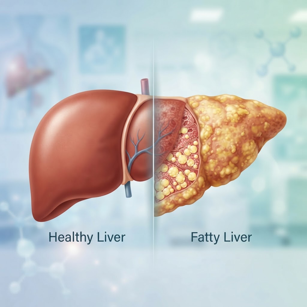
  • Chronic Acid Reflux: Causes, Triggers, and Natural Relief

    Chronic Acid Reflux: Causes, Triggers, and Natural Relief

    Key Takeaways

    • Acid reflux occurs occasionally, while chronic GERD represents a persistent, systemic issue often requiring long-term lifestyle management.
    • Symptoms of GERD extend well beyond chest pain to include respiratory issues, dental erosion, and chronic throat irritation.
    • Identifying personal heartburn triggers—whether dietary, postural, or stress-related—is the first step toward lasting symptom control.
    • Dietary modifications focusing on portion control and acid-neutralizing foods provide the foundation for improved digestive health.
    • Sustainable relief relies on a combination of medical consultation and evidence-based natural interventions rather than quick fixes.

    For millions of individuals, the simple act of eating can feel like a precursor to discomfort, turning mealtime into a source of anxiety. Chronic acid reflux and its more persistent counterpart, Gastroesophageal Reflux Disease (GERD), impact the quality of life on a daily basis, manifesting as a burning sensation that can be both painful and disruptive. While many people reach for over-the-counter solutions, truly mastering your digestive health requires a deeper dive into the underlying mechanisms of why acid escapes the stomach. By exploring the root causes, learning to decode your specific triggers, and implementing science-backed lifestyle changes, you can begin to regain control over your well-being. This guide, compiled by the healthauthoritylife Editorial Team, is designed to help you navigate the complexity of digestive health through actionable, sustainable strategies.

    Understanding the Difference Between Acid Reflux and GERD

    To effectively manage digestive discomfort, it is essential to first understand the distinction between casual acid reflux and the more complex condition known as chronic GERD. Acid reflux is a physical event that occurs when the lower esophageal sphincter (LES)—a muscular valve at the entry to the stomach—fails to close properly or relaxes at the wrong time. This allows digestive juices, which contain stomach acid, to travel backward into the esophagus. Almost everyone experiences this sensation occasionally, perhaps after a particularly heavy holiday meal or while lying down immediately after consuming a high-fat snack. If it happens rarely and does not cause significant damage to the esophagus, it is generally considered a manageable part of life.

    Gastroesophageal Reflux Disease, or GERD, is defined by the frequency and severity of these reflux events. When acid reflux happens regularly—typically defined by healthcare professionals as two or more times per week—it is classified as GERD. This is not merely an inconvenience; it is a chronic condition where the persistent exposure of the esophageal lining to acidic contents leads to inflammation, irritation, and, if left untreated, potential long-term damage. Unlike simple reflux, which often resolves quickly on its own, GERD typically requires a more structured approach to management, involving both lifestyle modifications and potential medical interventions.

    The primary concern with chronic GERD is the cumulative effect on the esophagus. The lining of the esophagus is not designed to withstand the highly acidic environment of the stomach. When it is subjected to constant acid exposure, the cells may begin to change in an attempt to protect themselves. This chronic inflammation can result in esophageal ulcers, strictures (narrowing of the esophagus), and a condition known as Barrett’s esophagus, which involves precancerous cellular changes. Consequently, moving from the perspective of “occasional indigestion” to “chronic management” is a critical shift in mindset. Understanding that GERD is a systemic issue rather than a one-off problem allows individuals to prioritize proactive digestive health tips that mitigate risk before damage occurs.

    Many people find that distinguishing between the two depends on the presence of “alarm symptoms.” While simple reflux might be triggered by a specific food choice, GERD often feels like a constant presence in the background of one’s health. Recognizing this difference helps in determining when it is time to seek professional evaluation rather than attempting to self-manage with dietary changes alone. If you find your symptoms are interfering with your ability to sleep, work, or consume a balanced diet, you are likely dealing with the chronic nature of GERD, which necessitates a comprehensive, long-term plan rather than a reactionary approach to occasional pain.

    Common Symptoms of Chronic Acid Reflux Beyond Heartburn

    While the sensation of a burning chest—commonly referred to as heartburn—is the hallmark sign of acid reflux, the clinical profile of chronic GERD is remarkably broad. Because the esophagus is connected to the throat and respiratory pathways, the escape of acid can cause “extra-esophageal” symptoms that many individuals do not immediately associate with their digestive health. Recognizing these atypical symptoms is a vital component of successful symptom management, as it prevents the misdiagnosis of other conditions, such as asthma or chronic allergies.

    One of the most frequent non-heartburn symptoms is a chronic, dry cough. When microscopic droplets of acid reach the upper airway, they can cause inflammation and a persistent tickle that leads to a dry, hacking cough, particularly at night or shortly after a meal. Similarly, many individuals experience “acid laryngitis,” which presents as a hoarse, raspy voice or a constant need to clear the throat. This occurs when stomach acid travels high enough to irritate the vocal cords. Because this is often worse in the morning, it is frequently overlooked, yet it is a classic sign of nocturnal reflux.

    Another subtle symptom is the sensation of a lump in the throat, known in medical circles as “globus sensation.” This feeling that something is stuck in the throat, even when you have not recently swallowed, is a common indicator of chronic irritation in the esophageal tissue. Some people also report experiencing regurgitation, which is the sensation of sour or bitter-tasting liquid rising into the back of the throat. Unlike vomiting, this is a passive experience and serves as a direct indicator that the LES is failing to maintain an airtight seal at the top of the stomach.

    Furthermore, dental health often reveals the silent damage caused by GERD. Stomach acid is potent enough to erode tooth enamel over time. Dentists may be the first to notice the signs of chronic reflux when they see thinning enamel, increased tooth sensitivity, or recurring decay on the back surfaces of the teeth. If you are experiencing unexplained chest pain that mimics cardiac issues, or if you find yourself waking up with a bitter taste in your mouth, these are significant indicators that your digestive health needs attention. Addressing these symptoms early is one of the most effective ways to prevent the progression of esophageal irritation and maintain overall health.

    Symptom Type Common Manifestation Best For
    Typical Burning chest pain after meals Standard heartburn assessment
    Respiratory Chronic dry cough or wheezing Identifying silent reflux (LPR)
    Throat/Oral Hoarseness, throat clearing, dental erosion Monitoring high-level acid exposure

    Major Causes and Risk Factors for Developing GERD

    Understanding why chronic GERD develops requires looking at the complex interplay between physical anatomy, lifestyle, and physiological function. While there is no single “cause” for everyone, the condition typically arises when the mechanisms designed to keep acid in the stomach become compromised. The primary culprit is often the lower esophageal sphincter (LES). In a healthy digestive system, this ring of muscle acts as a one-way gate, opening to allow food to enter the stomach and closing tightly to prevent acid from backing up. In individuals with GERD, this valve may be weak or experience inappropriate relaxation, allowing for the consistent backflow of gastric juices.

    One of the most prominent physical risk factors is a hiatal hernia. This occurs when the upper part of the stomach pushes through the diaphragm—the muscle that separates the chest from the abdomen. The diaphragm helps the LES stay shut, but when a hiatal hernia is present, this anatomical support is disrupted, making it significantly easier for acid to migrate upward. While many people have small hiatal hernias without experiencing severe symptoms, they are a frequent contributor to chronic reflux in those who are symptomatic.

    Lifestyle and behavioral factors play a substantial role in the development and worsening of GERD. Obesity is perhaps the most significant modifiable risk factor. Excessive weight, particularly around the abdomen, increases pressure on the stomach. This mechanical pressure can force the LES to open, facilitating the movement of acid into the esophagus. Even modest weight loss has been shown to significantly reduce the frequency of reflux episodes in many patients, making weight management a cornerstone of most digestive health plans.

    Dietary habits also act as major catalysts. Consuming large meals, eating late at night, or reclining immediately after consuming high-fat or acidic foods creates a scenario where the stomach remains full and under pressure for an extended period, increasing the likelihood that the valve will fail. Furthermore, certain substances are known to relax the LES. Caffeine, chocolate, alcohol, and peppermint can all diminish the muscle’s resting pressure, acting as “openers” for the esophageal valve. When these dietary choices are combined with smoking—which inhibits saliva production (a natural acid neutralizer) and impairs the clearing of acid from the esophagus—the environment becomes primed for chronic discomfort.

    Finally, we must consider the impact of pregnancy and certain medications. Pregnancy induces both hormonal changes that relax smooth muscles throughout the body (including the LES) and physical pressure from the growing uterus on the stomach. Simultaneously, medications such as certain blood pressure drugs, anti-inflammatories, or asthma inhalers can exacerbate reflux as a side effect. By recognizing these risk factors, individuals can begin to isolate which components of their daily lives are most likely contributing to their symptoms, providing a clear roadmap for how to stop acid reflux through targeted interventions.

    Identifying Your Unique Food and Lifestyle Triggers

    The journey toward managing chronic GERD is deeply personal because what triggers an episode for one person may be perfectly tolerable for another. This variability is why “one-size-fits-all” advice often fails to provide lasting relief. To regain control, you must become an investigator of your own health. The most effective tool in this process is the “Reflux Log,” a simple but powerful method to track your intake, behaviors, and symptom patterns over time. By documenting what you eat, when you eat it, and how you feel in the hours afterward, you can reveal hidden patterns that were previously obscured by the daily noise of life.

    When tracking triggers, look beyond the obvious. While citrus fruits, tomato-based sauces, and spicy foods are widely recognized as common heart burn triggers, your specific reaction may depend on the *combination* of foods. For instance, a small amount of dark chocolate might be fine on its own, but when paired with an alcoholic beverage or a high-fat coffee drink, it may cause a significant flare-up. Keep an eye on “latency”—the time delay between eating and the onset of symptoms. Some triggers act quickly, while others, particularly those that delay stomach emptying, may cause discomfort hours after the meal is finished.

    Lifestyle triggers are just as critical as dietary ones. Posture is a major, often overlooked factor. If you find that your symptoms spike while you are working at a desk or sitting on a couch for extended periods, you may be experiencing “postural reflux.” The slouching position compresses the abdomen and puts strain on the diaphragm, which can facilitate acid movement. Conversely, if your symptoms are worse at night, your trigger is likely related to the timing of your final meal. Experts generally suggest a gap of at least three hours between finishing your last meal and lying down to sleep, as this allows gravity to assist the stomach in emptying its contents before you are horizontal.

    Physical activity can also be a trigger, especially activities that involve inversions, heavy lifting, or high-impact movements. If you notice reflux during yoga or circuit training, consider modifying your routine to avoid exercises that require lying flat or bearing down heavily on your core. Additionally, stress management is a non-negotiable factor. The gut-brain axis is powerful; high levels of psychological stress can increase stomach acid production and hypersensitize the esophagus to even minor amounts of reflux. Tracking your stress levels alongside your physical symptoms can help you determine if emotional factors are driving your digestive issues.

    Once you have gathered two to four weeks of data, look for the “common denominators.” Are your symptoms consistently appearing after meals that feature a specific fat content? Do they occur when you are working late? This data-driven approach allows you to replace guesswork with a tailored plan. By eliminating the top two or three verified triggers, you can often see a significant improvement in your daily comfort levels, providing the relief needed to focus on long-term lifestyle changes.

    Evidence-Based Dietary Changes for Acid Reflux Relief

    Crafting a sustainable acid reflux diet plan is not about deprivation; it is about choosing foods that minimize the burden on your stomach and support the function of your lower esophageal sphincter. The goal is to choose items that are easy to digest, lower in acid, and unlikely to linger in the stomach for long periods. By focusing on these categories, you can manage your condition while still enjoying satisfying, nutrient-dense meals.

    The first principle is to prioritize lean proteins. Fatty proteins, such as fried meats or high-fat cuts of beef, take longer to digest, keeping the stomach full and increasing the internal pressure that leads to reflux. Instead, integrate chicken breast, turkey, fish, and plant-based proteins like lentils or tofu. These options are much lighter and less likely to trigger the LES to relax. When preparing these proteins, opt for baking, steaming, or grilling rather than frying, which adds unnecessary fats that can slow down gastric emptying.

    Complex carbohydrates and high-fiber vegetables are also your allies. Foods such as oatmeal, brown rice, whole-grain bread, and root vegetables like sweet potatoes and carrots are generally well-tolerated. Oatmeal, in particular, is highly recommended as a breakfast choice because it can absorb some of the acid in the stomach and provides a steady source of energy without causing the rapid spikes in insulin that can sometimes disrupt digestion. Green, non-acidic vegetables like broccoli, asparagus, and green beans are excellent, provided they are not cooked with excessive amounts of butter or oil.

    Regarding fruits, it is essential to look for low-acid varieties. While citrus is a common irritant, fruits like bananas, melons (such as cantaloupe and honeydew), and pears are usually very safe. Bananas have a naturally low acidity and a high pH, which can help coat the esophagus and protect it from stomach acid. Incorporating these into your snack rotation is a simple way to soothe your digestive tract throughout the day. It is also wise to keep a supply of ginger or chamomile tea, which are well-regarded GERD natural remedies known for their ability to calm digestive inflammation and reduce nausea.

    Portion management is perhaps the most crucial dietary habit. Eating large meals expands the stomach, which physically puts more pressure on the LES. By switching to smaller, more frequent meals, you can prevent the stomach from ever becoming overly full, which significantly reduces the mechanical likelihood of acid moving upward. This also keeps blood sugar levels stable, which can indirectly help in managing the stress and hormonal responses that exacerbate GERD. When eating, aim to chew thoroughly and eat slowly; this reduces the amount of air you swallow and gives your digestive system a head start on breaking down the food.

    Lastly, pay close attention to your beverages. Water is always the best choice for hydration. If you struggle with the taste, you can infuse it with cucumber or a small amount of non-acidic fruit. Avoid common triggers such as caffeinated sodas, alcohol, and very hot or very cold drinks, which can irritate the sensitive lining of the esophagus. By building your diet around these evidence-based choices—prioritizing lean nutrition, low-acid produce, and smaller portions—you create a foundation that fosters healing and long-term digestive stability.

    Natural Remedies to Soothe an Inflamed Esophagus

    When the delicate lining of the esophagus is subjected to frequent acid exposure, the resulting inflammation—often referred to as esophagitis—can lead to chronic pain, difficulty swallowing, and a persistent sensation of a lump in the throat. While medication provides a pathway for suppression, many individuals find that integrating natural, soothing agents helps provide immediate relief and supports the body’s intrinsic healing process.

    One of the most effective ways to manage inflammation is through the use of mucilaginous herbs. Slippery elm bark and marshmallow root are classic examples of botanical agents that, when prepared as a tea or taken in powdered form, create a protective gel-like coating along the esophageal wall. This physical barrier can act as a shield against the corrosive nature of gastric acid, allowing the tissue underneath to recover. For those looking for a cooling effect, aloe vera juice—specifically the kind prepared for internal consumption and stripped of laxative compounds like aloin—can offer a calming sensation that reduces the burning intensity after a meal.

    Furthermore, managing the pH level of the stomach without suppressing acid production entirely is a delicate balance. Some find that sipping ginger-infused water acts as an anti-inflammatory tonic. Ginger contains gingerols and shogaols, compounds that help calm the digestive tract and improve gastric motility, ensuring that food moves through the system at an appropriate pace rather than stagnating, which is a primary culprit in acid reflux symptoms. Honey, particularly high-quality, raw varieties like Manuka, may also provide a soothing, antimicrobial layer that assists in maintaining a healthy environment for tissue repair.

    Comparison Table: Natural Soothing Agents for Digestive Comfort

    Remedy Primary Benefit Best For
    Slippery Elm Bark Forms a protective mucilage barrier Immediate relief from burning sensations
    Ginger Root Speeds gastric emptying and reduces inflammation Preventing bloating and slow digestion
    Aloe Vera Juice Cooling, hydrating, and soothing properties Chronic throat irritation and esophagitis
    Chamomile Tea Relaxing to the nervous system and GI tract Reducing stress-induced acid production
    Licorice (DGL) Supports healthy mucus membrane function Long-term esophageal lining protection

    How Sleep Position and Habits Impact Nighttime Reflux

    Nighttime reflux is a significant challenge for many because of the simple physics of gravity. When we are upright, gravity assists the lower esophageal sphincter (LES) in keeping gastric contents firmly within the stomach. Once we lie flat, that natural barrier is diminished, and if the LES is weakened or overly relaxed, stomach acid can easily track back into the esophagus. Nocturnal reflux is often the most damaging form of the condition because the esophagus lacks the benefit of gravity to clear the acid, leading to prolonged exposure and severe inflammation.

    The most widely recommended modification for those suffering from night-time symptoms is to elevate the upper body. It is important to note that simply stacking pillows is rarely sufficient and can sometimes worsen the issue by inducing a sharp bend in the spine or neck, which puts pressure on the abdomen. Instead, experts suggest using a wedge pillow that creates a gentle incline from the hips to the head, or using blocks to elevate the head of the bed frame itself. By sleeping at a slight angle—typically 6 to 8 inches of elevation—gravity can help keep acid in the stomach.

    Habits surrounding the pre-sleep routine are equally vital. Eating a large meal within three hours of bedtime is one of the most common heart burn triggers. The stomach needs time to break down food; when we lie down with a full stomach, the volume of contents puts massive pressure on the LES. Developing a “dinner cutoff” policy is a highly effective digestive health tip. Additionally, choosing the left side for sleeping may be beneficial. Anatomically, the stomach sits below the esophagus when you lie on your left side, making it more difficult for acid to leak upward compared to lying on the right side or flat on the back.

    When to See a Doctor for Persistent Acid Reflux Issues

    While lifestyle changes and natural remedies can be transformative, there are instances where chronic GERD requires clinical intervention. Self-management should never delay the diagnostic process if you are experiencing “red flag” symptoms. If you find that your reflux is accompanied by unintended weight loss, chronic coughing, hoarseness that does not resolve, or a persistent sensation that food is sticking in your chest, it is imperative to schedule an appointment with a gastroenterologist.

    A medical professional can perform diagnostic tests, such as an endoscopy, to evaluate the structural integrity of your esophagus. This is critical for ruling out complications like Barrett’s esophagus, a condition where the chronic acid exposure leads to changes in the cells of the esophageal lining. Additionally, persistent reflux that remains unresponsive to dietary changes might suggest an underlying mechanical issue, such as a hiatal hernia, where a portion of the stomach pushes up through the diaphragm. In these cases, anatomy, rather than diet, is the primary driver of the problem, and a surgical or clinical approach may be necessary to provide lasting relief.

    The Role of Stress and Gut Microbiome in Digestion

    The connection between the brain and the gut, often referred to as the “gut-brain axis,” is a burgeoning field of research that holds significant implications for those managing acid reflux. Stress is not merely an emotional experience; it causes tangible physiological changes in the body. When you are under chronic stress, your body enters a “fight or flight” mode, which diverts blood flow away from the digestive tract. This slows down peristalsis (the wave-like movement of food) and can lead to increased stomach acid sensitivity and a delayed gastric emptying rate.

    Furthermore, the health of the gut microbiome—the ecosystem of bacteria residing in your digestive system—plays a fundamental role in how we process food. An imbalance of bacteria, sometimes called dysbiosis, can lead to increased gas production and bloating. This internal pressure pushes against the stomach, which can force the LES open and trigger reflux. Integrating fermented foods like kefir, sauerkraut, or kimchi, and prioritizing a diverse diet rich in prebiotic fibers, helps foster a robust microbiome. By supporting the beneficial bacteria in your gut, you improve your body’s overall digestive efficiency and reduce the likelihood of the mechanical triggers that lead to acid reflux.

    Safe and Effective Long-Term Management Strategies

    Long-term management of chronic GERD is about creating a sustainable lifestyle rather than relying on short-term “quick fixes.” The goal is to lower the overall burden on the LES so that it can regain its tone. A successful acid reflux diet plan is not about deprivation but about consistency. It involves identifying your specific triggers—whether they be chocolate, caffeine, spicy foods, or fatty items—and minimizing them while maximizing high-fiber, easily digestible meals.

    Weight management is often cited as a cornerstone of long-term success. Even a modest reduction in body mass can significantly reduce the amount of intra-abdominal pressure, which is the physical force pushing the stomach contents upward. Paired with consistent meal timing, stress management techniques like mindful breathing or yoga, and the use of natural aids when needed, individuals can often reach a point where reflux is not an active, daily disruption to their quality of life. The key is to remain proactive, monitoring how different foods and stressors affect your body and adjusting your routine to maintain the equilibrium that prevents symptoms before they start.

    Frequently Asked Questions

    Can drinking water help neutralize stomach acid?

    Drinking a small amount of water can help rinse the esophagus and push residual acid back into the stomach, providing temporary relief. However, drinking a large amount of water immediately after a meal may increase stomach volume and potentially increase pressure, which could trigger reflux in some individuals. It is best to sip water slowly between meals rather than gulping large amounts during or immediately after eating.

    Is it possible for acid reflux to lead to long-term health complications?

    Yes, if left unmanaged for a prolonged period, chronic acid exposure can cause structural damage. This includes esophageal ulcers, esophageal strictures (narrowing of the esophagus that causes difficulty swallowing), and Barrett’s esophagus, which is a condition where the tissue of the esophagus changes to resemble intestinal lining. Regular monitoring and proactive lifestyle management are essential to prevent these developments.

    Does chewing gum help with acid reflux?

    Chewing gum, particularly sugar-free varieties that do not contain mint (a known trigger), can increase saliva production. Saliva is naturally alkaline and acts as a buffer against acid, helping to neutralize the esophagus and promote the clearance of acid back into the stomach. Many people find this a helpful, simple trick to employ shortly after a meal.

    Why do spicy foods trigger such intense heart burn?

    Spicy foods often contain capsaicin, a compound that can irritate the lining of the esophagus and slow down digestion. By lingering in the stomach for a longer duration, food is more likely to trigger acid production. Furthermore, spicy foods can directly irritate the sensitive tissue already inflamed by previous reflux episodes, leading to that characteristic, intense burning sensation.

    Are there certain foods I should absolutely avoid?

    While triggers are highly individual, common offenders include coffee, chocolate, peppermint, alcohol, citrus fruits, tomatoes, and fried or high-fat foods. These items are known to either directly irritate the lining of the esophagus or cause the lower esophageal sphincter to relax, allowing acid to leak upward. It is recommended to keep a food diary to identify your personal list of “no-go” foods.

    How does smoking affect acid reflux?

    Smoking is a significant factor in chronic GERD because nicotine has been shown to relax the lower esophageal sphincter, making it easier for acid to escape the stomach. Additionally, smoking reduces saliva production, which is necessary to neutralize acid, and can decrease the overall efficiency of the esophageal muscles. Quitting smoking is often one of the most effective lifestyle changes for those looking to resolve persistent symptoms.

    Conclusion

    Managing chronic acid reflux and GERD requires a multifaceted approach that addresses both the physical mechanics of the digestive tract and the lifestyle habits that influence them. From mindful eating practices and strategic sleep positioning to the incorporation of natural remedies and stress-reduction techniques, you have more power over your digestive health than you might realize. By identifying your specific heart burn triggers and adopting a consistent, long-term strategy, you can minimize symptoms and protect your long-term health. Start by implementing one or two of the changes discussed here and monitor your progress; small, consistent adjustments often lead to the most significant improvements in comfort and quality of life. If symptoms persist, do not hesitate to reach out to a healthcare professional for a personalized assessment.

    By healthauthoritylife Editorial Team

    This article is for informational purposes only and is not a substitute for professional medical advice. Always consult your doctor before making changes to your diet, exercise routine, or treatment plan.

  • NAFLD Symptoms: Early Signs, Causes, and Natural Reversal Tips

    NAFLD Symptoms: Early Signs, Causes, and Natural Reversal Tips

    Key Takeaways

    • NAFLD is a metabolic condition where excess fat accumulates in liver cells, often without initial symptoms.
    • Early detection of NAFLD symptoms is crucial for preventing progression to serious liver damage.
    • Insulin resistance, obesity, and dietary habits are the primary drivers of fatty liver accumulation.
    • Transitioning from NAFLD to NASH involves chronic inflammation, which can lead to permanent scarring or fibrosis.
    • Reversing fatty liver is primarily achieved through sustainable dietary changes and metabolic support rather than quick-fix supplements.

    The human liver serves as the body’s ultimate filtration system, working tirelessly to process nutrients, metabolize fats, and detoxify the bloodstream. However, in our modern environment—characterized by sedentary habits and highly processed food intake—this vital organ is increasingly under siege. Non-alcoholic fatty liver disease (NAFLD) has emerged as one of the most common metabolic conditions globally, often referred to as a “silent epidemic” because it frequently develops quietly over many years. Understanding the intersection of liver health and metabolic function is no longer just a concern for those with pre-existing conditions; it is a necessity for anyone looking to optimize their long-term vitality. By recognizing the early signals your body provides and understanding the root causes of fat buildup, you can take proactive, evidence-based steps to protect your health and potentially reverse the course of liver disease before it leads to irreversible damage.

    What is Non-Alcoholic Fatty Liver Disease (NAFLD)?

    At its core, non-alcoholic fatty liver disease (NAFLD) is an umbrella term used to describe a spectrum of conditions where excessive fat builds up in the liver cells of individuals who consume little to no alcohol. While the term “fatty” might sound like a simple description of weight, in medical terms, it denotes a significant shift in how the organ functions. Under normal circumstances, the liver does store small amounts of fat; however, when that fat content exceeds a certain threshold—typically identified as five percent or more of the liver’s weight—the organ begins to struggle with its standard metabolic duties.

    The progression of NAFLD is closely tied to metabolic health. Many experts view the condition as the hepatic manifestation of metabolic syndrome, which includes a cluster of conditions such as high blood pressure, high blood sugar, excess body fat around the waist, and abnormal cholesterol levels. When the body becomes insulin resistant—meaning your cells stop responding efficiently to the hormone insulin—the liver begins to convert excess energy from the diet into triglycerides. These triglycerides then accumulate within the liver cells, a process known as hepatic steatosis.

    It is important to differentiate NAFLD from other forms of liver disease. Because the “non-alcoholic” component is central to the diagnosis, it distinguishes this condition from liver damage caused by chronic, heavy alcohol consumption. Despite the difference in origin, the physiological burden on the liver can be remarkably similar. NAFLD exists on a spectrum; it begins as simple steatosis, which is the accumulation of fat without significant inflammation or cellular damage. In this stage, the condition is often considered reversible if lifestyle interventions are implemented promptly. The danger lies in the lack of overt symptoms during these early phases, which often leads people to overlook their liver health until more significant metabolic disruptions occur.

    Because the liver is a resilient organ, it often manages to maintain “normal” blood work markers for years even while undergoing internal changes. This is why many clinicians emphasize that metabolic health should be monitored holistically. By focusing on systemic inflammation and insulin sensitivity, you are indirectly supporting your liver. Understanding that NAFLD is essentially a “storage error” within the body’s metabolic machinery helps clarify why dietary and lifestyle choices are the most powerful tools in your arsenal. Unlike many other chronic diseases, NAFLD provides a clear window of opportunity for correction, provided one understands the underlying mechanism of fat sequestration and its relationship to our daily nutritional habits.

    Early Symptoms and Warning Signs You Should Not Ignore

    One of the most challenging aspects of NAFLD is that it is frequently asymptomatic in its early stages. Many individuals live with a fatty liver for years, completely unaware that their metabolic health is declining. Because the liver does not contain pain receptors in its parenchyma—the functional tissue of the organ—you will not typically experience “liver pain” early on. However, the body often sends subtle, systemic signals that indicate the liver is struggling to process the metabolic load it is being asked to handle.

    One of the most common, albeit vague, indicators of liver stress is persistent fatigue or an overall feeling of malaise. When the liver is burdened by excess fat, its ability to efficiently convert stored energy into glucose or regulate the blood sugar levels needed for consistent daily energy is compromised. This can manifest as an afternoon “crash,” difficulty waking up in the morning, or a general lack of stamina during physical activities that previously felt manageable. While fatigue is a symptom of many health conditions, when accompanied by metabolic markers like a rising waist-to-hip ratio, it warrants closer investigation by a healthcare professional.

    Another subtle sign involves changes in the skin or overall body composition. Some people report unexplained dark patches of skin, particularly around the neck or in the armpits—a condition clinically known as acanthosis nigricans. This is often a visual marker of significant insulin resistance, which is the primary engine behind NAFLD. Additionally, many individuals with early-stage liver fat accumulation notice difficulty losing stubborn belly fat. Even when caloric intake is restricted, the liver’s inability to metabolize fats effectively can keep the body in a state of fat storage rather than fat mobilization.

    Digestive discomfort is another sign that should not be dismissed. While often misattributed to acid reflux or general indigestion, persistent bloating or feelings of fullness, particularly in the upper right quadrant of the abdomen, can occur if the liver becomes enlarged due to fat infiltration. Because the liver is located just under the ribcage on the right side, an enlarged organ may cause a dull, aching sensation, especially after a heavy or high-fat meal. This is not necessarily intense pain, but rather a sensation of pressure or “fullness” that signals the organ is working under strain.

    Finally, blood work can provide the earliest objective warnings, even before physical symptoms appear. While standard liver function tests (LFTs) might appear normal in the early stages, clinicians often look at secondary markers. Elevated levels of liver enzymes like ALT (alanine aminotransferase) and AST (aspartate aminotransferase) can act as red flags. However, because these levels can fluctuate, experts often recommend looking at the bigger picture, including triglycerides, HDL cholesterol, and fasting glucose levels. If these markers are trending in the wrong direction, it is a sign that your liver health is being impacted by your metabolic environment. Recognizing these signs early is the primary defense against the progression toward more permanent damage.

    Primary Causes and Risk Factors Behind Liver Fat Accumulation

    To effectively manage and reverse fatty liver, one must look at the “why” behind the accumulation. It is a misconception to think that dietary fat is the sole culprit of NAFLD. In reality, the liver creates fat through a process called *de novo lipogenesis*, which is heavily driven by the overconsumption of refined carbohydrates and fructose. When the body takes in more sugar than it can use for immediate energy, the liver is tasked with converting that excess into triglycerides for storage. This is why a high-sugar, high-processed-food diet is the most significant driver of NAFLD.

    Insulin resistance stands as the most critical risk factor. When your muscles and fat cells stop responding to insulin, the pancreas compensates by secreting more of it. High circulating levels of insulin tell the liver to prioritize fat storage over fat burning. This explains why obesity, particularly visceral obesity (fat stored around the abdominal organs), is so tightly correlated with NAFLD. Even “skinny-fat” individuals—those who may appear thin but have high amounts of internal, visceral fat—are at significant risk because the liver is the first place where that metabolic imbalance manifests.

    Approach Focus Best For
    Whole Food Diet Removing processed sugars and grains Long-term insulin sensitivity
    Intermittent Fasting Restricting the feeding window Reducing hepatic fat storage
    Strength Training Building lean muscle mass Improving glucose uptake
    Cardiovascular Exercise Improving aerobic capacity Overall metabolic efficiency

    Beyond diet, genetics play an undeniable role. Certain genetic variations can make individuals more susceptible to fat storage in the liver. However, genetics are rarely the sole cause; they typically act as a “loaded gun,” while environmental factors like diet and inactivity “pull the trigger.” Sedentary behavior is a massive contributor. Physical movement—especially muscle contraction—is the body’s most effective way of “emptying” the glucose from the bloodstream, thereby reducing the workload on the liver. Without this mechanical stimulus, the liver remains in a state of high-alert, constantly processing excess blood sugar and converting it into liver fat.

    Certain medical conditions also exacerbate the risk. Type 2 diabetes is perhaps the most significant comorbid condition. Because diabetes is essentially a state of chronic hyperglycemia and insulin resistance, the liver is under constant stress. Other factors include high blood pressure and abnormal cholesterol profiles, particularly low levels of HDL (the “good” cholesterol) and high levels of triglycerides. These markers are not just symptoms; they are indicators of a metabolic environment that is highly conducive to fat storage in the liver. Medications, particularly those that impact metabolism or are processed by the liver, can also play a role, though this should always be discussed with your healthcare provider.

    Ultimately, the primary cause of NAFLD is a mismatch between the energy we ingest and the energy we expend, combined with a metabolic system that has lost its flexibility. When we eat ultra-processed foods that spike our blood sugar, we are effectively hitting the “on” switch for liver fat creation. By understanding that this is a manageable metabolic problem rather than a static genetic defect, we regain agency over our health. The goal is to lower systemic insulin levels and provide the liver with the breathing room it needs to perform its primary function: cleaning, filtering, and repairing the body.

    How NAFLD Progresses to NASH and Liver Fibrosis

    When NAFLD persists, it is not merely a static “extra fat” issue. For many people, the continued presence of fat in the liver cells triggers a second hit: chronic inflammation. This stage of the disease is known as Non-Alcoholic Steatohepatitis, or NASH. In NASH, the liver cells are not only fatty but also inflamed and damaged. This inflammation acts as a signal for the liver to initiate a repair process, which involves the deposition of collagen fibers in an attempt to protect the tissue. This scarring process is known as fibrosis.

    The progression from simple fatty liver (NAFLD) to NASH can happen silently. Because the liver has such a massive capacity for regeneration, it can continue to function while slowly accumulating these fibrous, scar-like tissues. However, if the underlying inflammatory stimulus—typically poor diet and insulin resistance—is not removed, this fibrosis can progress through several stages. Over many years, the accumulation of scar tissue replaces healthy, functional liver cells. This is a critical threshold; once significant fibrosis has occurred, the structural integrity of the liver is permanently altered, which can severely impact its ability to perform its filtration and metabolic functions.

    If the scarring becomes extensive and widespread, the condition may reach the stage of cirrhosis. In cirrhosis, the liver becomes hard and shrunken, and its ability to filter toxins from the blood is severely compromised. This is the stage where serious systemic complications arise, such as fluid buildup in the abdomen, jaundice, or cognitive issues resulting from the liver’s inability to clear toxins. Understanding this path—from simple fat storage to inflammation, then to scarring—highlights exactly why early intervention is non-negotiable. Reversing fat accumulation is relatively straightforward, but repairing damaged, fibrotic tissue is significantly more difficult, if not impossible, in the late stages.

    The transition from NAFLD to NASH is driven by oxidative stress. When the liver is overloaded with fat, it produces molecules called free radicals as a byproduct of metabolism. These free radicals damage the liver cells’ membranes and DNA, triggering an immune response that leads to inflammation. This is why antioxidant-rich diets and minimizing sources of inflammation are so frequently recommended as part of a strategy for liver health. By reducing the metabolic load, you decrease the production of these damaging molecules, effectively halting the cascade that leads to fibrosis.

    Most importantly, the progression is not linear. Many people hover in the early stages of NAFLD for decades without ever progressing to NASH. The key difference between those who progress and those who stay in the early stage is often the intensity and duration of metabolic dysfunction. By actively managing blood sugar levels and reducing inflammation through diet and movement, you are essentially preventing the “second hit” that turns simple fat storage into a more serious, inflammatory liver condition. The power lies in your ability to control the inputs into your system, thereby preventing the oxidative stress that drives this harmful progression.

    Effective Dietary Changes to Support Liver Health

    When it comes to reversing fatty liver, the dietary strategy is not about starvation or extreme caloric restriction; it is about metabolic efficiency. The objective is to stabilize blood sugar, improve insulin sensitivity, and give the liver the nutrient density it needs to heal. The most effective liver health tips center on removing the primary sources of liver stress—namely, refined sugars, processed carbohydrates, and industrial seed oils—and replacing them with whole, anti-inflammatory foods.

    The cornerstone of a fatty liver diet is the dramatic reduction of added sugars, particularly high-fructose corn syrup. Fructose is metabolized exclusively by the liver, and in high amounts, it is almost exclusively converted into fat. By eliminating sodas, juices, sweetened snacks, and sugary baked goods, you remove the primary substrate the liver uses to create new fat. Simultaneously, focusing on high-fiber vegetables is vital. Fiber slows the absorption of glucose, which prevents the sharp spikes in insulin that drive fat storage. Leafy greens, cruciferous vegetables like broccoli and cauliflower, and fibrous legumes provide the fuel and support needed for the liver’s detoxification pathways.

    Healthy fats, surprisingly, are a necessary part of the recovery process. The goal is not a “low-fat” diet, but rather a “right-fat” diet. Substituting processed vegetable oils (like soybean, corn, or canola oil) with heart-healthy monounsaturated fats like olive oil and avocado can significantly improve liver inflammation. Omega-3 fatty acids, found in fatty fish like salmon, sardines, and mackerel, are particularly beneficial because they have been shown to help reduce liver fat content and systemic inflammation. These healthy fats help improve satiety, reducing the need for the refined carbohydrates that trigger insulin spikes.

    Protein quality also matters. Lean proteins like pasture-raised chicken, eggs, and wild-caught fish provide the essential amino acids required for the liver to repair itself. Protein also has a higher thermic effect than carbohydrates, meaning it requires more energy to digest, which can assist in overall metabolic management. For those following plant-based diets, incorporating high-quality sources like lentils, chickpeas, and fermented soy can provide similar benefits. The key is to avoid processed meats, which are often packed with additives and nitrates that may add an unnecessary burden to the liver’s filtration systems.

    Finally, the timing of meals is as important as the content. Many people find that intermittent fasting or narrowing their eating window—giving the body 12 to 16 hours of rest from digestion—can be a game-changer for liver health. During these fasting periods, insulin levels drop, allowing the liver to shift from “fat storage mode” to “fat burning mode.” By limiting the number of times you trigger insulin release throughout the day, you provide the liver with a consistent, reliable period to focus on maintenance and repair. Combining this timing strategy with a whole-food, high-fiber, and high-protein intake is the most proven, sustainable approach to reclaiming your metabolic health and reversing the effects of NAFLD.

    The Role of Exercise in Reducing Liver Fat

    When discussing how to reverse fatty liver, exercise is often cited as the most powerful non-pharmacological intervention available. While dietary adjustments provide the building blocks for recovery, physical activity acts as the metabolic engine that burns off excess triglycerides stored within hepatocytes (liver cells). The primary mechanism by which exercise benefits liver health is through the improvement of insulin sensitivity. As muscles become more active, they require more glucose for energy, which prevents the liver from having to convert excess blood sugar into fat stores.

    Research into metabolic health consistently indicates that a combination of aerobic exercise and resistance training yields the most significant improvements in hepatic fat fraction. Aerobic exercise—such as brisk walking, cycling, or swimming—promotes lipid oxidation. When you sustain a moderate heart rate, your body shifts from relying solely on glycogen to utilizing fatty acids as a primary fuel source, effectively “clearing out” stored fat from the liver.

    Resistance training, however, provides a different set of advantages. By increasing lean muscle mass, you effectively increase your resting metabolic rate. Muscle tissue is metabolically expensive to maintain, meaning a body with more muscle mass will burn more energy even during sedentary periods. Furthermore, muscle tissue acts as a “glucose sink,” soaking up excess blood sugar and keeping systemic insulin levels low—a critical factor in stopping the progression of non-alcoholic fatty liver disease.

    For those just starting, consistency outweighs intensity. Experts generally suggest aiming for at least 150 minutes of moderate-intensity aerobic activity per week, complemented by two sessions of full-body resistance training. It is important to note that you do not need to train like an athlete to see results; even small increases in daily movement, such as walking for ten minutes after each meal, can help manage post-prandial blood glucose spikes, thereby reducing the stress placed on the liver.

    Essential Supplements and Nutrients for Liver Support

    While no supplement can replace the foundational impact of a fatty liver diet and consistent movement, certain nutrients play a supportive role in reducing inflammation and promoting hepatic cellular repair. When selecting supplements, focus on those that support detoxification pathways and antioxidant status.

    The following table outlines common supportive nutrients often considered by those seeking to optimize their liver health.

    Supplement/Nutrient Mechanism of Action Best For
    Milk Thistle (Silymarin) Promotes protein synthesis and acts as an antioxidant Overall liver cell protection
    Omega-3 Fatty Acids Reduces systemic inflammation and lowers triglycerides Reducing hepatic fat accumulation
    N-Acetyl Cysteine (NAC) Precursor to glutathione, the body’s master antioxidant Enhancing liver detoxification pathways
    Vitamin E Protects cell membranes from oxidative stress Managing inflammation in non-diabetic individuals
    Choline Essential for lipid transport and fat metabolism Preventing fat buildup in the liver

    Milk thistle, specifically the extract known as silymarin, has been used for centuries to support liver function. It works primarily by inhibiting the entry of toxins into liver cells and stimulating the production of new liver tissue. Meanwhile, Omega-3 fatty acids, found in high-quality fish oils, are vital because they help regulate the genes involved in fat oxidation. By lowering the concentration of triglycerides in the blood, Omega-3s prevent the liver from being overwhelmed by circulating fats.

    Vitamin E is another potent nutrient that acts as a fat-soluble antioxidant. Because non-alcoholic fatty liver disease is often characterized by oxidative stress, Vitamin E helps neutralize the free radicals that can lead to further liver damage. However, it is essential to source these nutrients through high-quality whole foods whenever possible. Fatty fish, walnuts, flaxseeds, leafy greens, and cruciferous vegetables like broccoli and Brussels sprouts naturally contain many of these compounds, offering the added benefit of fiber and phytonutrients that work synergistically.

    Common Lifestyle Mistakes That Worsen Liver Health

    In the journey toward reclaiming metabolic health, the “what you do” is often just as important as the “what you don’t do.” Many individuals unknowingly engage in habits that put unnecessary strain on the liver, hindering even the best efforts to reverse fatty liver.

    The most pervasive mistake is the reliance on “hidden” sugars. Many processed foods marketed as “healthy” or “low fat” are often loaded with high-fructose corn syrup or refined sweeteners to improve palatability. Fructose is particularly taxing on the liver because it is processed almost exclusively in the liver cells. When consumed in excess, it is directly converted into fat, leading to a condition sometimes described as metabolic overload.

    Another frequent oversight is the assumption that the liver can handle any level of environmental toxicity if one is “eating clean.” Chronic exposure to endocrine-disrupting chemicals found in plastics, pesticides, and certain household cleaning products can place an added burden on the liver’s detoxification systems. While the liver is resilient, it can become sluggish when forced to process an overwhelming cocktail of synthetic chemicals alongside poor dietary choices.

    Finally, chronic stress is a major, often ignored factor. When the body is under persistent stress, it releases cortisol. Prolonged elevation of cortisol promotes the storage of visceral fat—the deep belly fat that is metabolically linked to fatty liver. Many people attempt to “diet away” their liver fat while remaining in a state of chronic sympathetic nervous system activation. Incorporating stress-management techniques such as deep breathing, quality sleep hygiene, and mindfulness is not just a mental health practice; it is a physiological necessity for managing liver-related metabolic inflammation.

    When to Consult a Specialist for Liver Function Testing

    Recognizing the early signs of liver damage is paramount, but knowing when to seek professional intervention is the bridge between proactive wellness and reactive medicine. If you are experiencing persistent fatigue, abdominal discomfort, or unexplained weight gain, it is prudent to move beyond general health optimization and consult with a healthcare professional, such as a gastroenterologist or hepatologist.

    Standard liver function tests (LFTs) primarily look at enzymes like ALT and AST, which indicate whether liver cells are inflamed or damaged. However, it is important to understand that these markers can sometimes remain within a “normal” range even if the liver is accumulating fat. If you have risk factors such as type 2 diabetes, obesity, or a family history of liver disease, a simple blood test might not tell the whole story.

    Specialists may recommend more advanced diagnostics if they suspect that simple steatosis (fatty liver) is progressing into non-alcoholic steatohepatitis (NASH). These may include specialized imaging, such as a FibroScan, which measures the stiffness of the liver. Liver stiffness is a key indicator of fibrosis, or scarring, which occurs when the liver attempts to repair chronic damage. The earlier these tests are conducted, the more effective lifestyle interventions will be, as early-stage liver damage is often reversible through rigorous diet and lifestyle changes. Do not wait for symptoms to become acute; if you have concerns regarding your metabolic markers, schedule a consultation to establish a baseline and monitor your progress over time.

    Frequently Asked Questions

    Is fatty liver disease reversible?

    Yes, for many individuals, fatty liver is reversible, especially in the early stages. Through consistent changes to diet, regular exercise, and weight management, the liver can reduce fat accumulation and heal itself. The key is sustained, long-term lifestyle adjustment rather than short-term “detox” trends.

    What should I avoid eating if I have fatty liver?

    You should aim to limit or eliminate refined carbohydrates, high-fructose corn syrup, sugary beverages, alcohol, and excessive saturated or trans fats. These foods contribute directly to insulin resistance and fat buildup within the liver cells.

    Are there any symptoms of fatty liver to look out for?

    Early stages of fatty liver are often asymptomatic. As the condition progresses, you might notice persistent fatigue, a dull ache in the upper right quadrant of the abdomen, or unexplained weight gain. Because these signs are non-specific, regular check-ups are essential.

    How long does it take to reverse fatty liver?

    The timeline varies significantly based on the severity of the fat buildup and an individual’s commitment to lifestyle changes. Some people see improvements in blood markers within a few months, while others may require a year or more of dedicated health habits to see significant physiological changes.

    Does water help with fatty liver?

    Yes, staying well-hydrated is essential. Water supports general metabolic health, helps the kidneys flush out toxins, and aids in digestion. Replacing sugary sodas, juices, and alcohol with water is one of the easiest and most effective ways to lower your daily caloric and sugar intake, which benefits the liver directly.

    Can a fatty liver diet be healthy for the rest of the body?

    Absolutely. A diet designed to reverse fatty liver is typically high in fiber, rich in antioxidants, and composed of whole, unprocessed foods. This approach naturally supports cardiovascular health, stabilizes blood sugar levels, and promotes healthy weight management, benefiting the entire body.

    Conclusion

    Non-alcoholic fatty liver disease is a silent indicator of your body’s overall metabolic health. While the prospect of a liver diagnosis can be concerning, it is important to remember that the liver is remarkably resilient and capable of repair when given the right environment. By shifting away from processed, sugar-laden diets, incorporating consistent movement, and supporting your system with the right nutrients, you are not just targeting the liver—you are revitalizing your entire metabolic framework. Take the first step today by auditing your pantry, scheduling a walk, or booking an appointment with your healthcare provider to discuss your metabolic markers. Your liver health is the cornerstone of your longevity; prioritize it with the consistency it deserves.

    By healthauthoritylife Editorial Team

    This article is for informational purposes only and is not a substitute for professional medical advice. Always consult your doctor before making changes to your diet, exercise routine, or treatment plan.
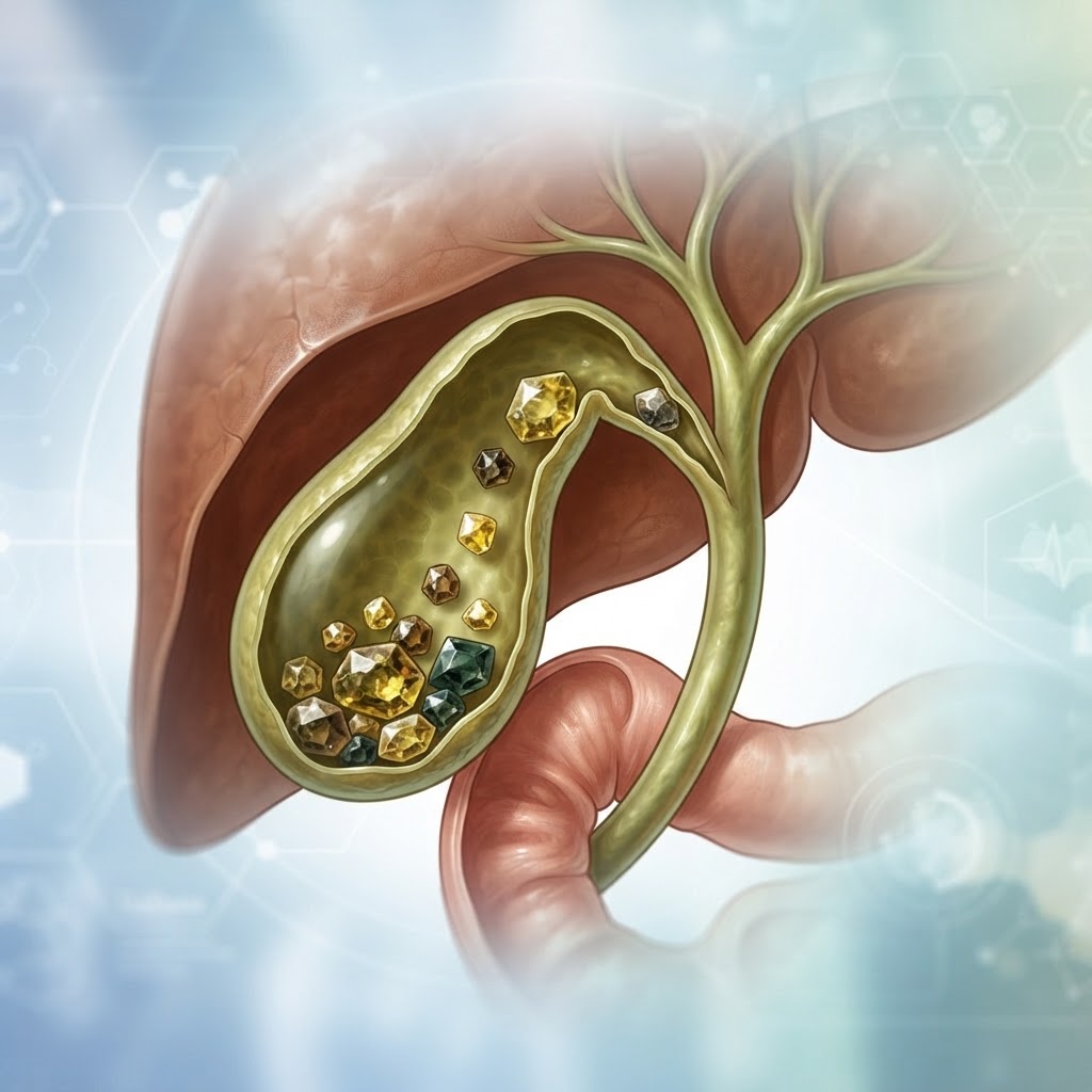
  • Chronic Nocturia: Causes, Symptoms, and How to Stop Waking Up

    Chronic Nocturia: Causes, Symptoms, and How to Stop Waking Up

    Key Takeaways

    • Chronic nocturia is defined by the consistent need to wake up one or more times during the night to urinate.
    • Frequent nighttime urination is often a symptom of underlying health issues rather than a standalone condition.
    • Lifestyle modifications, such as fluid timing and dietary adjustments, are often the first line of defense.
    • Bladder and prostate health play a critical role in how the body stores and releases urine overnight.
    • Consulting a healthcare provider is essential if sleep disruption becomes severe or affects your daily quality of life.

    There are few things more frustrating than being jolted awake by the sudden, urgent need to use the restroom, only to find yourself unable to drift back into a restful sleep. While waking up once occasionally is rarely cause for alarm, chronic nocturia—the persistent need to wake up to urinate—can strip away the restorative sleep your body desperately needs to recover. Over time, this cycle of broken rest can impact your cognitive function, mood, and long-term health. Understanding why this happens requires looking beyond the bathroom door and examining the complex interplay of your kidneys, bladder, hormonal regulation, and overall lifestyle. In this guide, the healthauthoritylife Editorial Team explores the root causes of excessive nighttime urination and provides actionable strategies to help you reclaim your nights.

    What Is Chronic Nocturia and When Should You Worry?

    At its core, nocturia is a clinical term used to describe the complaint that an individual has to wake up at night one or more times to void. However, simply waking up once does not necessarily signify a chronic health issue. Many experts generally agree that the clinical definition shifts toward a problematic concern when the frequency of waking up consistently disrupts your ability to achieve restorative sleep. For most adults, a healthy bladder should comfortably hold urine for six to eight hours, allowing for an uninterrupted night of rest.

    When you start experiencing frequent nighttime urination every single night, the impact extends far beyond mere annoyance. Chronic sleep fragmentation caused by nocturia has been linked to fatigue, irritability, and an increased risk of accidents due to daytime sleepiness. The primary concern is not just the act of urinating, but the underlying reason why your body is producing or signaling the need to void while you are supposed to be in a period of deep, restorative sleep.

    You should begin to consider nocturia as a potential health priority when the frequency becomes a regular pattern, or when it begins to interfere with your ability to function the next day. If you find yourself waking up two or more times a night, it is often a signal from your body that there is a shift in your physiological balance. It is important to differentiate between nocturnal polyuria—where your body produces an excessive volume of urine at night—and a reduction in bladder capacity, where your bladder cannot physically hold the amount of urine produced.

    To determine if you should worry, keep a bladder diary for a few days. Note exactly what you drink, when you drink it, and how many times you wake up. If you notice a consistent trend of high-volume urination at night, this might suggest a hormonal issue or a reaction to specific substances. If the volume is low but the urgency is high, it could point toward localized bladder sensitivity. In either case, if the behavior is new, persistent, and causing daytime exhaustion, it is time to move from self-observation to professional investigation. Waking up to pee is not just a sign of “getting older”; it is often a manageable symptom that can be addressed once the root cause is identified.

    Common Physiological Causes of Frequent Nighttime Urination

    The human body employs a sophisticated hormonal system to manage fluid balance, primarily through the production of antidiuretic hormone (ADH). Under normal circumstances, your brain increases the secretion of ADH as you prepare for sleep. This hormone signals your kidneys to concentrate urine, effectively slowing down production so that your bladder does not fill up while you are lying horizontally. When this process is interrupted or insufficient, it can lead to frequent nighttime urination.

    One of the most common physiological drivers is the age-related decline in nocturnal ADH production. As many people enter their middle and later years, the body’s ability to regulate this hormone can diminish, leading to increased urine production while the body is at rest. This creates a scenario where the kidneys continue to filter blood and produce urine at a rate similar to daytime, filling the bladder quickly and forcing an awakening.

    Another major factor involves fluid distribution. During the day, gravity causes fluids to pool in the lower extremities, such as the legs and ankles. When you lie down to sleep, that fluid moves back into the bloodstream, where it is processed by the kidneys. If you have any degree of fluid retention, this nocturnal shift can result in a significant surge in urine volume once you are in bed. This is why individuals who suffer from mild edema or cardiovascular conditions often report that their nocturia worsens specifically when they lie down.

    Furthermore, the kidneys’ efficiency changes throughout the 24-hour cycle. Some individuals have a naturally shifted circadian rhythm regarding their filtration rate. If your kidneys are more active at night due to metabolic or dietary factors, your bladder will reach its threshold much faster than it would during the day. Understanding these physiological shifts is crucial for developing a strategy to manage the condition. By observing your own body’s patterns, you can begin to see whether the issue is related to how much fluid you intake, how your body redistributes that fluid, or how effectively your kidneys are working with your endocrine system to manage your nighttime hydration status.

    Lifestyle Habits That Trigger Nocturnal Polyuria

    Many cases of frequent nighttime urination can be significantly mitigated by adjusting daily habits that place undue stress on the bladder and kidneys. Often, we are unaware of how seemingly benign choices—such as the timing of an evening cup of tea or a salty dinner—can dictate the quality of our sleep. The following table provides a breakdown of common lifestyle triggers and how they contribute to nocturia.

    Lifestyle Habit How It Triggers Nocturia Best For
    Late-Evening Fluid Intake Fills the bladder shortly after reclining. Reducing overall volume.
    High Sodium Consumption Causes fluid retention that releases at night. Managing fluid redistribution.
    Alcohol Consumption Suppresses ADH, causing rapid urine production. Improving hormonal regulation.
    Caffeine Late in the Day Acts as a natural diuretic and bladder irritant. Preventing bladder urgency.
    High-Protein Evening Meals May increase solute load for kidneys to filter. Stabilizing kidney filtration.

    To stop peeing at night, the most immediate step is “fluid mapping.” This does not mean you should deprive yourself of hydration; dehydration can actually lead to more concentrated, acidic urine that irritates the bladder lining, causing a false sense of urgency. Instead, the focus should be on front-loading your fluid intake. Aim to consume the majority of your water, herbal teas, or other beverages earlier in the day, tapering off significantly about three hours before your bedtime. This allows your kidneys enough time to filter the bulk of that liquid while you are still upright, moving about, and likely visiting the restroom naturally.

    Dietary sodium is another major culprit that is frequently overlooked. Salt binds to water in the body. If you eat a high-sodium dinner—often found in processed foods, restaurant meals, or cured meats—your body holds onto that excess water throughout the evening. Once you lie down to sleep, your body begins to shed this extra fluid. Reducing your sodium intake during the final meal of the day can substantially lower the amount of fluid that needs to be processed by your kidneys at night.

    Finally, consider the nature of the fluids you consume. Caffeine and alcohol are well-documented diuretics, but they also act as direct irritants to the bladder wall. Alcohol, in particular, inhibits the release of antidiuretic hormone, forcing your kidneys to work in overdrive. Eliminating these substances in the late afternoon and evening can drastically calm an overactive bladder, providing a more stable environment for uninterrupted rest. These lifestyle changes are often the most accessible nocturia treatment options, and for many, they provide the relief necessary to restore a regular sleep cycle without the need for medical intervention.

    Underlying Medical Conditions Linked to Nighttime Urination

    While lifestyle habits play a large role, it is imperative to acknowledge that chronic nocturia is often a symptom of an underlying medical condition. Because the kidneys, bladder, and endocrine system are interconnected with heart health and metabolic function, frequent nighttime urination can be a “canary in the coal mine” for broader health concerns. Recognizing these patterns is essential for anyone seeking long-term relief.

    Diabetes is perhaps the most well-known medical cause of excessive urination, both day and night. When blood sugar levels are high, the kidneys are forced to work harder to filter and absorb the excess glucose. When they cannot keep up, the glucose is excreted in the urine, dragging extra water with it. This creates a state of polyuria—a high volume of urine production. If you find that your nighttime awakenings are accompanied by unusual thirst, fatigue, or unexplained weight changes, it is critical to consult a healthcare provider to screen for blood sugar irregularities.

    Cardiovascular issues, including hypertension and congestive heart failure, also share a strong link with nocturia. When the heart struggles to pump blood efficiently, fluid naturally accumulates in the body’s tissues, particularly in the lower legs and ankles. This fluid remains trapped throughout the day due to gravity. As soon as you lie down, the pressure difference allows this fluid to re-enter the bloodstream. This surge of fluid increases the volume of blood the kidneys must process, leading to a spike in urine production overnight. In these cases, the nighttime urination is actually a functional attempt by your body to manage fluid balance, and it is a symptom that should be addressed by a medical professional to ensure heart health is being monitored.

    Sleep apnea is another often-overlooked contributor. During apnea episodes, the body experiences a drop in oxygen levels, which triggers the heart to release a hormone called atrial natriuretic peptide (ANP). This hormone signals the kidneys to produce more urine to lower blood pressure, effectively causing you to wake up with the urge to void. If you snore, wake up gasping, or feel perpetually unrefreshed, your nighttime urination might be a symptom of a breathing disorder rather than a bladder issue. By treating the sleep apnea—often through CPAP therapy or positional changes—many individuals find that their nocturia resolves entirely. Addressing these systemic issues is far more effective than trying to manage the bladder symptoms in isolation, as the bladder is merely responding to the signals and pressures created by the rest of the body.

    The Role of Bladder and Prostate Health in Nocturia

    When the cause of nocturia is not systemic—meaning it is not caused by kidney, heart, or hormonal issues—the source of the problem is often localized within the lower urinary tract. The bladder and, in men, the prostate gland, are the final gatekeepers of the urinary system. If these structures are not functioning optimally, even small amounts of fluid can trigger an urgent need to wake up.

    For men, the prostate gland is a frequent point of concern. As men age, the prostate often undergoes benign prostatic hyperplasia (BPH), or enlargement. Because the prostate surrounds the urethra, an enlarged gland can constrict the tube, making it difficult to fully empty the bladder. If the bladder is never truly empty, its functional capacity is reduced. You might have a bladder that is theoretically large enough to hold a night’s worth of urine, but because it is partially obstructed, it reaches its “full” signal much sooner. This creates a cycle where the bladder feels urgent even when it contains only a small volume of fluid. Symptoms like a weak stream, hesitation, or the feeling that you haven’t finished after urinating are common signs that the prostate may be contributing to your nocturia.

    For everyone, bladder sensitivity—sometimes referred to as an “overactive bladder”—can play a major role. This occurs when the muscles in the bladder wall (the detrusor muscles) become hypersensitive or begin to contract involuntarily before the bladder is actually full. These contractions send an urgent signal to the brain, waking you from a deep sleep, even if the actual volume of urine is minimal. This sensitivity can be exacerbated by dietary irritants, chronic inflammation, or even pelvic floor dysfunction. If the pelvic floor muscles are too tight or, conversely, too weak, they can fail to support the bladder correctly, leading to urgency that disrupts sleep.

    Strengthening the pelvic floor through targeted exercises is often a recommended approach for improving bladder control. For many, simply retraining the bladder and strengthening the muscles that support it can allow the bladder to hold urine more comfortably throughout the night. It is also worth noting that chronic constipation can put physical pressure on the bladder, further reducing its capacity. Maintaining good bowel health and ensuring the pelvic floor is functioning with the right balance of tension and relaxation are vital, often-ignored pillars of bladder health. By working to improve the structural health of the lower urinary tract, you can increase your “holding capacity,” giving you a greater buffer and allowing for a more restful, uninterrupted night of sleep.

    Diagnostic Tests to Identify the Root Cause

    Because frequent nighttime urination is often a symptom rather than a standalone condition, identifying the precise underlying trigger is the most critical step in successful management. When you present with chronic nocturia, healthcare providers typically follow a structured diagnostic pathway to differentiate between bladder issues, metabolic imbalances, and systemic health conditions.

    The process usually begins with a comprehensive medical history and a voiding diary. A voiding diary is an essential tool where you record the timing and volume of your fluid intake alongside the timing and approximate volume of your urine output over the course of 48 to 72 hours. This provides the medical team with an objective view of whether you are experiencing nocturnal polyuria (producing too much urine at night) or if your bladder capacity is simply too small to hold the volume throughout the sleep cycle.

    Physical examinations often include an abdominal and pelvic exam to check for bladder distention. Following this, laboratory testing is standard. A urinalysis is conducted to rule out urinary tract infections (UTIs) or the presence of glucose, which could indicate undiagnosed diabetes. Blood tests may also be ordered to assess kidney function, electrolyte balance, and levels of blood sugar or A1C, which reveals long-term glucose control. In men, a prostate-specific antigen (PSA) test may be recommended to investigate potential prostate enlargement.

    Advanced diagnostic imaging or specialized procedures might be necessary if initial tests are inconclusive. A bladder ultrasound is a non-invasive way to measure “post-void residual” urine—that is, how much urine remains in the bladder immediately after you attempt to empty it. If you have significant residual urine, it suggests your bladder is not emptying effectively, which could point to an obstruction or weak detrusor muscles. In some cases, a cystoscopy, where a thin, lighted tube is inserted into the urethra, may be performed to visualize the internal structure of the bladder for abnormalities, stones, or tumors.

    Effective Dietary Adjustments to Reduce Nighttime Trips

    The foods and beverages you consume in the hours leading up to bedtime have a profound impact on how often you wake up to pee. Implementing targeted dietary shifts is often the first line of defense in managing nocturia. The most effective approach involves a combination of fluid timing and the elimination of bladder irritants.

    Fluid Scheduling: Most experts recommend a “front-loading” hydration strategy. By drinking the majority of your daily fluids before 4:00 PM or 5:00 PM, you ensure that the bulk of the diuretic process occurs during the day. As the evening approaches, restrict intake to only what is necessary to quench thirst, ideally stopping all fluid consumption two to three hours before sleep.

    Eliminating Bladder Irritants: Many substances have diuretic properties or act as irritants to the lining of the bladder, causing it to contract prematurely even when it is not full. The most notorious culprits include:

    • Caffeine: Coffee, tea, and soda act as potent diuretics and bladder irritants. Caffeine increases blood flow to the kidneys and suppresses the hormone that tells your body to conserve water.
    • Alcohol: Alcohol inhibits the release of vasopressin, a hormone that regulates fluid balance. This suppression tells your kidneys to flush water out into the bladder much faster than usual.
    • Artificial Sweeteners: Ingredients like aspartame and saccharin are known to trigger urgency in sensitive individuals.
    • Acidic Foods: Spicy dishes, tomatoes, citrus fruits, and chocolate can irritate the bladder wall, leading to a constant sensation of needing to void.

    Consider keeping a log of your meals for one week alongside your voiding diary. Often, patients discover that a specific habit, such as an evening glass of wine or a late-night cup of herbal tea, is the primary driver of their nocturnal awakenings. By replacing these triggers with water earlier in the day, you reduce the physiological pressure on your bladder at night.

    Behavioral Strategies to Improve Your Sleep Quality

    Behavioral interventions focus on “training” the bladder and managing peripheral physiological factors that contribute to urine production while lying down. One of the most effective behavioral strategies is the use of compression stockings. If your nocturia is caused by edema—the pooling of fluid in your lower extremities during the day—that fluid returns to your bloodstream when you lie down to sleep. The kidneys then filter this excess fluid, creating a surge in urine production during the night. Wearing graduated compression stockings during the day helps keep that fluid in circulation, preventing it from overwhelming your bladder at night.

    Another powerful strategy is leg elevation. If you spend most of your day on your feet, taking time to elevate your legs above the level of your heart for an hour in the late afternoon can help the body redistribute that fluid while you are still awake. This allows your kidneys to process the excess volume during the day rather than while you are trying to rest.

    Bladder training is also vital. If your bladder has become “hyperactive,” it may be accustomed to voiding at small volumes. Bladder retraining involves slowly increasing the time between trips to the bathroom during the day, which helps the bladder muscle become more relaxed and increases its functional capacity. This “stretching” of the bladder helps it become more tolerant of higher volumes, effectively reducing the number of times it sends a “full” signal to your brain.

    Strategy Mechanism of Action Best For
    Compression Stockings Reduces peripheral fluid pooling Individuals with lower leg edema
    Bladder Retraining Increases functional bladder capacity Those with an overactive bladder
    Afternoon Elevation Redistributes fluid to central circulation Active individuals with evening swelling
    Voiding Diary Tracking Identifies intake-to-output patterns Identifying specific dietary triggers

    When to Consult a Specialist About Your Symptoms

    While minor changes in sleep patterns can be managed with lifestyle adjustments, there are distinct “red flags” that indicate your nocturia requires the expertise of a urologist or a nephrologist. You should seek a professional consultation if your frequent nighttime urination is accompanied by any of the following warning signs:

    First, if you notice hematuria (blood in the urine), this must be investigated immediately. While sometimes linked to benign conditions like stones, it can also be an indicator of bladder cancer or other serious structural issues. Second, if you experience significant pain or burning during urination (dysuria), it suggests a lingering infection or inflammation that requires antibiotic intervention.

    Third, if your nocturia is accompanied by unintended weight loss, extreme thirst, or excessive fatigue, these are systemic symptoms that warrant an investigation into metabolic disorders like diabetes or kidney disease. Fourth, if you are a man experiencing a weak or intermittent urinary stream, this is a classic sign of benign prostatic hyperplasia (BPH) or other prostate-related obstructions that require medication or surgical management.

    Finally, if you have already attempted lifestyle changes—such as reducing evening fluid intake and eliminating bladder irritants—for four to six weeks with no measurable improvement, it is time to see a specialist. Chronic sleep deprivation caused by nocturia has long-term health consequences, including increased risk for cardiovascular issues and cognitive decline. There is no need to simply “live with” the exhaustion when modern medical interventions can often restore a full night of restorative sleep.

    Evidence-Based Treatments for Managing Nocturia

    Once a root cause is established, medical professionals can offer targeted treatments. For individuals with nocturnal polyuria, synthetic analogs of the hormone vasopressin, such as desmopressin, are sometimes prescribed. This medication helps the kidneys concentrate urine, thereby reducing the volume produced overnight. Because this medication can affect sodium levels, it is typically used under strict medical supervision, especially in older adults.

    If the nocturia is due to an overactive bladder (OAB), anticholinergic medications or beta-3 adrenergic agonists may be recommended. These drugs work by relaxing the smooth muscle of the bladder, effectively calming the “spasms” that signal the brain that the bladder needs to be emptied. For patients whose bladder muscles are unresponsive to medication, newer therapies such as sacral neuromodulation (a small implant that sends electrical impulses to the nerves controlling the bladder) have shown significant success in clinical settings.

    In cases where BPH is the primary culprit, alpha-blockers are the standard pharmaceutical approach. These drugs relax the muscles at the neck of the bladder, allowing for more complete voiding and reducing the frequency of the urge to go. If medications are insufficient, minimally invasive surgical procedures, such as UroLift or laser therapies, can open up the urinary passage and relieve the obstruction, often leading to a dramatic reduction in nighttime voids.

    Frequently Asked Questions

    Is waking up once a night to pee considered chronic nocturia?

    Generally, medical professionals define nocturia as waking up two or more times per night to void. Waking up once is often considered within the range of normal, depending on your age and fluid intake, though if it consistently disrupts your ability to return to sleep, it is worth discussing with a doctor.

    Can stress or anxiety cause me to wake up to use the bathroom?

    Yes, psychological stress can trigger physiological changes. Anxiety stimulates the sympathetic nervous system, which can increase urine production and heighten your sensitivity to bladder sensations. Stress-related insomnia also makes you more “aware” of a slightly full bladder, leading to more frequent trips.

    Should I stop drinking water entirely after dinner?

    You should not stop drinking fluids entirely, as chronic dehydration can actually lead to more concentrated, acidic urine that irritates the bladder. Instead, focus on fluid restriction: shift your intake to the earlier hours of the day and limit your evening consumption to small sips if you are thirsty.

    Does sleep apnea contribute to nighttime urination?

    There is a strong connection between obstructive sleep apnea (OSA) and nocturia. When you struggle to breathe during sleep, the pressure changes in your chest signal the heart to release a hormone that increases urine production. Treating your sleep apnea with a CPAP machine often results in a significant reduction in nightly bathroom trips.

    Are there natural supplements that help with nighttime urination?

    Some people find relief with supplements like pumpkin seed extract or magnesium, which may support bladder muscle health or reduce prostate-related issues in men. However, these are not universal cures, and you should always consult with your doctor before starting any supplement to ensure they do not interact with your current medications.

    How can I tell if my nocturia is related to diabetes?

    Excessive thirst (polydipsia) and frequent daytime urination, alongside nighttime frequency, are common markers of undiagnosed diabetes. If you find you are drinking large volumes of water and still feel thirsty, or if you experience unexplained fatigue, a blood sugar test is a vital step in ruling out glucose-related nocturia.

    Conclusion

    Chronic nocturia is more than just a nuisance; it is a significant drain on your quality of life, productivity, and long-term health. By moving from a state of frustration to one of proactive management, you can regain control over your sleep. Whether your situation requires simple dietary tweaks, the use of compression stockings, or a formal medical treatment plan for underlying conditions like sleep apnea or BPH, remember that you do not have to settle for fragmented rest. Start by tracking your symptoms with a voiding diary, eliminate common bladder irritants, and don’t hesitate to consult a specialist if the problem persists. Reclaiming your sleep is an essential investment in your overall wellness.

    By healthauthoritylife Editorial Team

    This article is for informational purposes only and is not a substitute for professional medical advice. Always consult your doctor before making changes to your diet, exercise routine, or treatment plan.

  • Gallstone Symptoms: Causes, Warning Signs, and Relief Tips

    Gallstone Symptoms: Causes, Warning Signs, and Relief Tips

    Key Takeaways

    • Gallstones are hardened deposits of digestive fluid that form within the gallbladder, often causing severe abdominal discomfort.
    • The most frequent gallstone symptoms include intense, sudden pain in the upper right abdomen or back.
    • A combination of genetics, diet, and metabolic health influences the primary causes of gallstones.
    • Effective gallbladder health management often requires a mix of medical intervention and long-term dietary adjustments.
    • Recognizing the warning signs of a gallbladder attack is critical for seeking timely medical attention and preventing complications.

    The gallbladder is a small, pear-shaped organ often overlooked until it begins to cause significant physical distress. Tucked discreetly beneath the liver, this organ serves as a storage reservoir for bile—a digestive fluid produced by the liver that helps your body break down fats. When the delicate chemical balance within this fluid is disrupted, however, it can solidify into pebble-like structures known as gallstones. For millions of individuals, these stones remain silent and undetected for years. For others, they trigger intense, debilitating episodes that demand immediate attention. Understanding the nuances of gallstone symptoms, identifying the root causes of gallstones, and implementing sustainable strategies for gallbladder health are essential steps for anyone looking to navigate this common digestive condition with confidence and clarity.

    What Are Gallstones and How Do They Form?

    To grasp the mechanism of gallstone formation, one must first understand the function of bile. Bile is a complex mixture consisting of cholesterol, bilirubin, and bile salts. These substances must remain in a precise ratio to maintain a liquid state. When this balance is compromised—either due to an excess of one component or a lack of another—the bile becomes supersaturated. Over time, these microscopic crystals begin to clump together, eventually forming solid masses that vary in size from the tiny grains of sand to stones as large as a golf ball. These deposits represent the physical manifestation of gallstones.

    There are two primary varieties of these stones that health experts typically identify. Cholesterol stones, which are the most common, appear yellow-green in color and form when the bile contains more cholesterol than it can dissolve. Because the liver produces cholesterol, and the gallbladder stores bile, the process is inherently linked to metabolic function. The second, less common type, are known as pigment stones. These are typically smaller, darker in color, and composed primarily of bilirubin—a pigment produced during the normal breakdown of red blood cells. Conditions that lead to an excess of bilirubin in the bile, such as certain blood disorders or liver cirrhosis, often create an environment conducive to the formation of these pigment stones.

    The formation process is not instantaneous. It is often a slow, cumulative development that occurs as the gallbladder fails to empty its contents completely. When the gallbladder becomes sluggish, or when bile is allowed to stagnate, the concentration of solutes increases, making crystallization significantly more likely. This stagnation can be influenced by hormones, dietary habits, or underlying physiological conditions that affect how the gallbladder contracts. Once a stone has formed, it may remain stationary within the organ, causing no noticeable issues. However, if a stone migrates into the cystic duct—the narrow tube that carries bile out of the gallbladder—it can cause a blockage. This obstruction prevents the gallbladder from emptying, leading to the rapid buildup of pressure and the onset of acute discomfort, commonly referred to as a gallbladder attack. For many, this is the first point at which they realize they have a condition, highlighting the importance of understanding why these stones form and how they can eventually manifest as persistent gallstone symptoms.

    Common Symptoms of Gallstones and Gallbladder Attacks

    The clinical presentation of gallstone issues is often paradoxical: many individuals possess gallstones without experiencing any sensation at all, while others suffer from recurring, severe distress. When symptoms do emerge, they are frequently characterized by biliary colic—a term used to describe the steady, intense pain that occurs when a gallstone obstructs the bile duct. This pain is typically localized in the upper right quadrant of the abdomen, just below the rib cage. However, the nature of this pain is often erratic. It may radiate toward the right shoulder or extend through to the back, between the shoulder blades, creating a sensation that many people initially mistake for back pain or indigestion.

    A gallbladder attack often follows a specific pattern. It frequently occurs after a meal, particularly one high in fat content, as the gallbladder is stimulated to contract and release bile into the small intestine. If a stone is obstructing the outlet, the gallbladder muscles must work harder against the blockage, triggering a rhythmic, cramping, or persistent ache. Unlike simple indigestion, this pain does not typically improve with antacids or by passing gas. In many instances, the pain reaches a crescendo and then slowly subsides as the stone shifts or the gallbladder relaxes, though the underlying blockage may remain a recurring threat.

    Beyond the primary sensation of pain, other symptoms of gallstones can serve as warning signs. Nausea and vomiting are common physiological responses to the stress of an obstruction. Furthermore, if the blockage persists or affects the common bile duct, it can lead to more systemic issues. Jaundice, characterized by the yellowing of the skin and the whites of the eyes, occurs when bilirubin cannot effectively exit the body and begins to circulate in the bloodstream. Another indicator is the appearance of dark-colored urine or pale, clay-colored stools, both of which suggest that bile flow into the digestive tract has been severely compromised. While mild symptoms might be manageable, the onset of a high fever or chills alongside abdominal pain is a significant warning sign that the condition may have progressed to a serious infection. Recognizing these signs early allows for proactive management, preventing the escalation of a simple stone into a medical emergency.

    Symptom Category Description Best For
    Biliary Colic Sudden, steady, intense upper-right abdominal pain Identifying acute blockages
    Post-Prandial Discomfort Pain triggered specifically by high-fat meals Pinpointing dietary triggers
    Systemic Signs Jaundice, fever, or pale, clay-colored stools Detecting serious complications
    Referred Pain Pain felt in the shoulder or back area Differentiating from typical acid reflux

    Primary Causes and Risk Factors for Gallstone Development

    The causes of gallstones are multifaceted, weaving together a complex tapestry of genetic predispositions, hormonal influences, and lifestyle choices. While no single factor guarantees the development of stones, certain demographics and biological conditions show a significantly higher susceptibility. Gender is a primary variable; statistical observations consistently show that women are more likely to develop gallstones than men. This is largely attributed to the influence of estrogen, which can increase the concentration of cholesterol in bile and concurrently slow down gallbladder motility. Conditions that elevate estrogen levels, such as pregnancy, hormone replacement therapy, or the use of oral contraceptives, are therefore recognized as risk factors that can accelerate stone formation.

    Weight management plays a critical role in gallbladder health as well. Being overweight or living with obesity is strongly associated with an increased risk of developing cholesterol gallstones. This is primarily because higher body mass often correlates with higher cholesterol production. Conversely, rapid weight loss—especially through extreme calorie restriction or restrictive fad diets—can also be problematic. When the body loses weight too quickly, it prompts the liver to release extra cholesterol into the bile, and the gallbladder may not empty properly during the process, creating a perfect environment for stones to coalesce. Finding a balanced, sustainable approach to weight maintenance is one of the most effective ways to mitigate this risk.

    Dietary patterns further exacerbate the development of these deposits. Diets high in refined carbohydrates and sugar, and low in fiber, have been associated with increased gallstone risk. Fiber is essential for keeping the digestive system moving and binding with bile acids, which helps maintain the fluid balance necessary to keep cholesterol dissolved. Furthermore, metabolic syndromes such as insulin resistance or diabetes can impact the way the body processes fats and secretes bile. As we age, our risk profile naturally shifts, with older adults typically showing higher incidences of gallstone development. Understanding these root causes allows individuals to take a more critical look at their own health habits. By addressing modifiable factors—such as dietary fiber intake and gradual weight management—one can effectively lower their risk, shifting the body from a state of stone formation back toward healthy biliary function.

    How Doctors Diagnose Gallstone Issues

    When an individual presents with suspected gallstone symptoms, healthcare providers typically rely on a systematic approach to confirm the presence of stones and assess the extent of any complications. The diagnostic process usually begins with a thorough medical history and physical examination. During the exam, a provider may check for “Murphy’s sign,” a clinical maneuver where they press on the area beneath the right rib cage while the patient takes a deep breath. If the gallbladder is inflamed, the patient will typically experience a sharp halt in breathing due to the sudden onset of pain. While useful, this physical finding is merely the starting point for a more comprehensive investigation.

    Imaging technology is the gold standard for diagnosing gallstones. An abdominal ultrasound is the most common first-line test because it is non-invasive, highly accessible, and exceptionally accurate at identifying stones within the gallbladder. The high-frequency sound waves provide a real-time visual representation of the organ, allowing professionals to see the number, size, and location of the stones, as well as any signs of wall thickening that might indicate chronic inflammation. If the ultrasound is inconclusive or if the provider needs to look deeper into the biliary tree—the system of ducts that carry bile—other imaging options might be utilized. A HIDA scan (cholescintigraphy) is a specialized test that tracks the flow of bile from the liver into the gallbladder and small intestine, proving particularly useful for assessing how well the gallbladder is actually functioning.

    Blood tests are equally essential in the diagnostic toolkit. While blood work cannot detect the presence of the stones themselves, it can provide critical information regarding the impact of those stones on overall health. Elevated white blood cell counts can signify an active infection, while markers such as bilirubin, alkaline phosphatase, and liver enzymes help identify if a stone has migrated into the common bile duct, causing a blockage or inflammation of the liver or pancreas. By combining these diagnostic layers—the physical assessment, the clear imagery from an ultrasound, and the biochemical profile from blood tests—doctors can create a comprehensive picture of the situation. This multi-pronged approach ensures that if a patient is experiencing a gallbladder attack, the root cause is correctly identified, and an appropriate path forward can be established, whether that involves conservative observation or more definitive clinical intervention.

    Lifestyle Changes to Reduce Gallstone Risk

    Maintaining gallbladder health is a long-term commitment that centers on stabilizing bile composition and supporting efficient digestive function. Because the gallbladder’s primary job is to release bile in response to the fat content in food, the dietary strategy should focus on moderation rather than total fat avoidance. Consuming modest amounts of healthy, unsaturated fats—such as those found in avocados, olive oil, and nuts—is actually necessary to encourage regular gallbladder contractions. If the gallbladder never contracts, the bile stays stagnant, which is a leading precursor to stone formation. The goal is to provide a consistent, moderate stimulus for the organ to empty its contents regularly throughout the day.

    A fiber-rich diet is perhaps the most significant nutritional intervention for gallbladder health. Soluble fiber, found in foods like oats, legumes, fruits, and vegetables, binds with bile acids in the digestive tract and helps clear them from the body, which reduces the amount of cholesterol available to solidify in the bile. Furthermore, fiber helps stabilize blood sugar and insulin levels, reducing the metabolic triggers that are known to contribute to gallstone development. By swapping out refined grains for whole grains and prioritizing plant-based proteins, individuals can create a digestive environment that is less prone to stagnation and crystallization.

    Hydration also plays a silent but vital role in keeping bile thin and free-flowing. Since bile is largely composed of water, chronic dehydration can lead to a more concentrated, viscous fluid that is far more likely to form microscopic crystals. Experts generally recommend maintaining adequate water intake throughout the day to support overall digestive fluidity. Beyond diet and hydration, regular physical activity is a cornerstone of metabolic health that directly influences how the body processes cholesterol. Exercise has been shown to improve gallbladder contractility and support healthy lipid profiles, both of which decrease the likelihood of stone recurrence. For those concerned about their gallbladder, these lifestyle adjustments are not just preventative measures; they are foundational habits that promote overall wellness and prevent the painful, disruptive episodes associated with gallbladder health issues. Through consistent, gentle shifts in daily behavior, it is possible to cultivate a healthy, functional system that serves the body’s needs effectively for years to come.

    Dietary Strategies to Manage Gallbladder Pain

    When dealing with the discomfort of gallstones, your diet becomes one of the most powerful tools in your arsenal for symptom management. Because the gallbladder’s primary job is to store and concentrate bile—a fluid used to break down fats—consuming high-fat meals often acts as a trigger for a gallbladder attack. To minimize biliary colic, the goal is to reduce the workload of the gallbladder by choosing foods that are easy to digest and low in inflammatory components.

    The first step in a gallbladder-friendly diet is to adopt a low-fat approach. This does not mean eliminating fats entirely, as healthy fats are essential for nutrient absorption, but rather choosing them strategically. Focus on lean proteins such as skinless poultry, white fish, legumes, and tofu, rather than red meats, sausages, or processed deli meats. Dairy products should ideally be fat-free or low-fat to prevent the gallbladder from contracting forcefully in response to excess lipids.

    Fiber is another cornerstone of gallbladder health. A diet rich in soluble fiber—found in oats, lentils, beans, apples, and carrots—can help keep the digestive system moving efficiently. Some evidence suggests that a high-fiber diet may help bind to bile acids, potentially reducing the concentration of cholesterol within the bile, which is a common precursor to stone formation. However, if you are currently in the midst of an active flare-up, start by incorporating fiber slowly to avoid excessive gas or bloating, which can exacerbate abdominal pressure.

    Hydration is frequently overlooked, yet it is essential for maintaining the fluidity of bile. When you are dehydrated, bile can become sluggish and concentrated, making the environment more hospitable for crystal and stone formation. Aim to drink adequate amounts of water throughout the day. Herbal teas, particularly those that are caffeine-free, can also contribute to your daily intake without irritating the digestive tract.

    Additionally, small, frequent meals are often better tolerated than three large meals. Large portions require the gallbladder to release a significant surge of bile all at once, which can trigger painful spasms if stones are present. By eating five or six smaller meals, you provide a steadier, more manageable demand on your digestive system.

    When to Seek Emergency Medical Attention

    While many individuals can manage mild symptoms of gallstones with dietary adjustments, there are specific warning signs that indicate a gallbladder issue has escalated into a medical emergency. Ignoring these symptoms can lead to severe complications, such as infection of the gallbladder (cholecystitis), blockage of the common bile duct, or inflammation of the pancreas (pancreatitis).

    You should seek immediate emergency medical care if you experience any of the following:

    • Persistent, severe abdominal pain: Pain that lasts for several hours and does not subside with rest or over-the-counter measures is a red flag.
    • High fever and chills: These are classic indicators that an infection may be present within the gallbladder or the surrounding biliary tree.
    • Persistent vomiting: If you are unable to keep fluids down, you are at risk for rapid dehydration and electrolyte imbalance.
    • Jaundice: If the whites of your eyes or your skin take on a yellow tint, it suggests that a stone has likely moved out of the gallbladder and is blocking the common bile duct, preventing bile from reaching the intestines.
    • Dark urine and pale stools: These changes in elimination are often associated with biliary obstruction.
    • Confusion or dizziness: In elderly individuals, these symptoms can be a sign of sepsis, which is a life-threatening response to an untreated infection.

    Do not wait for an appointment if the pain is so intense that you cannot find a comfortable position or if you feel faint. When in doubt, it is always safer to be evaluated in an emergency department, where diagnostic imaging such as an ultrasound can quickly determine if an obstruction is occurring.

    Natural Approaches to Support Gallbladder Health

    While natural treatments cannot dissolve gallstones that have already formed, many people turn to integrative approaches to support overall liver and gallbladder function. These strategies are best viewed as preventive measures to improve digestive health rather than as cures for existing stones.

    Peppermint oil is frequently used in traditional wellness circles for its antispasmodic properties. It may help relax the muscles of the gallbladder and bile ducts, potentially easing the cramping sensations associated with biliary colic. Similarly, some favor the use of turmeric (curcumin) due to its well-documented anti-inflammatory benefits. Curcumin may stimulate bile production, which aids in the digestion of fats and helps keep bile flowing, though it should be used with caution if you have known duct obstructions.

    Magnesium is another mineral often discussed in the context of gallbladder health. Magnesium deficiency can contribute to muscle spasms, and by supporting proper muscle relaxation, it may help keep the bile ducts clear. Incorporating magnesium-rich foods like pumpkin seeds, spinach, almonds, and black beans is a gentle way to support the body’s natural processes.

    Beetroot juice is often cited in holistic health literature as a “liver tonic.” While it is not a direct treatment for stones, the betaine found in beets supports the liver and improves the consistency of bile, making it easier for the gallbladder to empty properly. As with any natural supplement or dietary change, it is vital to discuss these with your primary care provider to ensure they do not interact with other conditions or medications.

    Strategy Mechanism of Action Best For
    Low-Fat Diet Reduces gallbladder contraction demand Immediate pain prevention
    Increased Fiber Helps bind bile acids and regulate digestion Long-term bile health
    Magnesium Intake Supports smooth muscle relaxation Reducing cramping/spasms
    Hydration Maintains bile viscosity/fluidity General prevention of sludge

    Surgical vs. Non-Surgical Treatment Options

    When lifestyle management is insufficient or when symptoms become recurrent and debilitating, medical professionals often look toward more definitive treatment options. The decision-making process typically involves weighing the risks of surgery against the risks of living with symptomatic gallstones.

    Surgical Intervention: The most common treatment for symptomatic gallstones is a cholecystectomy, or the surgical removal of the gallbladder. Because the gallbladder is not a vital organ—meaning you can survive and digest food perfectly well without it—this is widely considered the gold standard for recurring gallbladder attacks. Most surgeons perform this laparoscopically, using a few small incisions and a camera, which leads to a faster recovery time and less postoperative pain compared to traditional open surgery.

    Non-Surgical Treatments: Non-surgical options are generally reserved for patients who are not candidates for surgery due to other underlying health complications. Oral dissolution therapy involves taking medication (typically ursodeoxycholic acid) that can slowly dissolve cholesterol-based stones over months or years. However, this method is often ineffective for large stones or calcified stones, and it carries a high risk of stone recurrence once the medication is stopped.

    Shockwave lithotripsy is another non-surgical technique that uses sound waves to break stones into smaller fragments. While this has been used in the past, it is much less common today because it often fails to address the underlying cause of stone formation, meaning new stones frequently form shortly after the procedure.

    Managing Gallbladder Health After Treatment

    If you have undergone a cholecystectomy, you enter a new phase of managing your digestive health. Without a gallbladder to store and concentrate bile, the bile flows continuously from the liver into the small intestine. For many, this adjustment is seamless, but for others, it can cause transient digestive issues, particularly after consuming high-fat meals.

    In the weeks following surgery, start with a bland diet. Gradually reintroduce fats to see how your digestive system responds. If you notice symptoms like diarrhea or bloating after eating fat, it is often helpful to keep a food diary to identify specific triggers. Some individuals find that taking digestive enzymes or bile salts as recommended by their medical team can assist the body in breaking down fats more effectively during this transitional period.

    Long-term health after gallbladder removal remains focused on the same principles as before: a diet rich in fruits, vegetables, and whole grains. Staying active is also crucial, as regular movement helps maintain overall metabolic health and keeps the digestive system regular, ensuring that bile flow continues to work efficiently despite the missing storage organ.

    Frequently Asked Questions

    Can gallstones disappear on their own?

    Gallstones typically do not disappear on their own. Once a stone has formed, it usually remains unless it is passed through the bile duct—which can be extremely painful and dangerous—or is surgically removed. While some very small “sludge” can sometimes be flushed through the system, actual stones rarely dissolve spontaneously.

    Is coffee good or bad for your gallbladder?

    Research suggests that regular, moderate coffee consumption may be associated with a lower risk of developing symptomatic gallstones. It is believed that caffeine may stimulate gallbladder contraction and improve bile flow. However, if you are currently experiencing an acute gallbladder attack, the acidity of coffee might irritate your stomach, so it is best to listen to your body.

    How long does a gallbladder attack last?

    A gallbladder attack (biliary colic) typically lasts between one and five hours. If the pain lasts longer than six hours, or if it is accompanied by fever, vomiting, or jaundice, it may indicate a complication, such as cholecystitis, and requires medical attention.

    Are certain people more prone to developing gallstones?

    Yes, risk factors for gallstones include being female, being over the age of 40, having a family history of gallstones, obesity, rapid weight loss, and certain medical conditions like diabetes or Crohn’s disease. Hormonal factors, such as those related to pregnancy or estrogen therapy, also play a significant role.

    Do I have to stop eating fat forever after my gallbladder is removed?

    No, you do not need to stop eating fat forever. After your body adjusts to the absence of the gallbladder—a process that can take a few weeks to a few months—you can usually return to a normal, healthy diet. However, it is always recommended to prioritize healthy, unsaturated fats over saturated and trans fats for overall heart and digestive health.

    What does gallbladder pain feel like?

    Gallbladder pain is often felt as a dull, steady ache or a sharp, cramping pain in the upper right side of the abdomen, just below the ribs. This pain often radiates to the center of the abdomen or through to the right shoulder blade or back. It is frequently triggered by eating a heavy or fatty meal.

    Conclusion

    Gallstone symptoms can range from mild, intermittent discomfort to severe, life-altering pain that demands immediate medical intervention. Understanding the triggers—namely high-fat meals—and recognizing the warning signs of complications are the first steps toward regaining control over your health. Whether you are managing symptoms through careful dietary choices or exploring surgical options, prioritizing your digestive health will lead to a better quality of life.

    Do not dismiss persistent abdominal pain as simple indigestion. If you suspect your gallbladder is the cause of your discomfort, schedule an appointment with your healthcare provider to discuss your symptoms and pursue an accurate diagnosis through imaging. Taking proactive steps today can prevent the escalation of symptoms and help you avoid the complications associated with untreated biliary disease.

    By healthauthoritylife Editorial Team

    This article is for informational purposes only and is not a substitute for professional medical advice. Always consult your doctor before making changes to your diet, exercise routine, or treatment plan.

  • Chronic Sinusitis: Causes, Symptoms, and Natural Relief Tips

    Chronic Sinusitis: Causes, Symptoms, and Natural Relief Tips

    Key Takeaways

    • Chronic sinusitis is defined by sinus inflammation lasting longer than 12 weeks despite medical intervention.
    • Recognizing chronic sinusitis symptoms early is essential for preventing long-term tissue damage and secondary infections.
    • Dietary choices significantly impact systemic inflammation, which directly correlates with sinus pressure and mucus production.
    • Environmental triggers like air quality, mold, and humidity levels often dictate the severity of persistent sinus issues.
    • Integrating natural remedies for sinus infection alongside conventional sinusitis treatment offers a comprehensive approach to long-term sinus health.

    For those living with the persistent, heavy pressure of congested nasal passages, the world can feel perpetually muffled. Chronic sinusitis is more than just a passing seasonal cold; it is a complex, long-term condition that disrupts sleep, diminishes productivity, and weighs heavily on overall quality of life. When the delicate lining of your sinus cavities stays inflamed for weeks or months on end, the body struggles to find balance, leading to a cycle of discomfort that often feels impossible to break. Whether you are navigating the complexities of chronic congestion relief or seeking to understand why your immune system remains on high alert, gaining a clear perspective on the root causes is the first step toward reclaiming your respiratory health. In this guide, the healthauthoritylife Editorial Team explores the intricacies of sinus inflammation, offering a research-backed roadmap to understanding your body’s signals and finding the relief you deserve.

    1. Understanding Chronic Sinusitis: What It Is

    To grasp the nature of chronic sinusitis, one must first understand the anatomy of the sinuses themselves. These are hollow, air-filled cavities located within your skull—around your nose, eyes, and forehead. Under healthy conditions, these cavities are lined with a thin layer of mucus that traps dust, allergens, and pathogens before they can enter the respiratory system. Tiny, hair-like structures called cilia then sweep this mucus toward the back of the throat, where it is swallowed and neutralized by stomach acid. It is a seamless, automated process that typically goes unnoticed.

    Chronic sinusitis occurs when this drainage pathway is disrupted for an extended period, typically lasting 12 weeks or longer, regardless of initial efforts to resolve the blockage. Unlike acute sinusitis—the type that usually follows a common cold or a brief bout of seasonal allergies and clears up within a few weeks—chronic sinusitis represents a persistent state of sinus inflammation. When the sinus ostia (the small drainage openings) become blocked, mucus becomes trapped. This stagnant environment provides the ideal breeding ground for bacteria, viruses, or fungi, which then colonize the cavity, perpetuate the swelling, and create a self-sustaining cycle of pressure and pain.

    The distinction between acute and chronic is critical because the treatment strategies differ significantly. While a short-term infection might be managed with rest and over-the-counter decongestants, chronic cases require a deeper look at the underlying physiology. Many individuals with chronic conditions exhibit structural anomalies, such as a deviated septum or nasal polyps, which physically impede the flow of air and mucus. However, it is not always a structural issue. Often, the lining of the sinuses becomes hypertrophic (thickened), meaning the tissue itself is permanently altered by chronic exposure to irritants. This leads to a scenario where the body is in a constant state of low-grade warfare, struggling to keep the airways clear.

    Understanding this condition requires moving beyond the “one-size-fits-all” approach to healthcare. It is important to view chronic sinusitis as a systemic issue rather than just a localized problem of the nose. When your sinuses are chronically inflamed, your entire immune system may be on high alert. This chronic stressor can deplete energy levels and contribute to a feeling of overall malaise. By identifying the specific nature of your congestion—whether it is driven by structural barriers, chronic infection, or persistent inflammation—you can better advocate for your own health and select the strategies that address the root of the problem rather than just masking the pain.

    2. Common Symptoms of Persistent Sinus Issues

    The hallmark of chronic sinusitis is a constellation of symptoms that refuse to fade. While patients may experience varying levels of severity, the clinical profile often includes a sense of “heaviness” behind the eyes, forehead, or cheeks. This is frequently described as a dull, throbbing pain that worsens when leaning forward or bending down, which increases blood pressure in the sinus cavities. It is essential to monitor these chronic sinusitis symptoms closely, as they often overlap with other conditions like tension headaches or migraines.

    One of the most debilitating aspects of this condition is chronic nasal congestion. Patients often report feeling like their nose is “stuffed” even when they can physically push a small amount of air through. This feeling is caused by the engorgement of the nasal turbinates—structures inside the nose that regulate airflow. When these are constantly inflamed, the sensation of blocked airflow remains persistent. This leads to a compensatory behavior: mouth breathing. Mouth breathing is far less efficient than nasal breathing; it bypasses the body’s natural filtration and humidification system, often leading to dry throat, bad breath, and poor sleep quality.

    Another prevalent symptom is the production of thick, discolored mucus. While clear mucus is common in mild irritation, chronic issues often result in yellow or green discharge, which can drip down the back of the throat—a phenomenon known as post-nasal drip. This, in turn, can cause a chronic cough, particularly at night when lying flat, as the excess mucus irritates the throat lining. Many individuals with chronic sinus issues also notice a significant reduction in their sense of smell (hyposmia) or even a total loss of taste. This happens because the inflammation prevents odor molecules from reaching the olfactory nerve endings at the top of the nasal cavity.

    Fatigue is an underrated but highly common symptom. Living with chronic inflammation is energy-intensive; the body is constantly mobilizing white blood cells and repair mechanisms to deal with the sinus tissue. Furthermore, the disrupted sleep caused by congestion and difficulty breathing can lead to “brain fog” and decreased cognitive performance during the day. It is common for these symptoms to wax and wane, leading many to believe they are recovering, only for the inflammation to flare up again when triggered by a change in air pressure, weather, or stress. If you notice these symptoms persisting for several months, it is time to shift your focus from acute symptom management to a long-term wellness strategy.

    Approach Primary Mechanism Best For
    Saline Irrigation Physical flushing of mucus and allergens Daily maintenance and clearing debris
    Steam Inhalation Moisturizing dry, irritated mucosal tissue Immediate relief from dry, crusty congestion
    Anti-inflammatory Diet Reducing systemic inflammatory markers Long-term prevention of chronic flare-ups
    Acupressure/Massage Improving blood flow and lymphatic drainage Relieving acute sinus pressure and tension

    3. Root Causes: Why Your Sinuses Won’t Clear

    To move toward effective sinusitis treatment, we must identify the “why” behind the inflammation. In many cases, it is not a single factor but a convergence of several triggers that keeps the sinuses in a state of turmoil. Understanding these root causes empowers you to move beyond symptomatic relief and start addressing the underlying barriers to your respiratory health.

    Structural impediments often serve as the primary hurdle. A deviated septum—where the thin wall separating your nasal passages is off-center—can significantly constrict the drainage pathway on one or both sides. When the airway is physically narrowed, the sinuses cannot drain effectively, even under normal conditions. Similarly, the presence of nasal polyps—soft, non-cancerous growths on the lining of your nasal passages or sinuses—can create a mechanical barrier that trap mucus. While medication can help shrink polyps or reduce the swelling around a deviated septum, the physical obstruction remains a persistent risk factor for recurrent bouts of sinusitis.

    Beyond physical structures, the immune system’s reaction to external threats is a massive driver of chronic inflammation. Allergic rhinitis, or persistent hay fever, is a leading culprit. If you are constantly exposed to allergens like pet dander, dust mites, or seasonal pollens, your immune system maintains an inflammatory response in the nasal cavity. This creates a state of “hyper-reactivity,” where the sinus lining becomes chronically thickened and sensitive. Over time, the constant swelling prevents normal ventilation, and the tissue becomes less effective at self-cleaning, leading to a vicious cycle of congestion that is frequently misdiagnosed as a persistent cold.

    Infectious etiology also plays a major role, specifically in cases where bacterial or fungal colonies have taken hold. While many assume sinusitis is always bacterial, chronic cases often have a complex microbiological profile. Fungal sinusitis, in particular, is a growing area of interest. In individuals with a compromised immune system or a hypersensitivity to airborne molds, fungal spores can colonize the sinus cavities. The body reacts by increasing inflammation to wall off the perceived threat, creating thick, sticky mucus that is notoriously difficult to clear. Conventional sinusitis treatment that focuses solely on antibiotics will fail in these scenarios, sometimes even making the situation worse by disrupting the natural bacterial flora that keeps fungal overgrowth in check.

    Finally, we cannot ignore the influence of systemic health. Issues such as GERD (Gastroesophageal Reflux Disease) have been linked to chronic sinusitis. The theory is that stomach acid or enzymes can reach the nasopharyngeal area, particularly when lying down, causing irritation and inflammation in the back of the nose and sinuses. Addressing the health of the digestive tract can sometimes result in surprising improvements in respiratory clarity. By viewing the body as an interconnected system, we see that the sinuses are often the “canary in the coal mine,” signaling that there is an inflammatory issue happening elsewhere in the body that requires attention.

    4. The Role of Diet and Inflammation in Sinus Health

    When searching for natural remedies for sinus infection, many overlook the power of the kitchen. While diet may not provide an instant “cure” for a blocked sinus, it serves as a powerful foundation for controlling the systemic inflammation that drives chronic sinus issues. If your body is in a pro-inflammatory state, your sinus tissue is likely to remain swollen and reactive to even the smallest environmental trigger. Shifting toward an anti-inflammatory nutritional profile is one of the most proactive steps you can take for your sinus health.

    The primary objective of an anti-inflammatory diet is to reduce the intake of foods known to trigger immune reactions and inflammatory markers. Dairy is a classic example. For many, particularly those with a history of sinus issues, dairy products can increase the perception of mucus thickness. While the science on whether dairy directly increases mucus production is still debated, many individuals find that eliminating dairy for a trial period significantly reduces their sense of congestion. Processed sugars and refined carbohydrates are other major offenders; these foods are linked to spikes in blood sugar and insulin, which are known to promote systemic inflammation. When blood sugar levels are unstable, the body’s inflammatory response is more easily triggered, making it harder for the sinuses to recover from any irritation.

    Conversely, focusing on whole, nutrient-dense foods can help soothe the inflammatory response. Omega-3 fatty acids, found in wild-caught fish, flaxseeds, and walnuts, are potent anti-inflammatories that help modulate the immune system. Incorporating these into your diet regularly can help “cool down” the inflammatory response throughout the body, including the delicate tissues of the nose and sinuses. Antioxidant-rich foods, such as berries, leafy greens, and colorful vegetables, provide the vitamins and minerals necessary to support mucosal tissue repair. Specifically, Vitamin C and Quercetin—a natural bioflavonoid found in onions, apples, and capers—have been shown to have antihistamine-like effects, which may help stabilize the mast cells that release histamines in response to allergens.

    Hydration is another critical dietary component that is often misunderstood. When you are suffering from chronic congestion relief, you need to keep your mucus thin and mobile. Proper hydration is essential for this. However, it is not just about drinking water; it is about the *type* of hydration. Herbal teas, especially those containing ginger, turmeric, or peppermint, provide a double benefit: they hydrate the body and provide therapeutic compounds that help thin secretions and soothe irritated airways. Ginger, for instance, contains gingerols, which have natural anti-inflammatory and decongestant properties. Turmeric, when combined with black pepper to improve bioavailability, is widely regarded for its ability to lower inflammatory cytokines.

    Finally, consider the timing and composition of your meals. As mentioned regarding GERD, eating late at night can cause reflux that irritates the sinuses. By finishing your last meal at least three hours before bedtime, you reduce the likelihood of acid reaching the upper respiratory tract while you sleep. By treating your diet as a core pillar of your sinus wellness strategy, you empower your immune system to function more effectively, reducing the “background noise” of chronic inflammation that keeps your sinuses blocked.

    5. Environmental Triggers and How to Avoid Them

    Your environment acts as the battlefield for your sinuses. If you are struggling with chronic sinusitis, the air you breathe every day is either helping or hindering your progress. Understanding how to manage your surroundings is a fundamental aspect of long-term sinus health. Many of the most common irritants are invisible, which makes them difficult to identify without a systematic approach to environmental control.

    Humidity is perhaps the most significant environmental factor for sinus health. Air that is too dry can parch the mucous membranes, causing them to crack and become susceptible to irritation and infection. On the other hand, air that is too humid—typically above 50%—can encourage the growth of mold and dust mites, both of which are major allergens and triggers for sinus inflammation. Investing in a reliable hygrometer to monitor the humidity levels in your home is a simple, inexpensive way to gain control. Using a humidifier in the winter, when heating systems dry out the air, and a dehumidifier in damp, basement-prone areas can help maintain the “Goldilocks” zone of moisture for your sinuses.

    Indoor air quality is another major concern. Modern homes are built for energy efficiency, which means they are well-insulated and often trap pollutants inside. Volatile organic compounds (VOCs) from cleaning products, synthetic fragrances, candles, and paint can act as chronic irritants to the nasal lining. If you suffer from persistent congestion, consider switching to fragrance-free cleaning products and minimizing the use of synthetic air fresheners. Furthermore, the role of dust and pet dander cannot be overstated. Regularly cleaning your HVAC filters, using HEPA-grade air purifiers, and washing bedding in hot water can significantly reduce the “allergen load” your sinuses are forced to deal with while you sleep—which is when you are most stationary and exposed to these particles.

    External environmental triggers, such as pollution and weather changes, are harder to control but can be managed. If you live in an area with high air pollution, check the air quality index before planning outdoor activities. On days with poor air quality, keep windows closed and rely on indoor air filtration. Rapid changes in barometric pressure, such as those associated with incoming storms, are a well-documented trigger for sinus pain. While you cannot change the weather, you can prepare for it. Many people find that performing a gentle saline rinse or using steam inhalation during these pressure shifts can mitigate the discomfort caused by the resulting changes in sinus cavity pressure.

    Lastly, pay attention to the “silent” triggers in your daily routine. Do you spend significant time in a workspace with poor ventilation? Do you exercise in a gym with dusty equipment? Being observant about where your symptoms flare up can provide clues about environmental triggers you might otherwise miss. Keeping a simple log of your symptom severity alongside your daily activities can help you identify patterns—perhaps you notice that your congestion is worse on days you work from a specific room or after visiting a certain type of environment. Once you identify these triggers, you can take deliberate steps to avoid or mitigate them, which is a powerful, proactive form of chronic congestion relief that requires no medication and provides lasting benefits for your overall sinus health.

    Natural Remedies to Reduce Sinus Congestion

    When dealing with chronic sinus inflammation, the primary objective is to thin the mucus and encourage it to drain from the sinus cavities. While pharmacological interventions exist, many individuals find substantial chronic congestion relief through targeted natural approaches that prioritize mucosal hydration and the reduction of local swelling.

    The most foundational natural remedy is nasal irrigation, specifically using a neti pot or a squeeze bottle filled with a sterile saline solution. The process physically flushes out allergens, dust, and excess mucus, while the moisture helps soothe irritated membranes. It is imperative to use distilled, sterile, or previously boiled water to ensure safety, as tap water can harbor dangerous pathogens.

    Another highly effective technique is the application of warm, moist heat. Using a warm compress over the nose, cheeks, and eyes for 10 to 15 minutes can increase circulation to the area, potentially loosening thick mucus and providing localized comfort. Similarly, steam inhalation serves as a classic remedy. By sitting in a bathroom filled with steam or inhaling from a bowl of hot water—taking care to avoid burns—the warm, humid air hydrates the nasal passages and helps break up congestion.

    Dietary adjustments also play a crucial role. Incorporating anti-inflammatory foods, such as those rich in omega-3 fatty acids or antioxidants like quercetin (found in onions, apples, and berries), may help modulate the inflammatory response in the nasal lining. Furthermore, maintaining high systemic hydration levels is essential. Water helps keep mucus thin; when the body is dehydrated, mucus becomes viscous and difficult to clear, which exacerbates the sensation of blockage.

    Remedy Type Primary Mechanism Best For
    Saline Irrigation Mechanical flushing of debris/mucus Daily maintenance and allergy clearance
    Steam Inhalation Hydration of nasal mucosa Immediate relief of thick, stubborn mucus
    Warm Compresses Vasodilation and localized comfort Reducing facial pain and sinus pressure
    Anti-inflammatory Diet Systemic reduction of cytokines Long-term prevention of flare-ups

    When to Seek Medical Attention for Sinus Pain

    While many cases of sinus discomfort can be managed at home, chronic sinusitis symptoms sometimes signal an underlying condition that requires professional medical intervention. It is vital to recognize the “red flags” that suggest a more serious complication or a persistent structural issue.

    You should consult a healthcare provider if your symptoms persist for more than twelve weeks despite consistent self-care efforts. Additionally, if you experience a “double sickening”—a phenomenon where you seem to be recovering from a respiratory illness, only to have symptoms suddenly return or worsen—this may indicate a secondary bacterial infection that requires prescription treatment.

    Emergency medical attention is necessary if you experience signs of infection spreading beyond the sinuses. Indicators of potential complications include high fever, severe headache that does not respond to over-the-counter pain relievers, swelling or redness around the eyes, vision changes, or neck stiffness. Any confusion, lethargy, or signs of neurological distress should be treated as a medical emergency, as these may suggest the infection has migrated toward the central nervous system.

    Diagnostic Tests for Chronic Sinusitis

    To move beyond symptom management and toward a definitive sinusitis treatment plan, healthcare providers often employ a variety of diagnostic tools. These tests are designed to visualize the sinus cavities and identify the root cause of your chronic inflammation.

    A physical examination typically begins with anterior rhinoscopy, where a provider uses a light and a small speculum to look into the nostrils. To get a deeper, more detailed view, they may perform a nasal endoscopy. This involves passing a thin, flexible tube with a camera on the end into the nasal passages, allowing for a clear view of the sinus openings and the ability to check for polyps, structural abnormalities, or signs of active infection.

    If physical inspection is inconclusive or if the provider suspects a more complex issue, a CT scan of the paranasal sinuses is often the gold standard. This imaging test provides a cross-sectional view of the sinuses, highlighting blockages, bone damage, or the extent of mucosal thickening. In cases where allergies are suspected as the primary driver of chronic congestion, a provider may order allergy testing, such as skin prick tests or blood panels, to identify environmental triggers that keep the sinus lining in a state of constant irritation.

    Lifestyle Changes for Long-Term Sinus Relief

    Achieving lasting sinus health requires more than just reactive measures; it demands a proactive change in your day-to-day environment. The air you breathe is the most significant factor in sinus health. High-quality HEPA air purifiers can drastically reduce the number of airborne particulates, such as pet dander, mold spores, and pollen, that contribute to sinus inflammation.

    Humidity control is equally important. Using a humidifier during winter months or in dry climates helps prevent the nasal mucosa from drying out, which can lead to cracking and increased vulnerability to infections. Conversely, if you live in a high-humidity environment that encourages mold growth, a dehumidifier may be necessary to keep indoor air quality in a healthy range.

    Lifestyle habits like tobacco cessation are non-negotiable for chronic sinusitis patients. Smoking, and even second-hand smoke exposure, paralyzes the cilia—the tiny, hair-like structures in your nose tasked with sweeping mucus out of your cavities. When cilia function is compromised, stagnant mucus becomes a breeding ground for inflammation and bacteria. Finally, prioritize stress management. Chronic stress is known to elevate cortisol levels, which can suppress the immune system and increase systemic inflammation, potentially worsening sinus issues.

    Supplement Strategies to Support Sinus Drainage

    Beyond traditional diet and lifestyle, certain evidence-informed supplements are frequently used to support sinus health. It is important to note that these should be used as adjuncts rather than sole treatments for chronic conditions.

    Bromelain, an enzyme complex derived from pineapples, is highly regarded for its potential to reduce nasal swelling and thin mucus. Research suggests it may help improve breathing by modulating the body’s inflammatory pathways. Similarly, N-acetylcysteine (NAC) is often utilized for its mucolytic properties; by breaking the disulfide bonds in mucus, it can help the body clear congestion more effectively.

    Herbal extracts such as stinging nettle are frequently used by those whose sinus issues are driven by seasonal allergies, as the plant acts as a natural antihistamine. Vitamin C and Zinc are also staples in sinus support protocols, serving as foundational nutrients that bolster immune function. Before adding these supplements to your regimen, always discuss them with your healthcare provider, particularly if you are taking blood thinners or other maintenance medications, to prevent adverse interactions.

    Frequently Asked Questions

    Is chronic sinusitis the same as a common cold?

    No, they are quite different. A cold is a short-term viral infection that typically resolves within a week or two. Chronic sinusitis is a long-term condition where the sinus cavities remain inflamed and swollen for 12 weeks or longer, despite various attempts at treatment.

    Can stress really cause sinus pressure?

    While stress does not physically “create” sinus congestion, it significantly impacts the body’s inflammatory response and immune function. High stress levels can exacerbate existing sinus issues, making the body less efficient at managing inflammation and more susceptible to recurring sinus symptoms.

    What is the role of nasal polyps in chronic sinusitis?

    Nasal polyps are soft, painless, noncancerous growths on the lining of your nasal passages or sinuses. They often occur due to chronic inflammation. By physically blocking the sinus drainage pathways, they trap mucus and contribute to the cycle of congestion and infection characteristic of chronic sinusitis.

    Are antibiotics always needed for sinus infection?

    No. Most sinus infections are viral in origin, meaning antibiotics are ineffective. Antibiotics are only appropriate when a provider confirms a secondary bacterial infection. Overuse of antibiotics can lead to resistance and disrupt the microbiome, which may ultimately complicate sinus health.

    How often should I use a saline nasal rinse?

    For most people struggling with chronic congestion, once or twice daily is standard. However, the frequency should be tailored to your specific symptoms and the guidance of your physician. Always ensure your device is cleaned thoroughly after every use to prevent introducing bacteria into your nasal cavity.

    Can diet truly influence sinus inflammation?

    Yes, diet can influence the systemic inflammatory load. Diets high in processed sugars and refined carbohydrates are known to promote inflammation, whereas an anti-inflammatory diet—rich in leafy greens, healthy fats, and antioxidants—can help the body manage the mucosal irritation associated with chronic sinusitis.

    Conclusion

    Managing chronic sinusitis requires a multifaceted approach that addresses both the immediate symptoms of congestion and the underlying causes of persistent inflammation. By integrating consistent saline irrigation, environmental modifications, and a supportive lifestyle, you can significantly reduce the frequency and severity of your flare-ups. Remember that while natural remedies provide excellent support for daily sinus health, they work best in harmony with professional medical oversight. If your sinus pain remains persistent or shows signs of progressing, reach out to a healthcare professional to create a personalized, long-term care plan. Take control of your sinus health today by identifying your unique triggers and committing to the habits that keep your respiratory system clear and functioning optimally.

    By healthauthoritylife Editorial Team

    This article is for informational purposes only and is not a substitute for professional medical advice. Always consult your doctor before making changes to your diet, exercise routine, or treatment plan.

  • Kidney Stones: Causes, Prevention, and Natural Relief

    Kidney Stones: Causes, Prevention, and Natural Relief

    Key Takeaways

    • Kidney stones are hard mineral and salt deposits that require medical evaluation when symptoms emerge.
    • Adequate hydration is the most effective lifestyle strategy for preventing recurring stone formation.
    • Understanding the chemical composition of a stone is essential for tailoring your long-term diet.
    • Chronic stone issues are often linked to systemic metabolic factors rather than just isolated dietary choices.
    • Natural kidney stone treatment approaches typically focus on pain management, hydration, and long-term preventative lifestyle adjustments.

    Few experiences in human health are as universally feared and physically demanding as passing a kidney stone. Often described by those who have endured it as a peak experience of discomfort, the development of these calcified deposits is a complex physiological process that bridges the gap between metabolic health, genetic predisposition, and daily habits. Whether you are currently navigating the acute phase of discomfort or seeking strategies for long-term prevention, understanding the mechanics of renal health is the first step toward reclaiming your well-being. This guide, compiled by the healthauthoritylife Editorial Team, explores the multifaceted nature of kidney stones, offering evidence-based insights into why they form, how to identify the warning signs, and the actionable lifestyle shifts required to support your kidneys for years to come.

    What Are Kidney Stones and Why Do They Form?

    At their core, kidney stones—medically known as renal calculi—are solid, pebble-like pieces of material that form within the kidneys when the urine contains higher-than-normal concentrations of minerals and salts. Under ideal physiological conditions, your kidneys act as sophisticated filtration systems, constantly balancing the volume of water and solutes in your blood. When this balance is disrupted, typically due to a lack of sufficient fluid or an excess of specific waste products, these substances begin to crystallize and aggregate.

    The process of stone formation, clinically referred to as lithogenesis, is essentially a failure of solubility. When the volume of urine is too low to dilute the minerals passing through the kidneys, or when the chemical composition of that urine favors crystallization, tiny particles begin to clump together. Over weeks or months, these particles can grow in size, potentially becoming large enough to cause a blockage within the ureter—the narrow tube that carries urine from the kidney to the bladder. This blockage is what triggers the classic, intense distress associated with the condition.

    Several factors contribute to this environment. The most common driver is persistent dehydration. Without enough water, urine becomes concentrated, allowing minerals like calcium, oxalate, and uric acid to cling to one another. However, dehydration is rarely the sole culprit for chronic sufferers. Genetics often play a significant role, as do metabolic conditions that affect how the body processes minerals. For instance, some individuals may naturally excrete higher levels of calcium in their urine, a condition known as hypercalciuria. Others may have an imbalance in urinary pH, which can make the environment more acidic or alkaline, thereby creating a “perfect storm” for specific types of crystals to flourish.

    It is important to recognize that while stones begin in the kidneys, they can migrate. Small stones might pass through the urinary tract without noticeable distress, but as they grow, their irregular shapes can scrape the delicate lining of the urinary tract. Understanding that these stones are not just “debris” but complex chemical structures is vital. They are a reflection of your body’s internal chemistry, signaling that your metabolic filtration system is under strain. By viewing kidney stones as a symptom of a systemic imbalance rather than a random event, you can better engage in natural kidney stone treatment and prevention strategies that address the root cause rather than just the acute manifestation.

    Common Symptoms of Kidney Stones You Shouldn’t Ignore

    Identifying kidney stone symptoms early can mean the difference between a manageable outpatient situation and an emergency room visit. The discomfort associated with a stone is notorious for its sudden onset, yet many people dismiss the subtle warning signs that precede the agony of a full-blown obstruction. One of the most classic signs is sharp, cramping pain in the back and side, often referred to as flank pain. This sensation occurs when the stone begins to move into the ureter, causing a muscular spasm and a buildup of pressure within the kidney.

    This pain is rarely static; it typically fluctuates in intensity, occurring in waves as the urinary tract attempts to move the stone forward. Many patients report that this pain radiates downward toward the lower abdomen and groin, a phenomenon caused by the shared nerve pathways between the kidneys and the lower urinary tract. This radiating nature is a hallmark indicator that the stone is migrating.

    Beyond pain, changes in urinary behavior serve as critical red flags. You might find yourself feeling a persistent, nagging urge to urinate, even when your bladder is nearly empty. If you do manage to pass urine, you may find the sensation painful or stinging—a symptom that is often mistaken for a urinary tract infection. In some cases, the urine itself will appear cloudy, dark, or tinged with pink or red, signifying the presence of blood caused by the stone abrading the interior wall of the ureter. It is also common to experience nausea or vomiting, as the intensity of renal pain can trigger a significant autonomic response in the body.

    It is crucial not to ignore systemic symptoms that suggest a potential infection. If the stone causes a complete blockage, urine can back up into the kidney, leading to a condition called hydronephrosis. Should you develop a fever or chills alongside your flank pain, this may indicate that the obstruction has led to an infection, which is a medical emergency. While many small stones pass on their own, the “wait and see” approach should never be applied to symptoms that include high fever, severe dehydration, or an inability to keep fluids down. Monitoring your body’s signals—the quality of your urine, the location of your discomfort, and the onset of systemic illness—is an essential part of being proactive with your renal health.

    Identifying the Different Types of Kidney Stones

    Not all kidney stones are identical, and understanding their composition is the foundation of an effective kidney stone diet. When a stone is retrieved or analyzed, the results categorize the stone into one of four primary types, each dictated by unique metabolic triggers. Knowing which type you are prone to forming allows you to target your natural kidney stone treatment strategies with precision, as a diet designed to prevent one type might inadvertently encourage the formation of another.

    Stone Type Primary Cause Best Strategy
    Calcium Oxalate High oxalate, low fluid, high salt Hydration and oxalate moderation
    Uric Acid Purine-rich diet, metabolic issues Lowering animal protein intake
    Struvite Chronic urinary tract infections Infection management and hygiene
    Cystine Genetic disorder (Cystinuria) Aggressive hydration and medication

    Calcium oxalate stones are by far the most common, accounting for the vast majority of cases. These form when calcium combines with oxalate, a compound found in many healthy vegetables and fruits. While it is tempting to think that cutting out calcium is the answer, research generally suggests that calcium is necessary to bind with oxalates in the digestive tract before they reach the kidneys. The key is balance, not complete elimination. If you restrict calcium too severely, your body may actually absorb more oxalate, which can then be excreted through the urine, increasing the risk of stone formation.

    Uric acid stones are the second most common variety. These occur when the urine is consistently acidic, often due to high intake of purines—compounds found in organ meats, shellfish, and certain alcohols. Unlike calcium stones, which are hard and jagged, uric acid stones can sometimes be dissolved through specific dietary modifications that shift the pH level of the urine. Struvite stones, meanwhile, are a different animal entirely. They are typically associated with persistent bacterial infections. The bacteria produce an enzyme that increases the ammonia levels in the urine, creating an alkaline environment where struvite crystals thrive. These stones can grow rapidly and become quite large, often requiring professional intervention to break apart.

    Finally, cystine stones are rare and are the result of a hereditary condition where the kidneys leak an excess of the amino acid cystine into the urine. This is a lifelong challenge that typically requires a consistent medical management plan alongside extreme hydration protocols. By determining the chemical makeup of your stones through diagnostic testing, you move from the realm of trial-and-error to a targeted approach for preventing recurrence. This knowledge is your most powerful tool in maintaining long-term renal health.

    Lifestyle Habits That Increase Your Risk of Kidney Stones

    While genetics and biology provide the baseline, lifestyle habits often serve as the catalysts that trigger the formation of kidney stones. In our modern, fast-paced world, several behavioral patterns have emerged as significant contributors to the rising prevalence of renal calculi. The most prominent of these is the habit of chronic mild dehydration. Many people go through their day barely noticing their thirst, only consuming liquids when they feel parched. By that stage, the body is already signaling that it is struggling to maintain fluid balance. When you are chronically under-hydrated, your urine remains concentrated for long hours, providing the perfect stagnant environment for crystal formation.

    Another major contributor is the modern “Western diet,” which is often high in processed sodium. Sodium intake is one of the most overlooked factors in renal health. When you consume too much salt, your kidneys are forced to work harder to filter out the excess, which inadvertently leads to higher calcium excretion in the urine. More calcium in the urine means more opportunity for that calcium to pair with oxalates to form stones. Furthermore, the high consumption of animal proteins—specifically red meat and processed meats—can increase the levels of uric acid in the body and decrease the levels of citrate, a natural substance in the urine that actually prevents stone formation.

    Physical inactivity also plays a role, albeit a less direct one. When you are sedentary, your body is less efficient at processing nutrients and regulating hormonal levels that influence kidney function. Some experts believe that limited mobility can contribute to higher rates of calcium loss from the bones, which then circulates through the bloodstream and ends up in the kidneys. Additionally, the prevalence of obesity has been strongly linked to an increased risk of stone formation, likely due to changes in urinary pH and systemic inflammation associated with metabolic syndrome.

    Finally, we cannot overlook the impact of environmental and stress-related factors. High-stress lifestyles can influence your dietary choices, often leading to a higher reliance on caffeine or sugar-sweetened beverages. While caffeine is a mild diuretic, its primary danger lies in its role as a potential dehydrating agent if it replaces water consumption. Similarly, excess sugar—particularly high-fructose corn syrup—has been associated with increased risk, as it can affect how the body metabolizes calcium. By identifying these habits—the salt shaker, the lack of water, the high-stress, high-protein routine—you can begin to implement systemic changes that significantly lower your risk of future episodes.

    Dietary Changes to Prevent Recurrent Kidney Stones

    If you have already experienced a stone, the focus must shift immediately to prevention. The goal of a kidney stone diet is not deprivation, but rather the creation of a stable chemical environment within the urinary tract. The most fundamental step is increasing your total fluid intake, with water being the primary choice. Aiming for a volume of urine that is clear or light yellow is a simple visual metric that indicates you are adequately hydrated. Adding a slice of lemon to your water is more than just a flavor enhancer; lemons contain citrate, a compound that binds with calcium in the urine and inhibits crystal growth, making it a valuable addition to your daily intake.

    Regarding calcium, the current consensus among experts is to avoid eliminating it. Instead, focus on consuming calcium-rich foods—such as low-fat yogurt, milk, or fortified plant-based alternatives—as part of your meals. When you consume calcium alongside foods high in oxalates (like spinach, beets, or almonds), the calcium binds with the oxalate in the stomach and intestines before it reaches the kidneys. This means the oxalate is excreted through the stool rather than the urine, effectively neutralizing its potential to cause damage. It is about the “pairing” of foods rather than the strict avoidance of any one category.

    Reducing sodium intake is perhaps the single most impactful dietary change you can make. By reading labels and avoiding processed, canned, or fast foods, you can significantly lower the amount of calcium your kidneys are forced to excrete. Aim to flavor your meals with fresh herbs, spices, citrus, and vinegars rather than relying on salt. This reduction not only supports your kidneys but is also excellent for blood pressure management, providing a “two-for-one” benefit for your cardiovascular health.

    For those prone to uric acid stones, limiting animal-derived purines is essential. This doesn’t necessarily mean a total move to a plant-based diet, but it does mean moderation. Swapping out a portion of red meat for plant proteins like beans, lentils, or tofu can have a profound effect on the acidity of your urine. Finally, consider the role of fiber. A diet rich in fruits, vegetables, and whole grains encourages a healthy gut microbiome, which can assist in the breakdown of oxalates before they enter the bloodstream. Consistency is the hallmark of a successful prevention strategy. These changes are not intended to be temporary fixes but are instead foundational habits that support long-term renal resilience, effectively reducing the likelihood of recurring stones and the subsequent need for kidney stone pain relief.

    The Role of Hydration in Flushing Out Toxins

    When discussing renal health tips, hydration is universally regarded as the cornerstone of preventative care. The kidneys function as the body’s sophisticated filtration system, processing approximately 120 to 150 quarts of blood daily to produce one to two quarts of urine. When fluid intake is insufficient, urine becomes concentrated, creating an environment where minerals like calcium, oxalate, and uric acid can precipitate and bind together to form crystals. Over time, these crystals coalesce into stones. Maintaining optimal hydration is the most effective way to dilute these substances and prevent them from settling in the renal tubules.

    The goal of proper hydration is to achieve a urine output of at least two liters per day. Achieving this requires more than just drinking water; it requires consistency. Throughout the day, the goal is to maintain a pale yellow or clear urine color. If your urine is dark amber, it is a primary indicator that your kidneys are struggling to process waste effectively and that you are at a higher risk of stone formation. For those living in warmer climates or those who engage in vigorous physical activity, fluid needs increase significantly due to perspiration, which removes water from the blood and concentrates the solute load in the kidneys.

    It is also important to consider the quality of fluids being consumed. While water is the gold standard, infusions like lemon water can be beneficial. Citrate, a chemical found in citrus fruits, inhibits stone formation by binding with calcium in the urine, thereby preventing it from bonding with oxalate. Incorporating fresh lemon juice into your daily water intake serves a dual purpose: it boosts fluid volume and provides a natural deterrent against crystal aggregation. Conversely, excessive intake of sugar-sweetened beverages, particularly those containing high-fructose corn syrup, has been linked to an increased risk of stone formation, as these sugars can alter the body’s calcium and phosphate metabolism. Staying hydrated is not merely about thirst; it is a clinical necessity for long-term renal health.

    Natural Remedies for Managing Kidney Stone Pain

    Kidney stone pain relief is often the primary concern for those experiencing an acute episode. While severe, agonizing pain—often referred to as renal colic—usually requires medical intervention, there are supportive natural methods that may help manage the discomfort while the body works to pass the stone. It is essential to recognize that “natural” does not mean “instant,” and these methods should be used with caution under the guidance of a healthcare professional.

    Heat therapy is perhaps the most accessible form of relief for renal colic. Applying a heating pad or a warm compress to the lower back or the side (flank) where the pain is localized can help relax the smooth muscles of the ureters. The intense pain associated with stones is often caused by muscle spasms as the ureter attempts to contract and move the stone toward the bladder. By promoting blood flow and soothing the affected musculature, heat can help mitigate the intensity of these spasms.

    Certain herbal teas have traditionally been used to support the body during the passage of a stone. Nettle leaf tea, for example, is often cited for its mild diuretic properties, which may encourage increased urine flow. Dandelion root tea is another common suggestion, as it acts as a kidney tonic. Additionally, some individuals find relief through magnesium supplementation. Magnesium is a crucial mineral that helps regulate nerve and muscle function; because it can help prevent muscle spasms, some evidence suggests it may assist in reducing the physical cramping that accompanies the passage of a stone. However, supplementation should always be discussed with a doctor to ensure appropriate dosing and to avoid interactions with existing health conditions.

    Ultimately, natural remedies should focus on comfort while ensuring that the body has the internal environment necessary to pass the stone. If pain becomes unbearable, or if it is accompanied by high fever, persistent vomiting, or an inability to urinate, these are clear signs that natural management is no longer sufficient and emergency care is needed.

    When to See a Doctor for Potential Kidney Issues

    Knowing when to transition from home management to professional clinical evaluation is critical. Kidney stones can occasionally lead to complications, such as urinary tract infections, severe obstruction of the urinary tract, or impaired renal function. You should prioritize seeing a physician if you experience any of the following warning signs:

    • Fever and Chills: These are significant indicators of an infection, which can become life-threatening if it spreads to the bloodstream (sepsis).
    • Persistent Vomiting or Nausea: If you cannot keep fluids or pain medication down, you risk dehydration and electrolyte imbalances.
    • Inability to Pass Urine: This is a medical emergency that suggests a complete obstruction of the urinary tract, preventing the kidneys from discharging waste.
    • Blood in the Urine (Hematuria): While common with stones, significant bleeding should always be evaluated to rule out other serious urological conditions.
    • Uncontrollable Pain: If over-the-counter or prescribed pain management fails to mitigate the discomfort, or if the pain comes in waves that are increasingly frequent and intense.

    Regular check-ups are also vital for those with a history of recurrent stones. A doctor can perform blood work and a 24-hour urine collection test to determine the chemical composition of your stones. Understanding whether your stones are calcium oxalate, uric acid, struvite, or cystine is essential for creating a targeted renal health plan. Without this diagnostic clarity, dietary and lifestyle changes may be ineffective.

    Supplements and Herbs That May Support Renal Health

    When exploring natural kidney stone treatment, it is important to distinguish between remedies that treat the stone itself and supplements that support overall renal function. Some botanicals and compounds may help maintain a healthy urinary environment, making it less hospitable to crystal formation.

    Supplement/Herb Mechanism of Action Best for
    Citrate (Potassium/Magnesium) Prevents crystal binding Calcium-based stone prevention
    Chanca Piedra Traditional stone-crusher herb Breaking down calcium stones
    Magnesium Relaxes smooth muscle Managing cramping and spasms
    Vitamin B6 Reduces oxalate production Preventing oxalate-heavy stones

    Chanca Piedra, known as “stone breaker” in several cultures, has been used traditionally for centuries. While research on its efficacy in humans is ongoing, many proponents suggest it helps to inhibit the formation of calcium oxalate crystals. Vitamin B6 is another significant supplement; low levels of this vitamin have been associated with increased oxalate production in the liver, which can lead to stone development. By ensuring adequate intake, you may influence the body’s metabolic pathway to produce fewer stone-forming compounds.

    It is important to note that supplements can have contraindications. For instance, high doses of Vitamin C can actually increase the amount of oxalate in the urine, which is counterproductive for those prone to calcium oxalate stones. Always consult with a healthcare professional regarding dosages to ensure that your supplement regimen aligns with your specific metabolic needs.

    How to Create a Long-Term Kidney Stone Prevention Plan

    Preventing the recurrence of kidney stones requires a shift from reactive care to proactive health maintenance. The most successful prevention plans are those that address the root causes of your specific stone type while being sustainable enough to become part of your daily life.

    First, obtain a formal analysis of any stone you pass. This is the single most important step in prevention. Once you know if your stones are primarily calcium oxalate, uric acid, or otherwise, you can build your kidney stone diet accordingly. For example, if you form calcium oxalate stones, you do not necessarily need to restrict calcium—in fact, calcium binds with oxalate in the gut, preventing it from reaching the kidneys. Instead, you should focus on limiting high-oxalate foods like spinach, beets, and nuts, while maintaining a healthy, moderate calcium intake through dairy or fortified foods.

    Second, prioritize consistent hydration. Invest in a reusable water bottle that makes it easy to track your intake throughout the day. Aim for a target volume, such as 2.5 to 3 liters, distributed evenly rather than consumed in large bursts. Add a slice of lemon or cucumber to increase palatability and provide citrates.

    Third, monitor your sodium intake. High sodium intake is one of the most common causes of kidney stones. When the body excretes excess sodium through the urine, it often pulls calcium along with it, increasing the risk of stone formation. By reducing processed foods, canned soups, and fast food, you can significantly lower the concentration of calcium in your urine.

    Finally, maintain a healthy body weight and engage in regular physical activity. Obesity is a known risk factor for various metabolic issues that contribute to stone formation. Exercise also aids in the regulation of hormones and metabolic processes that keep the kidneys functioning at their peak. By combining dietary vigilance, smart supplementation, and a commitment to hydration, you can significantly lower the probability of future episodes.

    Frequently Asked Questions

    What are the primary early kidney stone symptoms to watch for?

    Early symptoms often start as a dull ache in the side or back, which can progress to sharp, stabbing pain. Other indicators include blood in the urine, a frequent urge to urinate, or a burning sensation during urination. Some individuals may also experience nausea or vomiting as the body reacts to the pain.

    How does diet specifically influence the risk of developing stones?

    Diet is the most significant environmental factor for stone formation. Consuming too much salt, excessive animal protein, and high-oxalate foods can increase the concentration of minerals in the urine. Conversely, a diet rich in fruits, vegetables, and fiber can help balance the urinary pH and decrease stone risk.

    Can I pass a kidney stone at home without medical intervention?

    Yes, smaller stones can often pass naturally with plenty of fluids and mild pain management. However, this depends entirely on the size, location, and shape of the stone. If a stone is too large or is causing an obstruction, attempting to pass it at home can lead to serious kidney damage or infection, so medical supervision is always recommended.

    Is it true that I should avoid calcium if I have kidney stones?

    This is a common misconception. In reality, most people with kidney stones need a normal amount of dietary calcium. Calcium is necessary for bone health, and when consumed with meals, it binds to dietary oxalate in the digestive tract before it reaches the kidneys. Restricting calcium too severely can actually increase the risk of stone formation.

    What is the most effective way to prevent future stones?

    The most effective strategy is a combination of consistent hydration, a diet low in sodium and animal protein, and medical management based on the specific type of stone you have previously formed. A 24-hour urine test performed by a specialist can reveal your specific metabolic profile, allowing for a highly targeted prevention plan.

    Are certain people genetically predisposed to kidney stones?

    Yes, family history plays a significant role. If a parent or sibling has had kidney stones, your statistical risk is higher. Additionally, certain metabolic conditions that run in families, such as hypercalciuria (excess calcium in the urine), can make individuals more prone to forming stones regardless of their diet.

    Conclusion

    Navigating the challenges of kidney stones requires a multifaceted approach that prioritizes understanding your body’s unique needs. From managing the intense pain of an acute episode to implementing the subtle, daily dietary shifts that prevent recurrence, your journey toward renal health is one of persistence and awareness. By focusing on hydration, reducing sodium intake, and working closely with healthcare professionals to identify the composition of your stones, you can reclaim control over your well-being. Do not wait for another episode to begin your journey toward better kidney health. Start by assessing your hydration habits today, and consider scheduling a conversation with your physician to discuss a personalized prevention strategy. Your kidneys perform a vital, life-sustaining service; ensuring they have the support they need is an investment in your long-term vitality.

    By healthauthoritylife Editorial Team

    This article is for informational purposes only and is not a substitute for professional medical advice. Always consult your doctor before making changes to your diet, exercise routine, or treatment plan.

  • Heavy Metal Toxicity: Symptoms, Causes, and How to Detox Safely

    Heavy Metal Toxicity: Symptoms, Causes, and How to Detox Safely

    Key Takeaways

    • Heavy metal toxicity occurs when metals like lead, mercury, arsenic, and cadmium accumulate in soft tissues, disrupting normal organ function.
    • Daily exposure often stems from contaminated water, industrial pollution, certain cosmetic products, and even dietary choices like high-mercury fish.
    • Early heavy metal poisoning symptoms are frequently non-specific, often mimicking chronic fatigue, digestive distress, or neurological fog.
    • Diagnostic heavy metal testing—typically involving blood, urine, or hair analysis—is essential for identifying the specific type and extent of accumulation.
    • Detoxing heavy metals safely requires a focus on liver support, gut health, and nutrient-dense foods rather than aggressive, unverified “miracle” cleanses.

    In our modern environment, we are surrounded by elements that, while naturally occurring in the earth, can pose a significant threat when they accumulate in our bodies. Heavy metal toxicity is a condition that often develops silently, weaving its way into our daily lives through the air we breathe, the water we drink, and the items we consume. Because the symptoms of heavy metal exposure are rarely acute in the early stages, many people carry a body burden of these toxins for years without realizing the underlying cause of their declining health. Understanding how these metals interact with our biology—and learning how to support our body’s innate ability to process and eliminate them—is a cornerstone of proactive health management.

    What Is Heavy Metal Toxicity and Why Is It Dangerous?

    Heavy metal toxicity, often referred to as heavy metal poisoning, occurs when the body absorbs too much of a particular metallic element. While some metals, such as iron, copper, and zinc, are essential for life in trace amounts, heavy metals like lead, mercury, cadmium, and arsenic serve no beneficial biological purpose. Instead, they act as metabolic disruptors. When these elements enter the bloodstream, they seek out soft tissues and organs, particularly the liver, kidneys, and brain, where they bind to cellular structures and disrupt biochemical processes.

    The danger of heavy metal accumulation lies in their ability to interfere with enzyme function and create oxidative stress. Every cell in your body relies on specific enzymes to perform tasks like energy production, DNA repair, and hormone regulation. Heavy metals can “imitate” essential minerals, tricking the body into allowing them into cellular pathways where they don’t belong. For instance, lead can replace calcium in the bones and nervous system, leading to long-term structural and neurological issues. Because these metals are not easily broken down, the body often struggles to excrete them, leading to a state of bioaccumulation.

    When heavy metals accumulate, they often trigger a cascade of inflammation. By inducing the production of free radicals, these toxins overwhelm the body’s antioxidant defense systems. Over time, this oxidative damage can lead to chronic cellular dysfunction. This is why heavy metal toxicity is frequently linked to broader health trends that modern medicine struggles to explain, such as persistent unexplained fatigue, autoimmune flare-ups, and cognitive decline. It is not necessarily the presence of a single atom of lead that causes the issue, but the cumulative effect of chronic exposure over months or years that eventually tips the body’s internal balance from a state of regulation into a state of systemic toxicity.

    Common Sources of Heavy Metal Exposure in Daily Life

    One of the most persistent challenges in identifying heavy metal toxicity causes is the ubiquitous nature of these elements. They are not merely isolated industrial hazards; they are present in various forms throughout our modern environment. Understanding these sources is the first step toward reducing your body burden.

    Water quality remains a primary concern for many. Aging municipal infrastructure, particularly older plumbing systems, can leach lead and copper into drinking water. Similarly, groundwater in certain agricultural or industrial regions may have naturally high levels of arsenic. While filtration systems can mitigate some of these risks, many households unknowingly consume low-level contaminants daily. Another significant, yet often overlooked, source is the food chain. Large predatory fish, such as swordfish, shark, and certain types of tuna, are known to bioaccumulate high concentrations of methylmercury, a potent neurotoxin.

    The beauty and personal care industry is another area where heavy metal exposure can occur. Some conventional cosmetics, including lipsticks, mascaras, and skin-lightening creams, have been found to contain trace amounts of lead, cadmium, or mercury as byproducts or intentional additives for pigment. Because these products are applied directly to the skin or lips, the absorption rate can be higher than once assumed. Furthermore, environmental pollution remains a constant factor. Living in close proximity to major industrial centers, coal-burning power plants, or heavy traffic areas can lead to the inhalation of fine particulate matter containing metallic compounds. Even within the home, dust can act as a repository for heavy metals that have tracked in from outside or settled from old household paints and building materials.

    Agricultural practices also contribute to the prevalence of heavy metals in our food supply. Pesticides and fertilizers, especially those containing phosphate, can lead to the accumulation of cadmium in soil, which is then absorbed by leafy greens and root vegetables. Tobacco smoke is another major vector, as tobacco plants are efficient accumulators of cadmium from the soil. By being aware of these pathways, individuals can make informed choices to minimize their exposure, such as choosing high-quality water filtration, opting for organic produce where possible, and vetting personal care products for ingredient safety.

    Exposure Source Primary Metal Concern Best For
    Municipal Water Lead, Copper, Arsenic Reverse Osmosis Filtration
    Deep-Sea Fish Methylmercury Low-mercury wild-caught fish
    Conventional Cosmetics Lead, Cadmium Third-party tested clean beauty
    Industrial/Traffic Dust Particulate metals HEPA-grade air purification

    Early Symptoms of Heavy Metal Accumulation

    The signs of heavy metal exposure are notoriously difficult to pin down because they rarely present as a single, unique symptom. Instead, they often masquerade as common, lifestyle-related ailments. This “diagnostic mimicry” is the primary reason why many individuals live with toxicity for extended periods before seeking assistance. The body is remarkably resilient, and in the early stages of accumulation, it often successfully sequesters these metals into tissues where they cause the least immediate harm. However, as the threshold of tolerance is exceeded, the body’s compensatory mechanisms begin to fail.

    Neurological and cognitive changes are frequently reported in the early stages of metal toxicity. Many patients describe “brain fog”—a sense of mental sluggishness, difficulty concentrating, and impaired short-term memory. This can feel like a persistent haze that does not lift with caffeine or sleep. Irritability, anxiety, and mood swings are also common, as metals like mercury and lead have a high affinity for brain tissue. When these metals disrupt neurotransmitter balance, the resulting psychological symptoms can be severe enough to be misdiagnosed as generalized anxiety or depression.

    Digestive issues are another significant marker. The gut is often the first site of contact for environmental toxins, and a high heavy metal load can disrupt the delicate balance of the gut microbiome. This can lead to persistent bloating, malabsorption, and an overall feeling of gastrointestinal distress that seems unresponsive to standard dietary adjustments. Furthermore, chronic fatigue is perhaps the most universal sign of systemic toxicity. Because heavy metals inhibit cellular energy production in the mitochondria, you may find yourself waking up exhausted despite getting enough hours of rest. This is not simply tiredness; it is a profound lack of vitality that impacts daily productivity and quality of life.

    Physical signs also manifest in the skin, hair, and nails. Because the body attempts to excrete heavy metals through its primary elimination pathways, you might notice brittle nails, hair thinning, or unexplained skin rashes and dermatitis that seem to flare up without a clear allergen. Muscle and joint pain, often described as an “aching” sensation in the body, is also common as the inflammatory response to these metals spreads systemically. When these symptoms appear in clusters—for example, experiencing both chronic fatigue and unexplained brain fog—it is often a signal that the body is struggling to manage a toxic load, and a deeper look into one’s environment and health history is warranted.

    How Doctors Diagnose Heavy Metal Poisoning

    Because symptoms are vague, clinical diagnosis relies on specialized heavy metal testing rather than a simple physical examination. Professionals typically start by taking a comprehensive health history, looking for patterns in your lifestyle, occupation, and residential environment. If heavy metal exposure is suspected, they may recommend a variety of tests to determine both the type and the level of metal accumulation.

    Blood tests are the most common starting point for identifying acute exposure. They are excellent for measuring current, recent, or high-level toxicity because they show what is currently circulating in the bloodstream. For example, blood lead levels are the standard for detecting recent lead ingestion. However, because heavy metals move out of the blood and into tissues relatively quickly, blood tests may not accurately reflect chronic, long-term accumulation that has already stored itself in your bones, fat cells, or organs.

    To assess long-term or “body burden” levels, healthcare providers often look toward urine testing. A provocation test—where a doctor administers a chelating agent to help pull metals out of tissue—is sometimes used. By comparing the urine collected before and after the administration of the agent, doctors can get a much clearer picture of what the body has been storing over months or even years. While powerful, this method must be performed under strict supervision to ensure it does not cause an uncomfortable “dumping” effect where too many metals enter the bloodstream at once.

    Hair and nail analysis is another option, though it is often considered more supplemental than diagnostic on its own. These tests can provide a historical record of exposure, as the metals are incorporated into the protein structures as the hair or nail grows. However, because external factors like hair dyes or swimming pool water can contaminate the sample, these results must be interpreted with caution. Many integrative health professionals advocate for a multi-faceted approach, combining these tests to create a complete picture of the patient’s toxic load. By understanding the specific profile of metals present—whether it is mercury, lead, arsenic, or cadmium—a targeted approach to detoxification can be developed, rather than relying on a “one size fits all” remedy.

    The Role of Nutrition in Supporting Natural Detoxification

    When considering how to detox heavy metals, many people are drawn to aggressive, rapid-fire protocols. However, the most effective and sustainable approach to reducing metal accumulation is actually rooted in nutrition and gentle, long-term physiological support. Detoxing heavy metals safely is not about “stripping” the body; it is about providing the liver, kidneys, and lymphatic system with the specific tools they need to process and excrete stored toxins efficiently.

    The first priority in nutritional support is the optimization of the liver’s detoxification pathways. The liver is the body’s primary filtration plant, and it requires a consistent supply of sulfur-containing compounds and antioxidants to function properly. Cruciferous vegetables—such as broccoli, kale, cauliflower, and Brussels sprouts—are rich in glucosinolates, which support the liver’s Phase II detoxification pathways. These pathways are essential for “tagging” toxins so they can be safely flushed from the body. Including these foods in your daily rotation provides the structural building blocks for healthy enzyme activity.

    Hydration and fiber intake are equally critical. Many heavy metals are excreted through bile, which is dumped into the digestive tract. If your fiber intake is low, those toxins can be reabsorbed back into the bloodstream rather than being eliminated. By consuming a diet high in soluble and insoluble fiber, you create a “binding” effect that traps these excreted metals and ensures they leave the body through bowel movements. Chia seeds, flaxseeds, legumes, and whole grains are excellent sources that keep the digestive transit moving, preventing the recirculation of toxins.

    Specific nutrients act as natural antagonists to heavy metals. For example, adequate intake of selenium and zinc can help prevent metals like mercury from binding to critical cellular receptors. Antioxidants, specifically glutathione and Vitamin C, are essential for neutralizing the oxidative stress caused by the presence of heavy metals. Glutathione is often called the “master antioxidant” because of its central role in cellular health. While the body produces it naturally, it can be supported through the consumption of sulfur-rich foods like garlic, onions, and asparagus. Furthermore, incorporating fresh herbs like cilantro and parsley into meals is a time-honored practice. While research is ongoing regarding their specific efficacy in chelating heavy metals, these herbs are widely recognized for their ability to support kidney function and promote overall detoxification, making them a safe and healthful addition to any supportive diet.

    Evidence-Based Supplements for Safe Heavy Metal Removal

    When addressing heavy metal toxicity, the goal of supplementation is not necessarily to force an immediate, aggressive removal—which can cause the metals to redistribute to sensitive organs like the brain—but to support the body’s natural elimination pathways. The liver, kidneys, and lymphatic system require specific nutrients to effectively bind and transport toxins. A gentle, step-by-step approach is generally considered the safest way to support long-term detoxification.

    Among the most researched supplements for managing toxic burdens is N-Acetyl Cysteine (NAC). NAC is a precursor to glutathione, the body’s “master antioxidant.” Glutathione plays a pivotal role in phase II liver detoxification, where it binds to heavy metals and renders them water-soluble for excretion through urine or bile. By maintaining high levels of glutathione, you provide the body with the chemical tools it needs to neutralize reactive oxygen species generated by metal exposure.

    Chlorella, a single-celled green alga, is another frequently cited agent in wellness literature. Its structural composition, which includes a complex cell wall made of cellulose and polysaccharides, allows it to bind to heavy metals in the gastrointestinal tract. By acting as a bulking agent, it can help carry metals out of the body through the stool, preventing them from being reabsorbed in the gut. Because chlorella can be contaminated during the harvesting process, it is essential to source products that are independently tested for purity.

    Silantro and modified citrus pectin (MCP) are also used to support the body. MCP, in particular, is derived from the inner peel of citrus fruits and is processed to have a lower molecular weight, allowing it to enter the bloodstream and bind to circulating metals without stripping the body of essential minerals like calcium or magnesium. Magnesium itself is crucial during a detox; it supports nerve function and helps mitigate the oxidative stress that heavy metal toxicity symptoms often exacerbate.

    Supplement Primary Mechanism Best For
    N-Acetyl Cysteine (NAC) Boosts glutathione production Overall liver health and antioxidant support
    Modified Citrus Pectin Chelates metals in the blood Systemic removal and gut health
    Chlorella Binds to metals in the GI tract Daily maintenance and binding ingested toxins
    Alpha-Lipoic Acid (ALA) Powerful antioxidant Protecting neural tissue from metal stress

    Lifestyle Habits to Prevent Future Metal Exposure

    Detoxing is only half the battle; the other half is minimizing your “toxic load.” Even if you successfully reduce current levels, persistent exposure will continue to burden your systems. Prevention involves a combination of dietary choices, environmental awareness, and domestic habits.

    In the kitchen, the primary source of heavy metal exposure often comes from the water supply and cookware. Lead and copper can leach into drinking water through aging plumbing. Investing in a high-quality, under-sink reverse osmosis system—which is specifically certified to remove heavy metals—is one of the most proactive steps you can take. When it comes to cooking, avoid old, chipped, or low-quality non-stick surfaces, as these can break down and release harmful compounds. Stainless steel, cast iron, and high-quality ceramic are safer alternatives.

    Dietary choices significantly impact your risk. Large predatory fish, such as swordfish, shark, tilefish, and king mackerel, are notorious for bioaccumulating high levels of mercury. If you enjoy fish, shift your focus to smaller, lower-on-the-food-chain species like sardines, wild-caught salmon, and anchovies, which are significantly less likely to carry heavy metal loads. Furthermore, wash all produce thoroughly to remove pesticide residues, which are sometimes sprayed with metal-based fungicides.

    Finally, your home environment is a major factor. Old paint is a common source of lead dust. If you live in an older home, ensure that window sills and floors are kept clean using wet-mopping techniques rather than dry dusting, which can aerosolize lead particles. Additionally, be cautious with hobby supplies like stained-glass soldering materials, certain paints, or art glazes that may contain cadmium, lead, or arsenic. Always prioritize ventilation and personal protective equipment (PPE) when working with these materials.

    Chelation Therapy: What You Should Know Before Considering It

    Chelation therapy is a medical procedure that involves the administration of chelating agents—usually intravenous (IV) or oral—designed to bind heavy metals in the blood and facilitate their excretion. Common agents include EDTA, DMSA, and DMPS. Because these substances are potent, they are typically reserved for cases of acute poisoning diagnosed by medical professionals.

    Before considering chelation therapy, you must recognize the risks. Chelating agents are non-selective; they may bind to essential minerals like zinc, copper, and calcium, potentially causing dangerous mineral deficiencies. Additionally, rapid mobilization of metals can cause “redistribution toxicity,” where heavy metals are pulled from deep tissues (like bone or adipose fat) and dumped into the bloodstream, where they can damage the kidneys or interfere with brain chemistry during transit.

    If you are exploring this route, do not seek out “quick-fix” IV clinics without a proper diagnosis. A responsible provider will always start with comprehensive laboratory testing (such as blood or provocative urine tests) to determine exactly which metals are present and at what concentrations. Never undergo chelation without regular monitoring of your renal (kidney) and hepatic (liver) function. For most people, a lifestyle-based approach is safer and more sustainable than medical chelation, unless specific clinical criteria for poisoning are met.

    When to Seek Professional Medical Help for Toxicity Symptoms

    Knowing when a “wellness” issue has escalated into a medical emergency is vital. Heavy metal toxicity symptoms can mimic many other conditions, including neurological disorders, autoimmune flare-ups, and chronic fatigue syndrome. However, there are specific “red flags” that indicate your exposure may be significant enough to require clinical intervention.

    Seek urgent medical care if you experience:

    • Sudden, severe neurological changes (e.g., tremors, loss of coordination, or significant cognitive decline).
    • Unexplained abdominal pain or severe cramping that does not respond to OTC remedies.
    • A metallic taste in the mouth accompanied by nausea or vomiting.
    • Darkening of urine or significant changes in urinary output.
    • Acute episodes of heart palpitations or respiratory distress.

    When visiting a doctor, be prepared to provide a detailed history. Mention if you have recently moved, started a new job involving manufacturing or chemical work, or have undergone any recent home renovations. Request a heavy metal panel if you have a high index of suspicion, but remain open to the fact that your symptoms may also stem from other environmental or metabolic causes. A doctor can help differentiate between acute exposure, which requires immediate attention, and chronic, low-level exposure that may be managed through diet and lifestyle changes.

    Avoiding Common Detox Pitfalls and Unsafe Practices

    The “detox industry” is rife with misinformation. One of the most dangerous trends is the promotion of aggressive, DIY “metal cleanses” found on social media. These often involve taking high-dose binders or chelators for extended periods without medical supervision. This can lead to severe nutrient depletion and, as previously mentioned, redistribution of metals into the brain.

    Another pitfall is relying on unvalidated testing methods. Hair mineral analysis, while useful for tracking long-term exposure, can be notoriously difficult to interpret correctly. Many websites offer “do-it-yourself” interpretation charts that claim to diagnose illness based on hair samples, which lack scientific rigor. These tests should always be interpreted by a professional who understands that hair composition can be influenced by hair dyes, shampoos, and even the type of water used to wash the hair.

    Avoid any “detox program” that asks you to stop eating real food in favor of restrictive juices or proprietary “magic pills.” A healthy body requires a broad spectrum of minerals and vitamins to support the very enzymes responsible for detoxification. Starvation or extreme restriction will leave you unable to manufacture the glutathione and antioxidants you need to clear your system safely. Trust the slow, consistent approach over the quick fix.

    Frequently Asked Questions

    Can I detox from heavy metals by just drinking water?

    While staying hydrated is critical for kidney function and overall health, drinking extra water is not enough to “flush out” heavy metals that have already stored themselves in your tissues. Proper detoxification requires binding agents and nutritional support to help the body effectively process and eliminate these toxins.

    How do I know if I have been exposed to heavy metals?

    Exposure often occurs gradually. You may notice subtle symptoms like brain fog, persistent fatigue, digestive distress, or skin rashes. If you suspect exposure, the only way to confirm it is through professional medical testing, such as blood, urine, or hair analysis performed by a qualified laboratory.

    Is it safe to do a metal detox while pregnant?

    Absolutely not. Detoxing during pregnancy can be dangerous, as it may mobilize stored metals from your own tissues into your bloodstream, potentially exposing the fetus to higher concentrations of toxins. If you are pregnant or breastfeeding and suspect metal toxicity, consult your obstetrician immediately.

    How long does it take to get heavy metals out of the body?

    The duration depends on the type of metal, the level of accumulation, and your individual health. Chronic, low-level exposure may take many months—or even years—of consistent lifestyle adjustments, nutritional support, and medical supervision to reduce to safe levels. It is a marathon, not a sprint.

    Are heavy metals always bad for you?

    Some metals are essential in trace amounts for biological function, such as iron, copper, and zinc. However, “heavy” metals like mercury, lead, cadmium, and arsenic serve no biological purpose in the human body and are toxic even at very low levels. The danger lies in these non-essential metals displacing essential ones in your cells.

    Do foot detox baths actually remove heavy metals?

    There is no credible scientific evidence to support the claim that foot baths can extract heavy metals through the soles of your feet. While these baths may be relaxing, they do not facilitate the systemic removal of toxins from your internal organs or bloodstream. Rely on internal methods, such as a nutritious diet and medically supervised supplementation.

    Conclusion

    Navigating the complexities of heavy metal toxicity requires a balanced, informed, and cautious approach. While it is natural to want a quick resolution, the reality is that protecting your health from environmental pollutants is a lifelong commitment. By focusing on identifying sources of exposure, nourishing your body with the right nutrients, and seeking professional guidance when symptoms arise, you can effectively manage your toxic load. Remember that your body is remarkably resilient; when you provide it with the right tools and remove the ongoing stressors, it is often capable of restoring balance and optimizing your well-being. Prioritize evidence-based practices, stay skeptical of quick-fix miracle cures, and remain vigilant about your environment to support a cleaner, healthier future.

    By healthauthoritylife Editorial Team

    This article is for informational purposes only and is not a substitute for professional medical advice. Always consult your doctor before making changes to your diet, exercise routine, or treatment plan.