
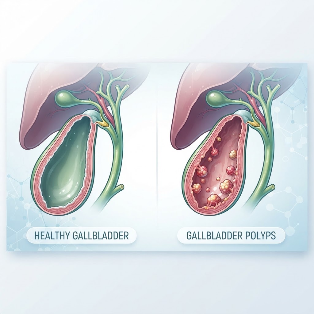
- Gallbladder polyps are abnormal growths that extend from the lining of the gallbladder wall into its interior.
- Most polyps are benign and asymptomatic, often discovered incidentally during routine medical imaging.
- Risk factors include age, history of gallstones, and underlying metabolic health markers.
- Diagnostic procedures primarily rely on high-resolution abdominal ultrasounds to assess size and morphology.
- While most polyps are harmless, size and growth rates are the primary factors doctors use to determine if surgical intervention is necessary.
Discovering that you have an abnormality in your gallbladder can be a source of immediate anxiety, but the reality for most individuals is far less alarming than it initially seems. Gallbladder polyps are surprisingly common findings in modern clinical practice, largely due to the increasing frequency and clarity of abdominal imaging. While the term “polyp” often sounds intimidating, these small tissue growths are frequently benign, requiring nothing more than periodic observation to ensure long-term gallbladder health. Understanding the nature of these growths, why they form, and when they require clinical attention is the first step toward managing your wellness proactively. In this comprehensive guide, the healthauthoritylife Editorial Team explores the nuances of these growths, helping you navigate the diagnostic process and dietary management strategies that support a healthy digestive system.
What Are Gallbladder Polyps?
Gallbladder polyps are localized elevations or projections of the gallbladder mucosa, the innermost lining of the organ that stores bile. Unlike gallstones, which are hardened deposits of cholesterol or bile salts that move freely within the gallbladder, polyps are attached to the inner wall. They can vary significantly in their composition and structural integrity. In clinical settings, they are classified into a few primary categories: cholesterol polyps, inflammatory polyps, and adenomas (true tumors). Cholesterol polyps are the most common type and are essentially accumulations of cholesterol-laden macrophages within the lining. These are generally considered non-neoplastic, meaning they lack the potential to become malignant.
The vast majority of these growths are discovered incidentally. A patient might visit their healthcare provider for unrelated symptoms like mild abdominal discomfort or digestive indigestion, leading to an ultrasound scan. It is during this scan that the technician notices small, fixed structures on the gallbladder wall. Because the gallbladder is a relatively small organ responsible for storing bile produced by the liver, any disruption to its interior surface can raise questions about overall digestive function. However, the presence of a polyp does not automatically imply that the gallbladder is malfunctioning or failing to process bile correctly.
Understanding the anatomy of the gallbladder is essential when discussing these growths. The gallbladder functions as a reservoir. When you consume fat-containing foods, the gallbladder receives a hormonal signal to contract, pushing bile through the bile ducts into the small intestine to aid in digestion. Polyps that remain small and do not obstruct the cystic duct typically do not interfere with this process. However, if a polyp grows to a significant size, it may theoretically act as a physical barrier or irritation point. Most clinicians distinguish between small polyps, often measuring less than 10 millimeters, and larger growths that require more rigorous surveillance. By maintaining a focus on gallbladder health through balanced nutrition and regular checkups, many individuals live their entire lives with stable, asymptomatic polyps without ever requiring surgery.
Common Symptoms of Gallbladder Polyps
One of the most frequently asked questions regarding these growths is whether they cause noticeable discomfort. The reality is that the vast majority of gallbladder polyps are completely asymptomatic. They do not cause pain, digestive distress, or systemic issues for the patient. Because they are often small and do not interfere with the flow of bile, they are “silent” findings that only manifest on diagnostic imaging. However, when gallbladder polyp symptoms do occur, they are often indistinguishable from the symptoms of gallstones or biliary colic, which can complicate the diagnostic process for both the patient and the physician.
When symptoms do arise, they typically manifest as a dull, aching sensation in the upper right quadrant of the abdomen, specifically just beneath the rib cage. This discomfort may be intermittent and is sometimes associated with the consumption of high-fat or greasy meals, which force the gallbladder to contract more vigorously. In some instances, this pain can radiate toward the right shoulder or the upper back, creating a sensation of tightness or pressure. It is important to distinguish this from the acute, sharp pain of a gallstone attack; polyp-related pain is generally less severe and less likely to be accompanied by systemic signs of infection, such as fever or chills.
Beyond localized pain, some individuals report nonspecific gastrointestinal distress. This may include bouts of nausea, occasional vomiting, or a feeling of bloating and fullness shortly after eating. While these symptoms are classic indicators of gallbladder dysfunction, they are not specific to polyps. For example, a person might experience these symptoms due to irritable bowel syndrome, gastritis, or dietary sensitivities. Therefore, the presence of these symptoms does not automatically confirm that a polyp is the culprit. If you are experiencing persistent pain or digestive changes, it is essential to undergo a clinical evaluation. Relying on self-diagnosis is rarely effective because the symptoms of gallbladder health issues often overlap with various other gastrointestinal conditions. Keeping a food and symptom diary can be a helpful tool to bring to your doctor, as it helps determine if the discomfort is truly tied to gallbladder contraction patterns.
Primary Causes and Risk Factors
The precise etiology of why the body develops gallbladder polyps is an ongoing subject of research. Unlike some conditions where a clear genetic or environmental trigger exists, these growths appear to result from a complex interaction of factors. The causes of gallbladder polyps are categorized based on their underlying biological mechanism. Cholesterol polyps, as noted, are closely linked to cholesterol metabolism. When there is an imbalance in the bile composition—specifically when bile is supersaturated with cholesterol—the mucosal lining of the gallbladder can accumulate these lipids, leading to the formation of cholesterol deposits that appear as polyps on imaging.
Several risk factors are associated with the development of these growths. Age is a primary factor, with incidence rates tending to increase in middle-aged and older adults. Gender also plays a role, as women are statistically more likely to develop gallbladder-related issues than men, likely due to hormonal influences on bile composition. Additionally, metabolic health markers, such as obesity, insulin resistance, and dyslipidemia, have been observed as common threads in patients with gallbladder polyps. These conditions can alter the chemistry of bile, making the gallbladder environment more conducive to the formation of both stones and mucosal irregularities.
Another significant factor is the presence of concomitant gallstone disease. Often, a patient with polyps will also have gallstones. This is likely because the environment that promotes stone formation—such as bile stasis or high cholesterol concentration—also supports the inflammatory processes that lead to polyp growth. Chronic gallbladder inflammation, or cholecystitis, can also contribute to the development of inflammatory polyps. When the gallbladder wall is chronically irritated, the body’s healing response may result in the proliferation of mucosal cells, creating these small, benign projections. While you cannot change your age or genetics, focusing on lifestyle factors—such as maintaining a healthy weight and following a diet low in refined sugars and saturated fats—can potentially support better biliary health and reduce the strain on the gallbladder. Experts generally agree that while these measures do not guarantee the prevention of polyps, they contribute significantly to overall digestive health and may prevent the progression of metabolic conditions that predispose individuals to gallbladder issues.
How Doctors Diagnose Gallbladder Polyps
Because these growths rarely cause symptoms, diagnosis is almost always achieved through imaging modalities. The gold standard for detecting and monitoring gallbladder polyps is the transabdominal ultrasound. This non-invasive procedure uses sound waves to create a real-time image of the gallbladder. Ultrasound is exceptionally good at identifying these growths because it can distinguish between stones and polyps; whereas gallstones are mobile and move when the patient changes position, polyps are fixed to the wall and remain stationary regardless of movement. Furthermore, ultrasound can determine the size of the polyp, which is the most critical metric for clinical decision-making.
In cases where the ultrasound findings are ambiguous, or if a doctor needs to see more detail to evaluate the nature of a larger polyp, they may recommend advanced imaging. Endoscopic ultrasound (EUS) is a more specialized, though invasive, procedure where an endoscope is passed through the esophagus and stomach to get a high-resolution view of the gallbladder from the inside. This provides much greater detail regarding the wall layers of the gallbladder. Another option is Magnetic Resonance Imaging (MRI) or MRCP (Magnetic Resonance Cholangiopancreatography), which provides detailed images of the biliary tree and the gallbladder structure without the need for radiation.
The diagnostic process is not just about finding the polyp, but assessing its characteristics. Clinicians use specific criteria—such as size, number, and appearance—to decide on a plan of action. For instance, a single, large, broad-based polyp requires a very different monitoring strategy than multiple, tiny, stalked (pedunculated) polyps. Below is a comparison of common diagnostic and management approaches for gallbladder concerns.
| Method | Purpose | Best For |
|---|---|---|
| Abdominal Ultrasound | Primary imaging of biliary tract | Initial screening and size tracking |
| Endoscopic Ultrasound | Detailed wall examination | Evaluating large or suspicious polyps |
| Wait and Watch | Routine monitoring protocol | Small, stable, benign-appearing polyps |
| Cholecystectomy | Surgical removal | Large, fast-growing, or symptomatic cases |
After the diagnosis, your doctor will likely establish a “monitoring gallbladder polyps” schedule. This often involves repeating an ultrasound every six to twelve months to check for changes. If the polyp remains stable in size over several years, the frequency of imaging may be reduced. This approach is highly effective because it ensures that if a polyp were to grow or change in character, the physician would detect it long before it poses any significant risk to the patient’s health.
Are Gallbladder Polyps Cancerous?
One of the most pressing concerns for patients upon receiving this diagnosis is the potential for malignancy. It is important to emphasize that the vast majority of gallbladder polyps are benign. They are often simply cholesterol crystals or localized inflammatory changes that have absolutely no link to cancer. However, clinicians maintain a cautious approach because a small subset of polyps, particularly adenomas, can show changes that are considered precancerous. This is why doctors prioritize the size of the polyp as the primary indicator for concern.
Generally, medical guidelines suggest that polyps measuring less than 10 millimeters in diameter have an extremely low risk of being malignant. These are almost universally managed with periodic surveillance rather than surgery. When a polyp exceeds 10 millimeters, the risk of it being an adenoma increases, and the likelihood of malignant transformation, while still relatively low in the grand scheme of medical statistics, becomes statistically significant enough for doctors to recommend removal. The growth rate of the polyp is another vital clue; a polyp that shows significant size increases between two ultrasound exams is viewed with much more suspicion than one that remains stable for years.
Are gallbladder polyps dangerous? In their early, small stages, they are generally not considered dangerous. The danger arises only if they remain unmonitored and are allowed to reach a size where they could theoretically transform or begin to cause obstruction. This is why strict adherence to the monitoring schedule set by your physician is crucial. By tracking the size, they are looking for “red flags” that warrant intervention. Factors that increase the suspicion of malignancy include sessile (flat-based) polyps, larger dimensions, and the presence of associated conditions like primary sclerosing cholangitis. For the average person with a tiny, pedunculated (stalk-like) polyp, the probability of it ever becoming a serious health threat is exceptionally low. The primary goal of medical management is to maintain awareness of these findings, ensuring they do not evolve into a clinical concern. Open communication with your gastroenterologist or surgeon will provide you with the most accurate assessment of your specific risk profile based on your imaging results and individual health history.
Monitoring and Surveillance Schedules
When gallbladder polyps are discovered during a routine abdominal ultrasound, the primary challenge for medical providers is distinguishing between benign cholesterol polyps and potentially malignant growths. Because most polyps remain asymptomatic and stable over long periods, the standard of care centers on a “wait and watch” approach. Surveillance schedules are not one-size-fits-all; they are highly customized based on the size of the polyp, the patient’s clinical symptoms, and the presence of underlying risk factors like primary sclerosing cholangitis or a family history of gallbladder malignancy.
Typically, if a polyp is smaller than 5 millimeters, it is often deemed low-risk. In many clinical settings, doctors may recommend a follow-up ultrasound in six to twelve months to ensure there has been no interval growth. If the follow-up scan shows stability, the frequency of imaging may be reduced or discontinued entirely, provided the patient remains asymptomatic.
For polyps that measure between 6 and 9 millimeters, the monitoring strategy often becomes more proactive. Because there is a broader range of uncertainty regarding the biological potential of these medium-sized polyps, clinicians frequently opt for more frequent surveillance intervals, such as every six months for the first year. This timeline is designed to catch rapid progression, which is a hallmark of atypical cells.
It is important to understand that imaging is the cornerstone of polyp management. During these appointments, the radiologist assesses not just the size of the polyp but its morphology—such as its attachment to the gallbladder wall (stalked versus broad-based) and its echogenicity. A sessile (broad-based) polyp is generally viewed with more caution than a pedunculated (stalked) one. If the surveillance images reveal that a polyp is increasing in size by 2 millimeters or more between scans, or if it reaches the critical threshold of 10 millimeters, the management plan typically shifts from observation to surgical intervention.
When Is Gallbladder Removal Necessary?
The transition from monitoring to surgical intervention—known as a cholecystectomy—is usually triggered by specific clinical criteria that suggest an increased risk of malignancy or severe symptomatic burden. The most widely accepted indicator for surgery is size. Once a polyp reaches or exceeds 10 millimeters in diameter, the statistical risk of it harboring neoplastic or malignant cells increases. At this juncture, experts generally agree that the risk associated with retaining the gallbladder outweighs the risks of the surgical procedure.
Another primary reason for surgical removal is the presence of symptoms. Even if a polyp is considered small, if a patient experiences frequent, recurrent biliary colic—a sharp pain in the upper right abdomen that may radiate to the shoulder—the polyp may be causing an obstruction or affecting the flow of bile. When polyps coexist with gallstones, the likelihood of symptomatic episodes increases significantly, often necessitating surgery to prevent complications such as cholecystitis (inflammation of the gallbladder) or pancreatitis.
Patient-specific risk factors also play a critical role. For example, individuals with underlying liver conditions, such as primary sclerosing cholangitis, are often advised to undergo surgery for smaller polyps because their risk of developing gallbladder cancer is higher than that of the general population. In these instances, the physician may prioritize preventative removal over the standard observation schedule.
Advances in surgical technology have made laparoscopic cholecystectomy the gold standard. This minimally invasive approach involves a few small incisions, allowing for faster recovery times and less postoperative pain compared to traditional open surgery. While the idea of organ removal can be daunting, the gallbladder is not essential for survival. After removal, the liver continues to produce bile, which flows directly into the small intestine. Most people return to a normal diet and activity level within a few weeks, making this a highly effective and definitive treatment for high-risk polyps.
| Polyp Characteristic | Standard Clinical Action | Best For |
|---|---|---|
| Under 5mm, asymptomatic | Annual observation or interval scan | Low-risk, stable patients |
| 6mm to 9mm, slow growth | Short-term surveillance (3-6 months) | Patients requiring strict monitoring |
| 10mm or larger | Cholecystectomy (surgery) | High-risk or suspected malignancy |
| Any size with acute pain | Surgical consultation | Symptomatic relief |
Dietary Changes to Support Gallbladder Health
While diet cannot dissolve a polyp in the same way it might theoretically influence cholesterol-based gallstones, maintaining a gallbladder-friendly diet is essential for reducing the workload on the biliary system and minimizing the inflammation that can exacerbate gallbladder-related discomfort. The goal is to eat in a way that promotes smooth bile production and flow, preventing the gallbladder from becoming “sluggish.”
A diet focused on whole, fiber-rich foods is one of the most effective strategies for overall digestive wellness. Soluble fiber, found in oats, beans, lentils, and fruits like apples and pears, binds to bile acids in the digestive tract and helps eliminate them, which can improve cholesterol balance. Many nutritionists recommend avoiding “yo-yo” dieting or rapid weight loss, which is a known catalyst for gallbladder stress. Instead, aim for gradual, consistent weight loss if needed, as sudden caloric restriction can lead to increased bile sludge and a higher risk of stone formation.
Fats are perhaps the most sensitive area of the gallbladder-friendly diet. While the gallbladder needs dietary fat to trigger its contractions, consuming large amounts of saturated or trans fats can overwhelm the system. Rather than eliminating fat entirely, focus on high-quality, anti-inflammatory fats. Incorporate omega-3 fatty acids found in fatty fish, walnuts, and flaxseeds, and lean into monounsaturated fats found in olive oil and avocados. These fats are easier for the body to process and help maintain a healthy inflammation response throughout the body.
Finally, consider the timing and size of meals. Eating large, heavy, or fatty meals can trigger intense gallbladder contractions, which may lead to discomfort if you have polyps or stones. Dividing your daily intake into smaller, more frequent, and balanced meals can allow the gallbladder to empty more gently and consistently, keeping the biliary system stable throughout the day.
Natural Lifestyle Habits for Gallbladder Wellness
Beyond nutrition, your daily movement and stress-management habits play an underappreciated role in gallbladder health. Sedentary behavior is often associated with a higher risk of biliary stasis, where bile sits too long in the gallbladder, thickening into sludge. Regular, moderate physical activity—such as brisk walking, swimming, or cycling—helps stimulate digestive motility and supports overall metabolic health. By keeping your body in motion, you encourage the physical movement of digestive fluids through the liver and gallbladder system.
Hydration is another pillar of gallbladder wellness. Bile is composed largely of water, and maintaining adequate fluid intake ensures that bile remains thin and free-flowing. Dehydration can lead to more concentrated bile, which may contribute to the formation of sludge or the irritation of polyps. Make it a habit to drink water consistently throughout the day rather than waiting until you are thirsty. Herbal teas, particularly those like ginger or peppermint, can also provide soothing benefits to the digestive tract and support calm, efficient digestion.
Stress management is arguably the most overlooked component of biliary health. The digestive system is intimately linked to the nervous system. Chronic stress triggers the release of cortisol and other hormones that can interfere with normal digestive function and motility. Techniques such as deep breathing, mindfulness meditation, or even simple restorative yoga can help shift the body from a “fight-or-flight” state to a “rest-and-digest” state, allowing the gallbladder to function in its optimal capacity.
Lastly, pay close attention to your body’s signals. If you notice persistent bloating, upper right quadrant pain after eating, or digestive distress, do not ignore these signs in hopes they will pass. Keeping a food and symptom journal can be an incredibly powerful tool to show your doctor. By tracking what you eat, when you feel pain, and how your body reacts to different types of fats, you provide your medical team with the concrete data they need to differentiate between simple digestive upset and issues requiring intervention.
Frequently Asked Questions
Are gallbladder polyps a precursor to cancer?
While most gallbladder polyps are benign—often made up of cholesterol deposits—some polyps can be precancerous. The likelihood of malignancy is strongly linked to the size of the polyp. Polyps larger than 10 millimeters are viewed with higher clinical suspicion, whereas very small polyps are rarely associated with cancer. Regular surveillance ensures that if a polyp begins to change in character or size, it can be addressed early.
Can you shrink gallbladder polyps naturally?
There is no scientifically validated evidence to suggest that polyps can be “shrunk” or dissolved through diet, herbal supplements, or cleanses. Because polyps are attached to the gallbladder wall, they do not behave the same way as mobile gallstones. While a healthy lifestyle can support overall gallbladder function, you should be wary of any claims promising to cure or eliminate polyps through natural remedies.
Do gallbladder polyps cause symptoms?
Most gallbladder polyps are asymptomatic and are discovered incidentally during ultrasounds performed for other reasons. However, some people may experience biliary colic, which involves pain in the upper right abdomen, nausea, or discomfort after eating, particularly if the polyp is large or located in a position that obstructs the flow of bile.
Is it possible to live normally after gallbladder removal?
Yes, most individuals lead entirely normal lives after a cholecystectomy. Because the gallbladder serves as a storage vessel for bile rather than the source, the liver continues to produce bile as needed. Most people find that they can return to a normal diet within weeks, though some may need to transition slowly back to high-fat foods as their digestive system adjusts to the new flow of bile.
How often should I have an ultrasound for a small polyp?
For small, asymptomatic polyps (typically under 5mm), doctors often recommend an annual ultrasound or an interval scan within 6 to 12 months to confirm stability. If the polyp remains stable and does not grow, your doctor may decide that further imaging is unnecessary. Always follow the specific timeline provided by your healthcare provider based on your unique risk factors.
What is the difference between gallstones and polyps?
The primary difference is their attachment and mobility. Gallstones are usually mobile—they can move around within the gallbladder and potentially block the bile ducts. Polyps are growths that are fixed, meaning they are attached to the inner wall of the gallbladder. While stones are often made of hardened cholesterol or pigment, polyps can be composed of various tissues, requiring different monitoring approaches.
Conclusion
Navigating a diagnosis of gallbladder polyps can feel overwhelming, but it is important to remember that most of these growths are benign and require nothing more than a watchful eye. By understanding the importance of size, symptoms, and professional monitoring, you can stay ahead of any potential complications. The key to maintaining gallbladder health lies in a balanced approach: supporting your digestion through mindful eating, staying active, managing stress, and adhering to the surveillance schedule recommended by your physician.
If you have recently discovered you have a gallbladder polyp, the best step is to have an open conversation with your healthcare provider about your personal risk profile and monitoring frequency. Taking proactive, informed steps today is the best way to ensure your long-term health and peace of mind. Remember, your body provides signals—listening to them is the first step toward effective health management.
By healthauthoritylife Editorial Team
This article is for informational purposes only and is not a substitute for professional medical advice. Always consult your doctor before making changes to your diet, exercise routine, or treatment plan.
Leave a Reply