
⚕️ Written by Dr. Sarah Mitchell, MD, MPH  •  📋 Evidence-Based Articles  •  🔍 Medically Reviewed

⚠️ Not a substitute for professional medical advice

Category: Children’s Health

Paediatric health, child nutrition, and family wellness articles.

  • Chronic Inflammatory Response Syndrome: Symptoms and Recovery

    Chronic Inflammatory Response Syndrome: Symptoms and Recovery

    Key Takeaways

    • Chronic Inflammatory Response Syndrome (CIRS) is a multi-system, multi-symptom illness caused by exposure to biotoxins in water-damaged buildings.
    • While often referred to as mold illness, CIRS can be triggered by various biological toxins including actinomycetes, Lyme disease co-infections, and cyanobacteria.
    • The hallmark of CIRS is a failure of the innate immune system to clear biotoxins, leading to persistent, systemic inflammation.
    • Diagnosis relies on a combination of specific clinical symptoms and blood-based biomarkers that reflect inflammatory dysregulation.
    • Effective recovery requires a multifaceted approach involving environmental remediation, binding agents, and nervous system regulation.

    For many individuals, a mysterious decline in health often begins with a seemingly benign event: a water leak in an apartment, a move into a new office, or a lingering illness that never quite resolves. When fatigue, brain fog, and chronic pain become the new baseline, the journey to find answers can be exhausting. Often, these patients are told their labs are normal, leaving them frustrated and searching for a root cause. This is where the framework of Chronic Inflammatory Response Syndrome (CIRS) becomes vital. CIRS represents a complex, multi-system health crisis rooted in an impaired immune response to environmental toxins. Understanding the mechanics of this syndrome is the first step toward reclaiming vitality and moving away from a life dictated by mysterious, persistent inflammation.

    What Is Chronic Inflammatory Response Syndrome (CIRS)?

    Chronic Inflammatory Response Syndrome (CIRS) is a systemic inflammatory condition defined by the body’s inability to effectively clear certain environmental biotoxins. In a healthy individual, the immune system encounters a foreign substance—such as mold spores or bacterial fragments—and clears it through a coordinated series of inflammatory responses. Once the threat is removed, the immune system returns to baseline. However, in individuals with a specific genetic susceptibility, this “off switch” fails to trigger.

    The core issue in CIRS is not just the presence of a toxin, but a prolonged, dysregulated immune reaction. When the body cannot neutralize these biotoxins, they remain trapped in the system, continuously stimulating the production of pro-inflammatory cytokines. This persistent “fire” within the body damages tissues, disrupts hormones, and alters cellular communication. It is a mistake to view CIRS as a simple allergy or an infection that can be cleared with standard antibiotics. Instead, it is an immune system dysfunction where the internal regulatory mechanism—meant to keep inflammation in check—has become permanently stuck in an “on” position.

    The term is often used interchangeably with mold illness, but CIRS is broader in scope. It encompasses the physiological aftermath of exposure to an environment that is biologically toxic. This may include not just mold, but also the “cocktail” of volatile organic compounds (VOCs), actinomycetes, and endotoxins found in water-damaged buildings. The cumulative effect of these triggers overwhelms the innate immune system, leading to the diverse array of systemic symptoms that define the syndrome. When this immune response remains unchecked, it creates a self-perpetuating cycle of inflammation that can persist for years if the underlying source of exposure is not identified and removed.

    Research into CIRS has evolved significantly over the last two decades. Experts generally agree that this condition is highly individualistic. Two people can live in the same water-damaged home, but only one may develop the debilitating symptoms of CIRS. This suggests a strong genetic component, often involving specific human leukocyte antigen (HLA) genotypes that influence how the body recognizes and processes biotoxins. For those affected, the impact is rarely limited to one organ system; instead, it is a whole-body manifestation that affects the brain, digestive tract, endocrine system, and neurological pathways, making it one of the most challenging conditions to diagnose and treat in modern clinical practice.

    Common Triggers of CIRS Beyond Mold Exposure

    While public awareness often highlights mold as the primary culprit for biotoxin illness, clinicians who specialize in CIRS are quick to point out that water-damaged buildings are complex ecosystems. Exposure to mold is usually just one piece of a much larger, toxic puzzle. A building that has suffered water damage provides a fertile breeding ground for a host of biological agents, many of which can be just as, if not more, inflammatory than the mold spores themselves.

    One of the most significant, yet frequently overlooked, triggers is the presence of actinomycetes. These are filamentous bacteria that grow alongside fungi in water-damaged environments. Actinomycetes produce potent toxins that can cause severe respiratory irritation and systemic inflammation. Because these bacteria are much smaller than mold spores, they are often easily inhaled and can penetrate deep into the tissues of the body, where they continue to drive the inflammatory cascade that characterizes CIRS. Ignoring these bacteria during the remediation process is a common reason why some individuals fail to see improvement even after clearing mold from their homes.

    Another major contributor is the group of endotoxins known as lipopolysaccharides (LPS). These are components found in the cell walls of Gram-negative bacteria. When these bacteria die, they release LPS into the air or surfaces. LPS is highly immunogenic, meaning it is exceptionally good at triggering an intense immune response. In a water-damaged building, high levels of LPS can be found in the dust and carpet, creating a constant, low-grade source of toxic exposure that the body struggles to eliminate. Additionally, cyanobacteria found in water sources or aerosolized from contaminated HVAC systems can contribute to the “toxic soup” that defines a CIRS-prone environment.

    Beyond built environments, certain tick-borne illnesses are closely linked to the CIRS model. Lyme disease, and particularly its co-infections like *Babesia* and *Bartonella*, can create an inflammatory state that mirrors the clinical presentation of mold-related CIRS. These pathogens also produce biotoxins that the body may be unable to clear due to the same underlying immune dysregulation. In clinical settings, it is often necessary to tease apart whether a patient is suffering from an active, recurring infection or if the lingering inflammation is a residual response to the biotoxins left behind by a past infection.

    Trigger Category Common Components Best For
    Water-Damaged Buildings Mold, Actinomycetes, Endotoxins General environmental assessments
    Tick-Borne Pathogens Lyme, Babesia, Bartonella Infection-related immune flares
    Cyanobacteria Blue-green algae, stagnant water Outdoor/Water-exposure scenarios

    Understanding these diverse triggers is essential for effective biotoxin illness treatment. Relying solely on mold remediation without addressing the bacterial components or potential co-infections may result in incomplete recovery. A comprehensive approach involves testing the environment for a broad spectrum of biotoxins and considering the patient’s history of exposure to various environmental and biological threats. This nuance is precisely why CIRS recovery tips often emphasize a thorough environmental audit as the foundational step of the entire healing process.

    Recognizing the Symptoms of Biotoxin Illness

    The clinical presentation of CIRS is notoriously vast, which often leads to misdiagnosis. Because the inflammation is systemic, it can manifest in virtually any organ system. Many patients report that their symptoms seem to migrate, shifting from joint pain one week to severe cognitive dysfunction the next. Recognizing these patterns is crucial for patients and practitioners alike, as the sheer breadth of symptoms often leads to patients being mislabeled with conditions like fibromyalgia, chronic fatigue syndrome, or even psychosomatic disorders.

    The neurological impact is frequently the most debilitating aspect of CIRS. Patients often describe a profound, persistent brain fog—a feeling as though they are “living in a fishbowl.” This cognitive impairment often includes difficulty with word retrieval, impaired short-term memory, and an inability to focus. Along with these cognitive shifts, mood disturbances such as anxiety, irritability, and uncharacteristic sadness are common. This occurs because the inflammatory cytokines crossing the blood-brain barrier interfere with neurochemical balance and cognitive processing speed.

    Physical symptoms are equally varied. A hallmark sign is muscle pain, often described as “lightning bolt” pains or a constant, heavy ache that does not respond to traditional anti-inflammatories. Many patients also experience chronic, unexplained fatigue that is not alleviated by rest. This is often accompanied by gastrointestinal distress, such as abdominal pain, bloating, and irregular bowel movements, as the gut-immune axis is heavily impacted by the systemic inflammatory state.

    Vision changes are another frequently reported symptom. Patients may notice increased light sensitivity, blurry vision, or difficulty focusing on near objects. This is not typically a problem with the eye itself, but rather a neurological processing issue where the brain struggles to adjust to visual input. Additionally, patients may experience what is often referred to as “electric shocks” or numbness and tingling in the extremities (paresthesia), which highlights the inflammatory impact on the peripheral nervous system.

    Beyond these neurological and physical signs, there are specific symptoms related to the body’s fluid regulation. Because CIRS can disrupt hormones like antidiuretic hormone (ADH) and melanocyte-stimulating hormone (MSH), many patients experience abnormal thirst, frequent urination, and altered sleep cycles. The body loses its ability to regulate its internal hydration levels and circadian rhythms, leading to a state of chronic physiological stress. Recognizing these specific symptom clusters—the combination of brain fog, muscle pain, visual changes, and thirst—is a major step toward clinical validation. When a patient presents with this constellation of symptoms, it serves as a strong clinical indicator that further investigation into a biotoxin-related cause is warranted.

    The Link Between CIRS and Nervous System Dysregulation

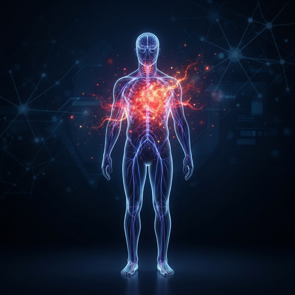
    At the intersection of CIRS and chronic illness lies the concept of nervous system dysregulation. The autonomic nervous system (ANS) acts as the body’s control center, balancing the “fight or flight” sympathetic response with the “rest and digest” parasympathetic response. In a healthy state, the body oscillates between these states as needed. In CIRS, however, the constant barrage of inflammatory cytokines forces the autonomic nervous system into a state of chronic sympathetic dominance.

    This state of hyper-arousal is a survival mechanism gone wrong. Because the body perceives the persistent inflammation as an ongoing threat, it stays in a constant state of high alert. This manifests as increased heart rate, shallow breathing, and a heightened startle response. Over time, this chronic stress on the nervous system depletes the body’s resources and hinders the recovery process. This is why many patients find that even after they have moved to a clean environment, their symptoms persist; the “threat detection” centers in the brain are still firing, keeping the inflammatory cascade alive.

    Furthermore, the inflammation associated with CIRS can directly impact the vagus nerve, which is the primary component of the parasympathetic nervous system. The vagus nerve is responsible for dampening inflammation throughout the body. When it becomes dysregulated due to toxic exposure, the body loses its most effective “brake” for inflammation. This creates a feedback loop: biotoxins cause inflammation, inflammation dysregulates the vagus nerve, and the now-impotent vagus nerve cannot signal the body to stop producing inflammatory cytokines. This cycle explains why CIRS recovery often requires more than just removing the source of the toxins.

    Addressing nervous system regulation is a critical component of any comprehensive recovery protocol. Simple interventions like breathwork, vagus nerve stimulation, and mindful movement can help bridge the gap between biological detoxification and physiological healing. By manually shifting the body from a sympathetic-heavy state back into a parasympathetic state, patients can create the necessary internal environment for the immune system to finally rest and reset. Clinicians often find that patients who engage in nervous system regulation strategies alongside detoxification protocols achieve better, more durable outcomes than those who focus on the biological triggers alone. It is about treating the body as an integrated whole, recognizing that the brain and the immune system are in constant, complex communication.

    How CIRS Is Diagnosed: Key Biomarkers to Watch

    Because CIRS is a systemic inflammatory condition, there is no single “magic” test that confirms the diagnosis. Instead, a proper CIRS diagnosis involves a synthesis of clinical symptom review and a specific panel of blood-based biomarkers. These biomarkers are chosen because they represent the molecular footprints of the inflammatory cascade that biotoxins trigger within the body. When these markers appear in a specific combination, it provides a high level of confidence that the patient is suffering from an environmentally acquired illness.

    One of the primary biomarkers monitored is Transforming Growth Factor-beta 1 (TGF-beta 1). This protein is an important regulator of the immune system, but in the context of CIRS, it is often chronically elevated. High levels of TGF-beta 1 are associated with systemic inflammation, tissue remodeling, and fibrosis. When this marker is high, it is a strong indicator that the patient’s immune system is currently over-activated and causing collateral damage to the body’s tissues. Similarly, Matrix Metalloproteinase-9 (MMP-9) is frequently tested. Elevated levels of MMP-9 indicate that inflammation is moving through the body and causing damage to blood vessels and other connective tissues.

    Another crucial biomarker is the C4a (Complement component 4a) level. This is a protein fragment that is a direct byproduct of the immune system’s activation. When the innate immune system identifies biotoxins it cannot handle, it triggers the complement cascade, of which C4a is a part. Extremely high C4a levels are a classic sign of an ongoing, active biotoxin-induced inflammatory response. Clinicians also keep a close eye on hormone-related markers, such as MSH (Melanocyte-Stimulating Hormone). MSH is often depleted in CIRS patients, which can lead to sleep disturbances, chronic pain, and an increased susceptibility to infection.

    Genetic testing for HLA-DR/DQ genotypes is also commonly used to understand a patient’s susceptibility. While this test doesn’t diagnose the illness, it helps identify why some individuals struggle to recover while others do not. Certain genetic variations make it much harder for the body to present antigens—the “identifying tags” of biotoxins—to the immune system. If a patient possesses these genetic markers, it helps clinicians set realistic expectations for the duration and intensity of the treatment required.

    Finally, visual contrast sensitivity (VCS) testing is an essential, non-invasive tool in the diagnostic process. VCS measures the brain’s ability to distinguish between shades of gray in various spatial frequencies. Biotoxins significantly impair the microcirculation in the brain and eyes, which in turn reduces the patient’s ability to detect contrast. While it is not a diagnostic test on its own, a low VCS score in a patient showing clinical symptoms of CIRS provides powerful, objective evidence that neurological inflammation is present. By combining these lab-based biomarkers with the VCS test and a rigorous symptom history, practitioners can build a clear, evidence-based picture of a patient’s health status, allowing for a targeted and effective treatment plan.

    The Role of Genetics in Your Inflammatory Response

    One of the most perplexing aspects of Chronic Inflammatory Response Syndrome (CIRS) is why some individuals become severely ill after exposure to a water-damaged building, while others living in the same environment remain asymptomatic. The answer often lies within the intricate dance between environmental biotoxins and human genetics, specifically regarding the Human Leukocyte Antigen (HLA) genes.

    The HLA complex is a group of genes that play a critical role in the immune system by helping the body distinguish between “self” and “non-self” invaders. In the context of CIRS, certain genetic haplotypes appear to predispose individuals to a failure in the adaptive immune response. When a healthy person inhales mold spores or other biotoxins, their immune system typically identifies the threat, tags it for removal, and clears it from the body. However, for those with specific genetic predispositions, this tagging process is hindered.

    If the immune system cannot recognize and properly mark the biotoxin for removal, the body enters a state of chronic, maladaptive inflammation. Instead of clearing the irritant, the body initiates a persistent release of inflammatory cytokines. This is why CIRS diagnosis often involves genetic testing; understanding your HLA status can provide clarity on why your body struggles to recover without specific, targeted interventions. It is important to note that having a genetic predisposition does not guarantee you will develop the syndrome, but it does mean your immune system may lack the “genetic blueprint” required to effectively process certain environmental toxins on its own.

    Furthermore, genetic variations also influence how the body handles detoxification pathways, such as the methylation cycle and glutathione production. If your genetics make it harder for your body to convert harmful substances into water-soluble compounds for excretion, the biotoxin load remains high, leading to a perpetuation of mold illness symptoms. Recognizing these genetic factors is not about feeling “doomed” by your DNA, but rather about empowering yourself with the knowledge needed to tailor your recovery plan to your specific biological needs.

    Environmental Remediation: Clearing Your Living Space

    Effective CIRS recovery is fundamentally impossible if you remain in the environment that triggered the inflammation. Environmental remediation is the process of identifying, containing, and removing the sources of biotoxins—most commonly mold—from your home or workplace. Remediation is not as simple as scrubbing a spot of visible mold; it requires a deep, systematic approach to indoor air quality.

    The first step in remediation is professional inspection. You should look for companies that specialize in mold assessment, ideally those who have experience with CIRS patients. These professionals often use moisture mapping, infrared cameras, and air sampling to detect hidden water damage behind walls, under floorboards, or within HVAC systems. It is vital to distinguish between a standard cleaning service and a certified mold remediator; the latter follows specific protocols to prevent the spread of mold spores during the cleanup process.

    Once sources are identified, containment is paramount. During the remediation phase, the affected areas must be sealed off with heavy-duty plastic sheeting to prevent cross-contamination of the rest of the house. Professionals will use Negative Air machines equipped with HEPA filtration to keep the air pressure lower in the affected room, ensuring that spores are drawn into filters rather than drifting into clean spaces.

    After physical remediation, the focus shifts to porous materials. Carpets, upholstered furniture, and soft textiles that have been exposed to high levels of mold are often impossible to clean thoroughly and may need to be discarded. Hard surfaces should be wiped down with specialized solutions, and the entire HVAC system should be cleaned and upgraded with high-quality filtration. Maintaining a low indoor humidity level—ideally between 30% and 50%—is the most effective way to prevent the mold from returning. If you are struggling to decide between various remediation strategies, the table below provides a quick comparison of standard environmental control methods.

    Method Description Best For
    HEPA Air Purifiers High-efficiency particulate air filtration. Reducing airborne spores in daily living areas.
    Dehumidification Maintaining humidity below 50%. Preventing future mold growth and microbial proliferation.
    Professional Remediation Containment and physical removal of mold colonies. Addressing significant water damage or hidden mold.
    Testing (ERMI/HERTSMI-2) DNA-based analysis of dust samples. Identifying the specific mold species and biotoxin load.

    Natural Strategies for Supporting Your Body Through CIRS

    While environmental remediation and clinical biotoxin illness treatment are the pillars of recovery, natural support strategies are essential for calming the systemic “fire” of inflammation. The goal is to modulate the immune system and support your body’s natural detoxification pathways.

    One of the most potent ways to support recovery is through the use of binders. Binders are substances that attach to biotoxins in the gut, preventing them from being reabsorbed and allowing them to be safely excreted through the digestive tract. Common natural binders include activated charcoal, bentonite clay, and certain types of dietary fibers. These should be taken under guidance, as they can also bind to medications and essential nutrients if not timed correctly.

    Reducing your “total toxic load” is another crucial strategy. Your body is already struggling to manage biotoxins; adding unnecessary stressors can overwhelm your systems. This means minimizing exposure to household chemicals, synthetic fragrances, pesticides, and heavy metals. Switching to non-toxic cleaning products, using glass or stainless steel for food storage, and filtering your drinking water are simple but profound ways to reduce the burden on your liver and kidneys.

    Additionally, supporting the lymphatic system is often overlooked in CIRS recovery. Unlike the blood, the lymphatic system has no pump of its own and relies on movement to circulate fluid and clear waste. Incorporating light, gentle movement—such as walking, rebounding, or lymphatic drainage massage—can help your body move toxins toward the exit points. Incorporating anti-inflammatory herbs, such as turmeric (curcumin), ginger, and boswellia, can also assist in naturally managing the cytokine storm that characterizes this syndrome.

    Nutritional Protocols to Reduce Systemic Inflammation

    Nutrition in the context of CIRS is not just about weight management; it is about providing the building blocks for tissue repair and immune regulation. Because CIRS creates a state of chronic inflammation, an anti-inflammatory diet is the gold standard for management.

    Experts generally recommend an “elimination-style” approach to identify food sensitivities. Many individuals with biotoxin illness find that their gut lining has become permeable, often referred to as “leaky gut,” which allows undigested food particles and bacteria to enter the bloodstream, further fueling the immune fire. By focusing on whole, unprocessed foods, you can begin to soothe the digestive tract.

    Key components of a CIRS-friendly nutritional protocol include:

    • High-Quality Fats: Prioritize Omega-3 fatty acids from wild-caught fish, flaxseeds, and walnuts. Omega-3s are natural anti-inflammatories that help balance the inflammatory response.
    • Cruciferous Vegetables: Broccoli, cauliflower, kale, and Brussels sprouts contain compounds like sulforaphane, which support the body’s phase-two detoxification pathways in the liver.
    • Gut-Healing Foods: Bone broth, fermented vegetables (like sauerkraut and kimchi), and prebiotic-rich foods support the microbiome, which is often severely disrupted in biotoxin illness.
    • Eliminating Triggers: Sugar, refined carbohydrates, and processed vegetable oils are pro-inflammatory and should be avoided or strictly minimized. Many patients also find relief by eliminating gluten and dairy, which are common inflammatory triggers.

    Staying hydrated is equally important. Water is the medium through which toxins are transported to the kidneys and liver for excretion. Adding a pinch of high-quality sea salt or a squeeze of lemon to your water can help with mineral absorption and cellular hydration, ensuring your body has the resources it needs to stay in “recovery mode” throughout the day.

    Working With Functional Medicine for Long-Term Recovery

    Chronic Inflammatory Response Syndrome is a complex, multi-system condition that rarely responds to the “quick fix” approach of conventional medicine. This is why working with a functional medicine practitioner—someone who looks for the root cause of the illness rather than just suppressing the symptoms—is often the most successful path to long-term health.

    A functional medicine provider will typically begin with a deep dive into your medical history and specific testing. This may include markers of inflammation like TGF-beta1, MSH (Melanocyte-Stimulating Hormone), and VEGF (Vascular Endothelial Growth Factor). These biomarkers provide an objective view of how your body is reacting to internal and external stressors.

    The therapeutic partnership in functional medicine involves a phased approach. The primary focus is almost always removal from the environment, followed by the use of specialized binders to clear the “biotoxin burden.” Once the toxin load is lowered, the focus shifts to addressing secondary issues that developed because of the illness, such as hormonal imbalances, mitochondrial dysfunction, or chronic fatigue.

    This journey requires patience and active participation. Recovery from CIRS is not linear; it is common to experience ups and downs. A functional medicine team acts as a guide, adjusting your treatment plan based on your progress and re-testing biomarkers to ensure the inflammation is actually receding. By fostering a collaborative relationship with a practitioner who understands the nuances of biotoxin illness, you gain an advocate who respects the complexity of your symptoms and supports your path back to full vitality.

    Frequently Asked Questions

    What is the primary cause of CIRS?

    The primary cause of CIRS is exposure to indoor water-damaged buildings containing a mixture of microbes, including mold, bacteria, fungi, and the chemicals these organisms produce. This exposure triggers an uncontrolled inflammatory response in genetically susceptible individuals.

    How can I tell the difference between CIRS and an allergy?

    While both can involve respiratory symptoms, CIRS is a systemic inflammatory condition that affects multiple body systems, including the neurological, digestive, and immune systems. Allergies are typically an IgE-mediated response to a specific allergen, whereas CIRS is a chronic, multisystemic illness resulting from biotoxin overload.

    Is CIRS considered a permanent condition?

    CIRS is not typically a permanent condition, but it is often a chronic one if the underlying environmental exposure and immune dysregulation are not addressed. With proper remediation and a targeted medical treatment plan, many individuals find significant relief and can return to a high quality of life.

    Does mold illness cause mental health symptoms?

    Yes, brain fog, anxiety, depression, and irritability are commonly reported mold illness symptoms. These occur because the inflammation in the body can also affect the brain, leading to what some refer to as “neuroinflammation,” which impacts cognitive function and mood regulation.

    How long does the recovery process usually take?

    Recovery time varies significantly depending on the duration of exposure, the severity of the biotoxin load, and the individual’s underlying health status. While some individuals experience improvement within a few months, full recovery often takes a year or longer as the body works to recalibrate its immune system and heal from systemic inflammation.

    Can CIRS be diagnosed with a simple blood test?

    There is no single blood test that confirms CIRS on its own. Diagnosis is generally based on a combination of a detailed patient history, specific symptom clusters, and a panel of laboratory biomarkers that measure inflammatory markers and hormones associated with the syndrome.

    Conclusion

    Navigating the complexities of Chronic Inflammatory Response Syndrome can feel overwhelming, but it is important to remember that your symptoms are real and there is a scientifically grounded path to recovery. By understanding the intersection of genetics and environment, taking decisive action to clear your living space of mold, and nourishing your body with targeted nutrition and functional medicine support, you can reclaim your health. CIRS is not a life sentence; it is a signal from your body that it needs a different environment and a specific type of support to thrive. If you suspect you are suffering from the effects of biotoxin exposure, begin by documenting your symptoms and seeking out a practitioner who understands the specialized nature of this condition. Your journey toward healing begins with the first step of creating a safe environment and supporting your innate capacity to heal.

    By healthauthoritylife Editorial Team

    This article is for informational purposes only and is not a substitute for professional medical advice. Always consult your doctor before making changes to your diet, exercise routine, or treatment plan.

  • Exocrine Pancreatic Insufficiency: Symptoms, Causes, and Relief

    Exocrine Pancreatic Insufficiency: Symptoms, Causes, and Relief

    Key Takeaways

    • Exocrine pancreatic insufficiency occurs when the pancreas fails to produce enough enzymes to properly digest food.
    • Common EPI symptoms often include unintended weight loss, chronic diarrhea, and significant abdominal bloating after meals.
    • Chronic pancreatitis, cystic fibrosis, and various surgeries are among the primary causes of EPI.
    • Effective management typically centers on pancreatic enzyme replacement therapy to restore nutrient absorption.
    • Early diagnosis is critical to preventing long-term complications related to severe malabsorption and nutrient deficiencies.

    Your digestive health serves as the foundation for your body’s overall vitality, acting as the complex mechanism that breaks down the food you eat into the fuel required for every biological process. At the center of this intricate system is the pancreas, a vital organ tasked with producing the essential enzymes needed to metabolize fats, proteins, and carbohydrates. When this process is interrupted, the result is a condition known as exocrine pancreatic insufficiency, or EPI. For those living with this condition, the simple act of eating can become a source of discomfort and physiological strain. Understanding the nuances of how your pancreas functions—and what happens when it falters—is the first step toward reclaiming your digestive comfort and nutritional stability. In this guide, the healthauthoritylife Editorial Team explores the intricacies of EPI, from its underlying biological roots to the modern therapeutic approaches designed to improve your quality of life.

    What Is Exocrine Pancreatic Insufficiency?

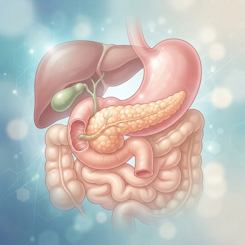
    Exocrine pancreatic insufficiency, commonly referred to as EPI, is a medical condition defined by the pancreas’s inability to secrete an adequate amount of digestive enzymes. Under normal physiological conditions, the pancreas secretes “exocrine” enzymes—specifically lipase, protease, and amylase—into the small intestine. These enzymes are the primary workhorses of your digestive system. Lipase is responsible for breaking down dietary fats, protease tackles proteins, and amylase processes carbohydrates. When the pancreas is compromised, it cannot produce or deliver these enzymes to the duodenum, the first part of the small intestine, in the volumes required to digest a standard meal.

    The clinical significance of this deficiency cannot be overstated. Without these enzymes, food passes through the digestive tract largely undigested. This state, known as maldigestion, inevitably leads to malabsorption, where the body fails to extract life-sustaining nutrients from the food consumed. It is a common misconception that EPI is only a digestive concern; rather, it is a systemic issue. Because fats are essential for the absorption of fat-soluble vitamins—specifically vitamins A, D, E, and K—a deficiency in these enzymes triggers a domino effect that can lead to severe nutritional gaps. Over time, the inability to process macronutrients deprives the body of the building blocks required for muscle repair, energy production, and immune function.

    Experts often categorize the condition based on the severity of enzyme loss. In many cases, the pancreas retains some function, but not enough to support high-fat or large-volume meals. In more advanced stages, the loss is significant enough that even small, frequent meals can result in uncomfortable symptoms. It is vital to note that EPI is not a disease in itself, but rather a functional consequence of various underlying health issues that damage the pancreas. This means that the treatment approach is rarely “one size fits all.” Instead, it is highly dependent on the root cause—whether that is long-standing inflammation, anatomical changes from surgery, or genetic factors. Recognizing that the pancreas is failing to meet the body’s metabolic demand is the gateway to understanding why your body may feel depleted, despite an adequate or even high caloric intake.

    Common Symptoms of EPI You Should Recognize

    Recognizing the symptoms of EPI can be difficult because they often mimic other common gastrointestinal disorders, such as irritable bowel syndrome (IBS) or food intolerances. However, there are hallmark signs that signal the pancreas is struggling to perform its exocrine duties. The most prominent of these indicators is steatorrhea—a medical term for loose, fatty, or oily stools. Because the lipase enzyme is necessary to break down fat, a lack of it means dietary fats pass through the digestive tract essentially untouched. This often results in stools that appear pale, bulky, foul-smelling, and difficult to flush, as they contain significant amounts of undigested fat.

    Beyond the characteristics of bowel movements, those living with EPI frequently report chronic abdominal discomfort. This is often experienced as a dull ache or significant bloating that intensifies shortly after eating. Because the body is struggling to break down complex food structures, these items remain in the gut longer than they should, leading to increased gas production and persistent feelings of fullness or distension. For many, this manifests as a cycle where the fear of triggering a painful or inconvenient digestive episode leads to a restrictive diet, further compounding the issue of unintentional weight loss.

    Weight loss in the absence of a restrictive diet is a major warning sign. When the body cannot access the calories it consumes, it begins to cannibalize its own energy stores—muscle mass and body fat. Individuals may notice their clothing fitting loosely or experience a general feeling of weakness and fatigue that does not resolve with sleep or improved dietary choices. Additionally, because malabsorption impacts the intake of fat-soluble vitamins, people may develop brittle nails, thinning hair, or skin changes associated with vitamin deficiencies. While these symptoms can be vague, when they occur in clusters, they serve as a critical prompt to seek a professional evaluation. Keeping a food and symptom journal can be an incredibly useful tool, as it helps you and your healthcare team identify whether your symptoms align with the specific timing of enzyme depletion post-meal.

    Primary Causes and Risk Factors for EPI

    The causes of EPI are diverse, reflecting the various ways the pancreas can sustain damage or obstruction. Chronic pancreatitis stands as the most common culprit in many clinical populations. This is a condition characterized by long-term inflammation of the pancreas, often resulting from alcohol consumption or recurring acute pancreatitis episodes. Over time, the repeated inflammation causes scar tissue to form within the pancreatic tissue. This fibrosis gradually replaces the functional cells responsible for enzyme production with non-functional tissue, leading to a steady decline in exocrine capacity.

    Cystic fibrosis is another significant cause, particularly in younger populations or those diagnosed early in life. In this genetic condition, the body produces thick, sticky mucus that clogs the ducts of the pancreas. These blockages prevent the digestive enzymes from ever reaching the small intestine, essentially trapping them inside the organ. Without intervention, individuals with cystic fibrosis often exhibit symptoms of malabsorption from an early age. Beyond these, surgical interventions play a major role in the development of EPI. Procedures like a Whipple procedure, which involves removing the head of the pancreas, or surgeries to treat gastric ulcers or cancers, can physically remove or bypass the structures that facilitate normal pancreatic function. In these cases, the insufficiency is considered a predictable outcome that requires active management.

    Furthermore, pancreatic cancer itself, or tumors that obstruct the pancreatic duct, can lead to localized insufficiency. Even without complete removal, the presence of a mass can block the flow of enzymes, resulting in clinical symptoms of malabsorption. Other risk factors include advanced age, smoking, and certain autoimmune conditions that trigger inflammation throughout the body. Understanding the specific cause is essential because it informs the long-term prognosis and the strategy for managing the insufficiency. While some causes, such as structural damage from surgery, are fixed, others, like chronic pancreatitis, may require ongoing lifestyle modifications to prevent further tissue degradation. Recognizing these risk factors early allows individuals to remain vigilant about their digestive health and engage in proactive discussions with their care team.

    Approach Mechanism Best For
    Pancreatic Enzyme Replacement Therapy (PERT) Replaces missing enzymes with pharmaceutical-grade capsules. Those with clinically diagnosed enzyme deficiency.
    Low-Fat Diet Modification Reduces the demand on the pancreas by limiting dietary fats. Early management and symptom reduction during flares.
    MCT Oil Supplementation Provides easily absorbed fats that bypass standard pancreatic digestion. Individuals struggling with severe weight loss and malnutrition.
    Frequent Small Meals Spreads the enzymatic workload evenly throughout the day. Patients experiencing post-meal bloating and discomfort.

    How Malabsorption Impacts Your Overall Health

    Malabsorption is the physiological end-point of untreated EPI, and its consequences ripple throughout every organ system. When the body cannot break down and absorb macronutrients—fats, proteins, and carbohydrates—it enters a state of perpetual “metabolic hunger.” Even when caloric intake is sufficient, the gut simply cannot extract the fuel. The immediate effect is a deficit in energy, which explains the profound fatigue many with EPI report. Without adequate protein, the body struggles to maintain muscle mass, leading to sarcopenia, which is particularly dangerous in older adults. Muscles are not just for movement; they are essential for metabolic regulation, and their loss can significantly impact physical independence.

    The secondary effects of malabsorption are perhaps more insidious. Because fat digestion is impaired, the body cannot absorb fat-soluble vitamins—A, D, E, and K—even if you are eating healthy, vitamin-rich foods. Vitamin D deficiency is a major concern, as it is vital for bone density. Over time, individuals with untreated EPI are at a significantly higher risk for developing osteopenia or osteoporosis, leading to brittle bones and an increased risk of fractures. Vitamin K is essential for blood clotting, and deficiencies can lead to unexplained bruising or bleeding tendencies. Vitamin A is crucial for vision and skin integrity, while vitamin E plays a vital role in immune health and protecting cells from oxidative stress.

    Furthermore, the systemic impact extends to the gut microbiome. When undigested food—especially carbohydrates and proteins—reaches the large intestine, it becomes fuel for the wrong type of bacteria. This can lead to small intestinal bacterial overgrowth (SIBO), which complicates symptoms by causing further gas, bloating, and systemic inflammation. The body is essentially in a state of stress, struggling to balance nutrient deprivation with the discomfort of chronic digestive distress. This creates a feedback loop: poor digestive health leads to poor nutrition, which weakens the immune system, making it harder for the body to manage the underlying cause of the EPI. Understanding this connection underscores why addressing EPI is not just about stopping diarrhea or bloating; it is about protecting your long-term health and preventing the secondary complications that arise from a body that is starving in the midst of plenty.

    Diagnostic Tests Used to Identify Pancreatic Issues

    Diagnosing exocrine pancreatic insufficiency requires a systematic approach, as there is no single test that can identify the condition with 100% certainty in all scenarios. Typically, the diagnostic process begins with a thorough medical history and physical examination to identify the classic signs of malabsorption. Once suspicion is raised, healthcare providers often utilize specific stool tests. The most common and reliable non-invasive diagnostic tool is the fecal elastase-1 test. Elastase is a digestive enzyme that, unlike other enzymes, is not degraded as it passes through the digestive tract. By measuring the concentration of this enzyme in a stool sample, experts can estimate how well the pancreas is functioning. Low levels of fecal elastase are a strong indicator that the pancreas is not producing enough digestive power to meet the body’s needs.

    In cases where the diagnosis is more complex, imaging studies are often employed to examine the anatomy of the pancreas. Magnetic resonance cholangiopancreatography (MRCP) is a specialized type of MRI that creates detailed images of the pancreatic and bile ducts. This is exceptionally useful for visualizing obstructions, scarring from chronic pancreatitis, or tumors that might be impeding the flow of enzymes. An endoscopic ultrasound (EUS) is another powerful diagnostic tool; it involves passing a thin, flexible tube with an ultrasound probe down the throat and into the stomach. This allows for high-resolution, internal imaging of the pancreas, enabling providers to see early structural changes that might not be visible on standard external scans.

    It is important to understand that testing is often done in stages. A provider might begin with non-invasive stool tests to establish a baseline of pancreatic function. If the results are inconclusive but clinical symptoms remain persistent, they may move toward imaging. In some academic or specialized centers, doctors may conduct a “secretin stimulation test,” which is considered the gold standard for assessing pancreatic capacity. This involves placing a tube into the small intestine and measuring how the pancreas responds to an injection of secretin, a hormone that stimulates enzyme production. While highly accurate, this test is invasive and time-intensive, so it is usually reserved for cases where the diagnosis is still unclear after other testing. Regardless of the route taken, these tests provide the data needed to create a tailored management plan, ensuring that the patient receives the correct dosage of pancreatic enzyme replacement therapy to effectively restore their nutritional balance.

    Managing EPI with Pancreatic Enzyme Replacement Therapy

    For individuals diagnosed with exocrine pancreatic insufficiency, the primary clinical treatment is Pancreatic Enzyme Replacement Therapy (PERT). Because the pancreas is no longer producing sufficient levels of lipase, protease, and amylase to break down fats, proteins, and carbohydrates, PERT acts as a bridge, providing synthetic enzymes that mimic natural digestive secretions. When taken correctly, these enzymes help mitigate the primary malabsorption signs associated with the condition, such as steatorrhea (oily, foul-smelling stools) and unintended weight loss.

    Success with PERT is highly dependent on timing and dosage. Enzymes are not “pills” to be taken once a day; they must be ingested with every meal and snack. If a patient consumes a meal without proper enzyme coverage, the food will pass through the gastrointestinal tract largely undigested, potentially leading to immediate discomfort, bloating, and nutrient deficiency. The objective is to ensure that the enzymes arrive in the duodenum at the same time as the food bolus. Often, gastroenterologists recommend taking half of the prescribed dose at the beginning of a meal and the remainder halfway through to ensure continuous coverage throughout the digestive process.

    It is important to understand that enzymes are formulated to be pH-sensitive. They are typically enteric-coated to prevent the stomach’s acidic environment from degrading them before they reach the small intestine. Because of this coating, capsules should never be crushed or chewed. For individuals who struggle with swallowing, many clinicians suggest opening the capsule and mixing the microspheres into a small amount of acidic food, such as applesauce or yogurt, which can help protect the integrity of the coating until it reaches the stomach.

    Adjusting to PERT requires patience. Dosage is rarely “one size fits all” and is typically titrated based on the fat content of the diet, the severity of the symptoms, and clinical markers of nutrient absorption. It is common for a patient to work closely with their medical team for several weeks, tracking food intake and stool consistency, to find the “sweet spot” for their specific physiology.

    Essential Dietary Changes to Support Digestion

    While PERT is the cornerstone of managing EPI, the quality and composition of your diet remain critical factors in your overall digestive health. When your pancreas struggles to produce enzymes, certain macronutrients become significantly harder to process. Consequently, nutritional strategy often shifts toward high-density, easily digestible foods that minimize the workload on the digestive system.

    Fats, while essential for brain and hormonal health, are the most difficult macronutrients for an EPI-affected system to process. Many patients find relief by opting for medium-chain triglycerides (MCTs). Unlike long-chain fats, which require extensive pancreatic lipase to be broken down, MCTs are absorbed directly into the portal vein and delivered to the liver for immediate energy, bypassing much of the standard pancreatic digestive process. Incorporating MCT oil into salad dressings or smoothies can be an effective way to maintain caloric intake without triggering severe digestive distress.

    Furthermore, portion control is vital. Large, heavy meals can overwhelm the available enzyme supply, even when taking PERT. Shifting to five or six smaller, nutrient-dense meals throughout the day can significantly improve absorption and prevent the “food coma” or bloating that often follows a standard, large dinner. The focus should be on lean proteins, such as poultry, fish, or plant-based proteins like tofu, and well-cooked vegetables that are easier to break down than raw, fibrous options.

    Dietary Strategy Primary Benefit Best For
    Small, frequent meals Prevents overwhelming the enzyme supply Reducing post-meal bloating
    MCT oil supplementation Easier fat absorption Maintaining weight and energy
    Peeling/Cooking vegetables Reduces insoluble fiber load Sensitive digestive tracts
    Protein-first eating Ensures muscle mass preservation Patients experiencing rapid weight loss

    Natural Lifestyle Strategies for Pancreatic Support

    Beyond clinical intervention and diet, adopting specific lifestyle habits can support your pancreas and optimize digestive function. Because digestion is heavily influenced by the autonomic nervous system—specifically the “rest and digest” parasympathetic branch—managing stress is more than just a mental health practice; it is a physiological necessity for anyone managing EPI.

    Stress causes the body to divert blood flow away from the gut to the extremities, which can exacerbate feelings of indigestion and slow gastric motility. Engaging in mindfulness practices, such as deep diaphragmatic breathing before a meal, can signal to your body that it is safe to prioritize digestion. Furthermore, staying hydrated is crucial. Enzymes rely on a fluid-rich environment to travel through the intestinal tract. Drinking water throughout the day, rather than large quantities during a meal, helps keep the digestive tract lubricated and supports the efficient movement of food.

    Moderate movement, such as walking after meals, is another highly recommended strategy. Gentle activity aids in gastric emptying and helps prevent the stagnation of food in the gut, which can reduce the frequency of bloating and constipation. However, it is important to avoid intense, high-impact exercise immediately after a meal, as this can divert blood away from the gut and impede digestion. Consistency in timing, hydration, and movement creates a predictable environment for the digestive system, which can help in managing symptoms over the long term.

    When to Consult a Specialist for Digestive Concerns

    Knowing when to transition from basic lifestyle adjustments to specialized medical care is the hallmark of proactive digestive health. If you notice persistent signs of malabsorption—such as stools that are consistently pale, oily, or difficult to flush—you should seek an evaluation from a gastroenterologist. These symptoms are objective indicators that your body is not extracting the nutrients it requires to function.

    Additionally, if you have already been diagnosed with conditions known to impact the pancreas, such as chronic pancreatitis, cystic fibrosis, or history of pancreatic surgery, regular screening is mandatory. Even if you are currently asymptomatic, EPI can develop gradually. Sudden, unexplained weight loss, persistent abdominal pain that radiates to the back, or new-onset diabetes should always be investigated promptly by a medical professional.

    A specialist will likely employ diagnostic tools like the fecal elastase test, a non-invasive stool test that measures the levels of elastase, an enzyme produced by the pancreas. Low levels of elastase are often definitive proof of pancreatic insufficiency. Once a diagnosis is made, a specialist will guide you through the process of balancing your enzyme doses and monitoring for fat-soluble vitamin deficiencies (specifically vitamins A, D, E, and K), which are common in those with chronic malabsorption. Do not attempt to manage severe weight loss or persistent digestive distress through self-diagnosis alone; professional medical guidance is necessary to prevent long-term systemic health complications.

    Frequently Asked Questions

    Can EPI be reversed with diet alone?

    Generally, EPI is caused by structural damage to the pancreas, meaning it is typically a chronic, irreversible condition. While diet and lifestyle changes are essential for managing symptoms and improving quality of life, most patients require ongoing Pancreatic Enzyme Replacement Therapy (PERT) to properly digest food and absorb nutrients.

    What happens if I forget to take my enzymes?

    If you forget to take your enzymes, you may experience digestive symptoms such as bloating, abdominal cramping, gas, or oily stools shortly after the meal. While missing a single dose is unlikely to cause long-term harm, chronic failure to take enzymes can lead to significant nutrient deficiencies, weight loss, and muscle wasting over time.

    Is alcohol consumption safe for those with EPI?

    Alcohol is generally discouraged for individuals with pancreatic issues. Alcohol can trigger inflammation in the pancreas (pancreatitis), which may further degrade its ability to produce enzymes. It is best to consult with your gastroenterologist regarding your specific condition, but most experts advise complete avoidance to prevent worsening pancreatic function.

    Are there plant-based alternatives to standard enzyme supplements?

    Most clinical-grade pancreatic enzymes are porcine-derived because their structure closely mimics human enzymes. While there are some plant-based or fungal-derived digestive enzymes available over the counter, they are often not potent enough to treat medically diagnosed EPI and are not regulated with the same clinical rigor as prescription PERT. Always discuss supplementation with your doctor.

    How do I know if my enzyme dose is correct?

    Your doctor will determine if your dose is correct by monitoring your clinical symptoms, such as the consistency and frequency of your stools, and your ability to maintain a healthy weight. They may also order blood tests to check for appropriate levels of fat-soluble vitamins (A, D, E, and K), which indicate whether you are successfully absorbing nutrients.

    Can stress trigger an EPI flare-up?

    While stress does not cause the structural damage underlying EPI, it can significantly worsen digestive symptoms. Stress affects gut motility and blood flow to the digestive organs, making it harder for your body to process food effectively. Reducing stress through mindfulness and physical relaxation can help minimize the severity of digestive discomfort associated with the condition.

    Conclusion

    Living with exocrine pancreatic insufficiency requires a thoughtful approach to health, but it does not mean sacrificing your quality of life. By understanding the critical role of pancreatic enzymes, adopting a gut-friendly dietary routine, and maintaining close communication with your medical team, you can effectively manage malabsorption and restore your vitality. Managing your health is an ongoing process of monitoring, adjusting, and advocating for the care you need to thrive. Whether you are in the early stages of diagnosis or seeking better management of your current symptoms, remember that consistency is your greatest ally in achieving digestive balance. If you are experiencing any of the symptoms discussed in this article, please reach out to your primary care physician or a gastroenterologist today to schedule an evaluation.

    By healthauthoritylife Editorial Team

    This article is for informational purposes only and is not a substitute for professional medical advice. Always consult your doctor before making changes to your diet, exercise routine, or treatment plan.
  • NAFLD Diet Plan: Foods to Eat and Avoid for Liver Health

    NAFLD Diet Plan: Foods to Eat and Avoid for Liver Health

    Key Takeaways

    • A structured NAFLD diet plan focuses on reducing liver inflammation through nutrient-dense, whole-food choices.
    • Eliminating high-fructose corn syrup and refined carbohydrates is a critical step in halting liver fat accumulation.
    • Healthy fats, particularly omega-3 fatty acids, are essential for reducing existing liver fat deposits.
    • Weight management through a liver healing diet significantly improves insulin sensitivity and metabolic health.
    • Consistency in dietary choices is more important for long-term liver health than short-term restrictive cleanses.

    Receiving a diagnosis of non-alcoholic fatty liver disease can feel overwhelming, but it is important to recognize that the liver is a remarkably resilient organ with an impressive capacity for regeneration. Unlike many other health conditions where management relies solely on medication, liver health is profoundly dictated by the daily decisions we make at the dinner table. By adopting a proactive approach to your nutrition, you are not just preventing further damage; you are actively providing your body with the tools it needs to repair cellular pathways, reduce inflammation, and reverse the accumulation of excess fat. This guide serves as your comprehensive roadmap to navigating the non-alcoholic fatty liver disease diet, helping you understand how the specific foods you consume act as either building blocks for recovery or catalysts for further stress on your liver.

    Understanding NAFLD and the Role of Nutrition

    Non-alcoholic fatty liver disease (NAFLD) is characterized by the accumulation of excess fat in the liver cells of individuals who consume little to no alcohol. While the term may sound intimidating, the condition typically exists on a spectrum. In its earliest stages, the liver simply stores extra fat. However, if left unmanaged, this accumulation can trigger a persistent inflammatory response, leading to more complex liver issues. The primary goal of any effective NAFLD diet plan is to address the underlying metabolic dysfunctions that cause this fat to settle in the liver in the first place.

    At its core, the liver is the body’s primary metabolic hub. It processes almost everything we consume, converting nutrients into energy and filtering toxins from the bloodstream. When we consistently provide the body with more fuel than it can utilize—particularly in the form of processed sugars and refined carbohydrates—the liver is forced to convert that excess glucose into triglycerides, which are then stored within the liver tissue. Over time, these fat droplets begin to crowd the liver cells, impairing their ability to function efficiently and sending signals of distress throughout the entire body.

    Nutrition acts as the first line of defense because the liver is uniquely sensitive to metabolic signaling. When you adjust your intake, you are essentially changing the instructions you give to your metabolism. A balanced fatty liver nutrition plan focuses on stabilizing blood sugar levels and improving insulin sensitivity. When insulin levels are kept within a healthy range, the liver is less inclined to store fat and more capable of oxidizing, or burning, the fat it currently holds. This transition is not about temporary deprivation, but rather about creating a sustainable environment where your liver can finally focus on its primary roles—detoxification and energy regulation—rather than acting as a storage unit for excess dietary intake.

    Furthermore, managing NAFLD is closely tied to systemic inflammation. A poor diet, heavy in processed ingredients, keeps the body in a state of low-grade, chronic inflammation, which exacerbates the stress on liver cells. Conversely, a nutrient-dense approach provides the antioxidants, vitamins, and minerals necessary to neutralize free radicals and promote cellular repair. This process is often referred to as a liver healing diet, emphasizing foods that reduce oxidative stress. By focusing on whole, unprocessed foods, you lower the workload for your liver, allowing it the “breathing room” it needs to purge excess fat. It is a fundamental shift from fueling a storage-oriented metabolism to one that is focused on regeneration, efficiency, and long-term metabolic health.

    Top Foods to Include for Liver Regeneration

    When selecting the best foods for fatty liver health, the focus should be on nutrient density and anti-inflammatory properties. Your goal is to choose items that provide essential vitamins and minerals without causing sharp spikes in insulin. Incorporating these foods into your NAFLD diet plan can help support your liver’s natural ability to detoxify and repair itself.

    Cruciferous vegetables such as broccoli, cauliflower, Brussels sprouts, and kale are among the most powerful allies for liver health. These vegetables are rich in fiber and contain compounds that have been shown to support the liver’s detoxification enzymes. Fiber, in particular, is essential because it binds to toxins in the digestive tract and helps regulate blood sugar levels, preventing the liver from receiving a sudden deluge of glucose. Similarly, leafy greens like spinach and arugula provide high concentrations of antioxidants, which combat the oxidative stress that often accompanies fatty liver disease.

    Healthy protein sources are also vital. While some believe that protein should be avoided, high-quality, lean proteins help stabilize blood sugar and prevent the intense cravings associated with carbohydrate-heavy diets. Options like wild-caught fatty fish (such as salmon, sardines, and mackerel) provide an excellent source of omega-3 fatty acids. Omega-3s are renowned for their ability to lower liver fat levels and reduce systemic inflammation. In addition, legumes and lentils offer a plant-based alternative that is high in both protein and fiber, making them a staple for those looking to improve liver markers.

    Berries and other antioxidant-rich fruits, such as blueberries, strawberries, and blackberries, are excellent additions to your plate. Unlike sugary snacks, these fruits offer high fiber content along with polyphenols, which may help slow down the accumulation of fat in the liver. However, moderation remains key, as fructose metabolism still occurs in the liver. Additionally, incorporating nuts like walnuts and seeds such as flax or chia adds necessary healthy fats and Vitamin E to your diet, both of which are protective against liver damage.

    Food Group Recommended Choices Best For
    Cruciferous Veggies Broccoli, Brussels sprouts Enzyme support
    Fatty Fish Salmon, Sardines Reducing fat deposits
    Whole Grains Oats, Quinoa Sustained energy
    Plant Fats Avocados, Walnuts Inflammation control

    Foods That Cause Inflammation and Fatty Deposits

    Understanding what to avoid with NAFLD is just as important as knowing what to include. Certain food groups act as direct fuel for liver fat storage and trigger inflammatory cascades that hinder the organ’s recovery. By identifying these triggers, you can make informed decisions that prevent the worsening of fatty deposits and protect the structural integrity of your liver cells.

    At the top of the “avoid” list are processed meats and foods high in saturated and trans fats. While fat is not inherently “bad,” the types of fat found in fried fast foods, pre-packaged snack foods, and processed meats like bacon, sausage, and deli meats can be highly taxing on the liver. These foods often contain high levels of sodium and synthetic preservatives, which increase the burden of detoxification. Furthermore, they are typically low in the essential nutrients the liver needs to repair itself, providing only empty, high-calorie fuel that is easily shunted toward fat storage.

    Another major culprit is the consumption of ultra-processed snacks. These items are engineered for “palatability,” which often means they are a combination of refined flour, cheap industrial seed oils, and hidden sugars. These oils—often high in omega-6 fatty acids—can tip the balance of inflammation in the body. While omega-6 fatty acids are necessary in small amounts, the modern diet is often disproportionately skewed toward them, which can promote an inflammatory state that complicates liver healing. When you read a label and see a long list of ingredients you cannot pronounce, it is generally a signal that the food is a poor choice for a liver-conscious lifestyle.

    It is also worth mentioning that common beverages can be a silent driver of NAFLD. Even drinks that are marketed as “healthier” alternatives often contain hidden sweeteners or lack nutritional value. Consuming calories through sugary sodas, energy drinks, and highly sweetened coffees creates an immediate surge in blood glucose, forcing the liver into a state of hyper-activity to manage the excess sugar. Because the liver processes fructose differently than glucose, high-fructose corn syrup in particular has been linked to the rapid development of liver fat. Removing these inflammatory triggers allows the liver to prioritize its secondary tasks, such as metabolic regulation, rather than constantly managing the fallout from a diet high in processed, inflammatory debris.

    The Impact of Sugar and Refined Carbs on Your Liver

    Few factors impact the liver as heavily as the intake of sugar and refined carbohydrates. When we discuss a non-alcoholic fatty liver disease diet, the conversation inevitably circles back to glycemic control. When you consume refined carbohydrates—such as white bread, pasta, pastries, and white rice—your body breaks them down into glucose very quickly. This causes a rapid spike in blood sugar, which triggers a significant release of insulin from the pancreas. While insulin is necessary for cells to take up energy, chronic surges in insulin levels tell the liver to stop burning fat and start storing it.

    The liver’s role in managing sugar is central to its dysfunction. When glucose levels are too high, the liver converts that excess energy into fatty acids through a process called de novo lipogenesis. These fatty acids are then stored within the liver, contributing to the development of fatty deposits. Refined carbs lack the fiber that usually slows down digestion; because they bypass this natural “brake” system, the liver receives a massive amount of energy all at once that it simply cannot process efficiently. Over time, this leads to a condition known as insulin resistance, where cells stop responding appropriately to insulin, further trapping the liver in a cycle of fat storage.

    Fructose, commonly found in high-fructose corn syrup and added sugars, is uniquely problematic. Unlike glucose, which can be used by cells throughout the body for energy, fructose is metabolized almost entirely by the liver. When the liver is bombarded with fructose, it has no choice but to convert it directly into fat. This is why many experts suggest that the rise in fatty liver cases correlates strongly with the widespread increase of added sugars in our food supply. By reducing your reliance on refined carbohydrates and eliminating added sugars, you are directly alleviating the metabolic pressure on your liver cells.

    Instead of refined grains, opt for complex carbohydrates that include fiber, such as whole oats, quinoa, and brown rice. Fiber acts as a crucial buffer; it slows down the rate of glucose absorption, which prevents the sharp insulin spikes that trigger the liver to store fat. When you choose whole, unrefined options, your liver receives a steady, manageable supply of energy rather than a chaotic influx. This simple shift in carbohydrate quality is one of the most effective ways to break the cycle of fat accumulation and provide your body with the stability it needs to begin the process of liver healing.

    Why Healthy Fats Are Essential for Fatty Liver Recovery

    There is a lingering misconception that in order to “lose fat” in the liver, one must follow a strictly low-fat diet. However, for those managing NAFLD, it is not the *presence* of fat that causes problems, but rather the *type* of fat consumed. High-quality, healthy fats are actually essential components of a liver healing diet. They provide necessary structural support for cell membranes, facilitate the absorption of fat-soluble vitamins (A, D, E, and K), and help regulate the hormones that govern satiety and blood sugar.

    Monounsaturated fats, found in foods like olive oil, avocados, and various nuts, are particularly beneficial. Research into the Mediterranean dietary pattern, which is high in these healthy fats, has consistently shown positive outcomes for liver health. These fats help improve insulin sensitivity, meaning your body becomes more efficient at using the energy it takes in, which in turn reduces the need for the liver to store excess fat. Including moderate amounts of high-quality fats can actually help you feel fuller for longer, reducing the temptation to reach for refined, carb-heavy snacks that we know contribute to liver fat accumulation.

    Omega-3 fatty acids, mentioned previously in the context of fatty fish, deserve specific focus regarding recovery. These fats are potent anti-inflammatories. Because NAFLD often involves a persistent state of low-grade inflammation, omega-3s act like a soothing balm for the liver. They work by inhibiting the pathways that lead to inflammation and helping to regulate the lipid metabolism within the liver itself. By replacing processed vegetable oils—which are often rich in inflammatory omega-6 fatty acids—with sources of omega-3s and monounsaturated fats, you are essentially changing the building materials available to your liver cells.

    It is important to emphasize that while these fats are healthy, they are still calorie-dense. A successful NAFLD diet plan is about balance. You should treat healthy fats as a way to enhance your meals—a drizzle of extra virgin olive oil on a salad, half an avocado added to a protein-rich lunch, or a small handful of walnuts as a snack—rather than as a main course. By incorporating these fats intentionally, you provide your liver with the essential components it needs to maintain cell structure and metabolic health, while keeping your overall caloric intake in check to support gradual weight management. This strategic inclusion of healthy fats is a far superior approach to the extreme fat-free diets that often lead to nutrient deficiencies and increased cravings.

    Hydration and Its Importance in Liver Detoxification

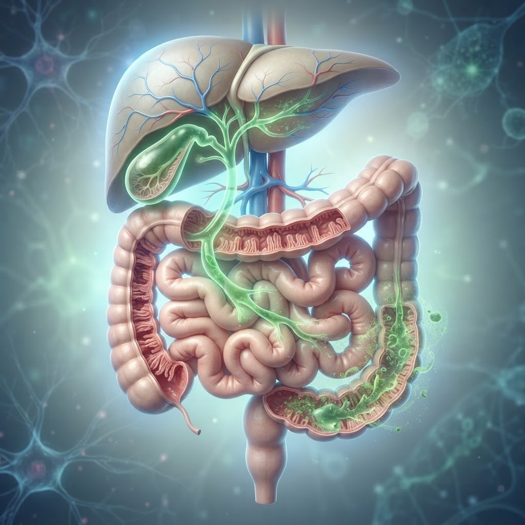
    While food choices provide the building blocks for metabolic health, water acts as the essential medium through which all liver detoxification processes occur. The liver is the body’s primary filtration system, responsible for neutralizing toxins, metabolizing fats, and processing nutrients. To perform these demanding tasks, the liver requires a constant supply of fluid to keep bile, the digestive fluid that breaks down fats, moving smoothly through the biliary tree.

    When an individual is chronically dehydrated, the bile can become thick and sluggish. This state, often referred to as biliary stasis, hinders the liver’s ability to clear away waste products and excess cholesterol. Over time, this inefficiency can contribute to the accumulation of fat within the liver cells, exacerbating the progression of non-alcoholic fatty liver disease. Furthermore, adequate hydration supports kidney function, which works in tandem with the liver to remove the byproducts of detoxification from the bloodstream.

    It is important to understand that not all fluids serve the liver equally. While water is the gold standard for hydration, many people inadvertently introduce stress to their liver by relying on sugary sodas, energy drinks, or excessive alcohol. These beverages force the liver to prioritize processing simple sugars and toxins rather than performing its restorative tasks. Replacing these drinks with filtered water, herbal teas, or lemon-infused water can significantly reduce the liver’s daily workload.

    To maximize the benefits of a liver healing diet, consider the following hydration strategies:

    • Morning Prime: Drink 16 ounces of room-temperature water upon waking. This kickstarts metabolic processes and helps flush out residual waste processed by the liver during the overnight fast.
    • Mindful Sipping: Rather than gulping large amounts of water at once, distribute your intake throughout the day. Consistent hydration ensures that the liver remains well-perfused and efficient.
    • Herbal Infusions: Certain herbal teas, such as dandelion root or milk thistle, are traditionally associated with liver support. While these are not magic cures, they can serve as hydrating, antioxidant-rich alternatives to caffeinated beverages.
    • Electrolyte Balance: If you are engaging in regular exercise to combat NAFLD, ensure you are replenishing electrolytes naturally through mineral-rich water or electrolyte-dense foods like cucumber and celery, rather than sugary sports drinks.

    Meal Planning Strategies for Sustainable Results

    The most effective non-alcoholic fatty liver disease diet is not a temporary “detox” but a lifelong shift in nutritional habits. Sustainability is the cornerstone of progress. Many individuals struggle because they attempt to overhaul their entire kitchen overnight, leading to burnout. Instead, focus on gradual, structural changes to your meal planning routine that prioritize nutrient density and blood sugar stability.

    A successful fatty liver nutrition plan revolves around the concept of “plate building.” Instead of counting every single calorie, aim to fill your plate in a way that stabilizes insulin—the hormone primarily responsible for driving fat storage in the liver. A balanced plate for liver health typically consists of fifty percent non-starchy vegetables, twenty-five percent high-quality lean protein, and twenty-five percent fiber-rich complex carbohydrates.

    Preparation is the primary deterrent against the temptation of processed foods. When you have pre-portioned, liver-friendly meals available, you are less likely to reach for convenient, low-quality snacks when hunger strikes. Consider these strategic approaches:

    1. Batch Cooking: Dedicate two hours each weekend to roasting large trays of cruciferous vegetables like broccoli, cauliflower, and Brussels sprouts. These contain sulfur compounds that support phase-two liver detoxification.
    2. Smart Staging: Keep washed greens, pre-cut vegetables, and hard-boiled eggs at eye level in your refrigerator. Make the healthy choice the path of least resistance.
    3. The “One-New-Recipe” Rule: Avoid the overwhelm of a complete pantry overhaul by trying only one new NAFLD-friendly recipe per week. This allows you to build a database of meals you actually enjoy.
    4. Label Awareness: When grocery shopping, prioritize single-ingredient foods. The fewer the ingredients on a label, the more likely the food is to support liver health.
    Liver-Supportive Food Replacements
    Current Food Choice Liver-Friendly Alternative Best For
    White Rice or Pasta Quinoa or Steel-Cut Oats Stabilizing Blood Glucose
    Commercial Salad Dressing Olive Oil and Apple Cider Vinegar Reducing Inflammatory Omega-6s
    Potato Chips Raw Walnuts or Pumpkin Seeds Healthy Fat and Mineral Intake
    Fruit Juice Whole Berries (Blueberries, Raspberries) High Fiber & Antioxidant Protection
    Beef or Processed Deli Meat Wild-Caught Salmon or Lentils Omega-3 Fatty Acids & Liver Repair

    Lifestyle Habits to Complement Your Liver Diet

    While diet is the primary lever in managing NAFLD, it does not act in a vacuum. Your liver’s health is deeply influenced by your broader lifestyle environment. Chronic stress, sedentary behavior, and sleep deprivation all create a metabolic landscape that encourages fat storage. To optimize your liver-healing efforts, consider integrating these supportive habits into your daily flow.

    The Role of Movement: Physical activity is perhaps the most significant secondary factor in liver health. Exercise increases the oxidation of fatty acids, essentially forcing the liver to burn its stored fat for energy. Resistance training, in particular, has shown promise in improving insulin sensitivity, which is a critical step in reversing liver fat accumulation. You do not need to train like an athlete; even consistent, low-intensity movement such as brisk walking or swimming for 30 minutes daily can improve metabolic efficiency.

    Stress Management: The liver is an endocrine-responsive organ. Chronic stress keeps the body in a state of high cortisol, which stimulates the release of glucose into the bloodstream. Constant high blood sugar levels force the liver to work overtime, often resulting in fat deposition. Techniques such as diaphragmatic breathing, yoga, or simple time in nature can help shift the body into a parasympathetic state, allowing the liver to focus on restoration rather than survival.

    Sleep Hygiene: Quality sleep is a non-negotiable component of metabolic recovery. During deep sleep, the body undergoes a series of repair processes. Poor sleep is directly linked to an increased risk of metabolic syndrome and insulin resistance, both of which are closely tied to NAFLD. Aim for seven to nine hours of restorative sleep by creating a cool, dark environment and limiting screen time before bed to protect your circadian rhythm.

    Monitoring Your Progress and When to See a Specialist

    Managing non-alcoholic fatty liver disease is a marathon, not a sprint. Because the liver is a remarkably resilient organ capable of regenerating its cells, progress is often possible, but it is rarely instantaneous. Monitoring your journey involves both subjective markers and objective medical data.

    Subjectively, you may notice improvements in your energy levels, mental clarity, and digestion within the first few weeks of adopting a liver-healing diet. You may also notice less bloating and more stable moods as your blood sugar fluctuations stabilize. However, these markers do not tell the whole story. You must partner with a healthcare professional to track your progress accurately.

    Regular check-ups are vital. Experts generally agree that individuals with NAFLD should be monitored through:

    • Liver Function Tests (LFTs): These blood panels measure specific enzymes that can indicate liver inflammation. While they don’t show the extent of fat, they are essential for tracking the liver’s overall stress response.
    • Imaging Studies: Ultrasound, FibroScan, or MRI-based tests are the gold standards for quantifying the actual amount of fat present in the liver. These are typically performed periodically to assess the effectiveness of lifestyle interventions.
    • Metabolic Panels: Tracking your blood pressure, lipid profile (cholesterol and triglycerides), and HbA1c (a marker for long-term blood sugar) is critical. As your metabolic health improves in these areas, your liver health will almost certainly follow.

    When is it time to see a specialist? If your liver enzymes remain elevated despite strictly adhering to a nutrition and exercise plan, you should consult with a hepatologist or a gastroenterologist. Additionally, if you experience persistent abdominal pain, jaundice (yellowing of the skin or eyes), unexplained bruising, or severe fatigue, seek medical attention immediately. These can be indicators that your condition is progressing toward more serious complications, such as fibrosis or cirrhosis, which require specialized medical intervention beyond dietary adjustments alone.

    Frequently Asked Questions

    Can NAFLD be completely reversed through diet alone?

    For many individuals, especially those in the early stages of the disease, significant improvement or even complete reversal of liver fat accumulation is possible through sustained lifestyle changes, including weight management and a balanced diet. However, because each person’s metabolic profile is unique, this must be assessed and monitored by a medical professional to ensure the condition is resolving appropriately.

    Is fruit bad for someone with a fatty liver?

    Whole fruits are generally beneficial because they contain fiber, water, and essential micronutrients that help regulate blood sugar. However, fruit juice and excessive amounts of concentrated fructose should be limited, as high intake of refined sugars can contribute to lipogenesis, or the creation of new fat, within the liver cells.

    How long does it take to see improvements in liver health?

    While everyone’s body responds differently, many people begin to notice improvements in metabolic markers, such as fasting blood sugar and energy levels, within a few weeks of consistent dietary changes. Significant reductions in liver fat as observed on medical imaging typically occur over the course of several months of sustained effort.

    Are there specific supplements I should take for my liver?

    While some supplements like Vitamin E or Omega-3 fatty acids are sometimes discussed in the context of liver health, there is no “magic pill.” Always consult with a doctor before starting any supplement, as the liver must process these substances, and an incorrect dose or low-quality product could potentially add unnecessary strain to an already compromised liver.

    Do I have to give up all fats to manage NAFLD?

    Absolutely not. The type of fat is what matters most. You should avoid trans fats and limit excessive saturated fats, but healthy monounsaturated and polyunsaturated fats—found in avocados, olive oil, walnuts, and fatty fish—are essential for overall health and can actually help improve metabolic outcomes when consumed in moderation.

    Is it safe to consume coffee while managing NAFLD?

    Research frequently suggests that moderate, unsweetened coffee consumption may have a protective effect on the liver, potentially lowering the risk of progression in liver disease. However, this assumes the coffee is not loaded with high-calorie syrups, creams, or sugars, which would negate any potential health benefits.

    Conclusion

    Taking control of non-alcoholic fatty liver disease is one of the most proactive steps you can take for your long-term health. By focusing on whole, unprocessed foods, prioritizing consistent hydration, and integrating movement into your daily routine, you provide your liver with the environment it needs to repair and function optimally. Remember that your liver is a resilient organ; every healthy meal and every consistent lifestyle choice is a building block toward a healthier, more vibrant you. Start small, stay consistent, and remember that you are not alone in this journey. For personalized guidance tailored to your specific metabolic needs, schedule a consultation with your primary care physician or a registered dietitian today to refine your path forward.

    By healthauthoritylife Editorial Team

    This article is for informational purposes only and is not a substitute for professional medical advice. Always consult your doctor before making changes to your diet, exercise routine, or treatment plan.

  • Bile Acid Malabsorption: Symptoms, Causes, and Natural Relief

    Bile Acid Malabsorption: Symptoms, Causes, and Natural Relief

    Key Takeaways

    • Bile acid malabsorption occurs when the intestines cannot properly reabsorb bile acids, leading to digestive disruption.
    • Bile acid diarrhea is a hallmark sign, often misdiagnosed as irritable bowel syndrome.
    • Structural changes in the gut, such as gallbladder removal or inflammatory conditions, are primary drivers of BAM.
    • Bile acids act as natural laxatives in the colon, which explains why malabsorption causes frequent, watery stools.
    • Managing symptoms often requires a multifaceted approach involving dietary modifications, lifestyle shifts, and targeted gut support.

    If you have spent months—or even years—dealing with unpredictable, chronic diarrhea, you know that the impact on your quality of life is profound. Many people struggling with these persistent digestive issues eventually receive a diagnosis of bile acid malabsorption (BAM). While the name may sound complex, the mechanism is relatively straightforward: your body is struggling to recycle the essential digestive fluids needed to break down fats. When this cycle is interrupted, the result is an uncomfortable and often embarrassing gastrointestinal cascade. Understanding BAM is the first step toward reclaiming your digestive health, as many people find that identifying the root cause allows them to implement effective, natural strategies for relief. In this guide, the healthauthoritylife Editorial Team explores the intricacies of this condition, from the underlying physiological causes to practical, holistic approaches for daily management.

    What Is Bile Acid Malabsorption (BAM)?

    Bile acid malabsorption, frequently referred to in medical literature as BAM, is a clinical condition where the distal ileum—the final segment of the small intestine—fails to absorb bile acids effectively. Under normal physiological circumstances, bile acids are produced by the liver, stored in the gallbladder, and released into the small intestine to emulsify dietary fats. This emulsification process is vital for the absorption of fat-soluble vitamins and fatty acids. Once the digestive process is complete in the small intestine, approximately 95% of these bile acids are reabsorbed in the terminal ileum, returned to the liver via the portal vein, and recycled. This intricate “enterohepatic circulation” is highly efficient.

    When BAM occurs, this recycling system breaks down. Instead of being reabsorbed back into the body, the excess bile acids continue their journey into the large intestine (the colon). Because bile acids are inherently irritating to the lining of the colon and act as potent stimulants for fluid secretion, their presence in the large bowel leads to rapid transit time and significant fluid accumulation. This process is the primary physiological driver behind bile acid diarrhea.

    For many individuals, BAM remains an “invisible” condition. It is frequently misdiagnosed as irritable bowel syndrome with diarrhea (IBS-D) because the symptoms are nearly identical to the naked eye. However, distinguishing between the two is critical because the management strategies differ significantly. While IBS-D is often managed through stress reduction and dietary avoidance, BAM specifically requires addressing the transit of bile through the gut. Chronic bile acid malabsorption can be categorized into three types: Type 1, which results from structural damage to the terminal ileum (often due to Crohn’s disease or surgical resection); Type 2, which is idiopathic, meaning it occurs without an identifiable underlying condition; and Type 3, which arises from other gastrointestinal disorders, such as gallbladder removal, celiac disease, or small intestinal bacterial overgrowth (SIBO). Understanding the specific type and mechanism of your BAM is the foundation of navigating long-term digestive health and finding sustainable relief.

    Common Symptoms of Bile Acid Malabsorption

    The hallmark of bile acid malabsorption is undoubtedly diarrhea, but the clinical presentation often goes beyond simple frequency. BAM symptoms are typically characterized by their urgency, unpredictability, and, in many cases, a watery consistency that feels impossible to control. Because bile acids are chemically aggressive substances, they draw water into the colon, leading to stools that are often described as explosive or “urgent.”

    Many patients report that these symptoms occur regardless of whether they have consumed a large meal. However, high-fat meals tend to trigger more severe episodes because they stimulate the release of higher volumes of bile from the gallbladder. This leads to a classic pattern: symptoms are often at their worst following a heavy meal, or occasionally, early in the morning when the digestive system begins its daily cycle. Unlike some other forms of digestive distress that may cause periodic bloating or constipation, the diarrhea associated with BAM is remarkably persistent and chronic.

    Beyond the diarrhea itself, individuals often report significant secondary symptoms related to the constant irritation of the intestinal lining. These include:

    • Abdominal cramping: Sharp, localized pain that often precedes a bowel movement.
    • Fecal urgency: A sudden, intense need to find a restroom, often causing significant anxiety in social or professional settings.
    • Bloating and gas: As the balance of the gut microbiome is disrupted by the high volume of bile acids, many experience uncomfortable distension.
    • Fatigue: Chronic diarrhea leads to poor nutrient absorption, particularly of fat-soluble vitamins (A, D, E, and K). Over time, this nutrient deficiency can manifest as persistent tiredness, skin issues, or weakened immune resilience.

    The psychological toll of these BAM symptoms is significant. The unpredictability often leads to a restrictive diet or avoidance of travel, exercise, and social gatherings. It is important to note that because the symptoms of BAM overlap with many other conditions, keeping a detailed digestive diary can be a powerful tool. Recording not only what you eat but also the timing, frequency, and consistency of your stools helps you identify patterns. If your diarrhea persists for more than four weeks or is accompanied by “red flag” symptoms—such as unexplained weight loss, blood in the stool, or fevers—it is essential to consult with a healthcare professional to rule out more severe underlying pathologies like inflammatory bowel disease or colorectal cancer before focusing on natural relief strategies.

    Primary Causes and Risk Factors

    The causes of bile acid malabsorption are as diverse as the patients they affect, but they generally fall into the categories of structural damage, metabolic dysfunction, or secondary effects of other illnesses. When looking for the root cause, clinicians look at the anatomical path the food takes through the gastrointestinal tract. If the “docking station” for bile reabsorption—the terminal ileum—is compromised, the system fails.

    Structural damage is one of the most common causes of bile acid malabsorption. This is frequently seen in patients who have undergone surgical procedures, such as an ileal resection (the removal of a portion of the small intestine). In these cases, the body literally loses the physical tissue required to grab and return the bile acids to the liver. Crohn’s disease is another major contributor; the inflammation that occurs in the ileum during an active flare can physically scar the tissue, rendering it unable to perform its absorption duties. Even when the Crohn’s is in remission, the damage left behind can cause long-term BAM.

    However, many people experience these symptoms without a history of surgery or IBD. This brings us to secondary causes, where another condition creates an environment that triggers BAM. For example, gallbladder removal (cholecystectomy) is a classic risk factor. Without a gallbladder to store and meter out bile, the liver dumps bile directly into the small intestine in a constant, uncontrolled drip. This can easily overwhelm the reabsorption capacity of the ileum, especially after a meal. Similarly, conditions like SIBO (Small Intestinal Bacterial Overgrowth) can complicate the picture. Bacteria in the small intestine can deconjugate bile acids, changing their chemical structure and making them more difficult for the body to reclaim. Other factors include:

    • Celiac disease: Chronic inflammation of the intestinal lining can interfere with normal bile absorption.
    • Radiation therapy: Patients who have received abdominal or pelvic radiation may experience lasting damage to the ileal mucosa.
    • Type 2 Diabetes: Some research suggests that metabolic changes associated with diabetes can alter bile acid signaling and movement.
    • Idiopathic BAM: In a surprising number of cases, testing reveals no identifiable structural cause. This suggests that there may be a genetic component or a functional disconnect in the bile acid feedback loop that is not yet fully understood by modern medicine.

    Identifying your specific risk factor is not just an academic exercise; it dictates the success of your management plan. For instance, if your BAM is caused by SIBO, you must treat the bacterial overgrowth to see improvement. If it is caused by gallbladder removal, you may need to focus on bile-binding agents or specific dietary modifications that reduce the workload on your system. By understanding the “why” behind your symptoms, you can avoid trial-and-error approaches that may not offer the relief you deserve.

    How Bile Acids Affect Your Digestion

    To truly grasp how to support your body, one must appreciate the incredible chemical power of bile. Bile is a complex mixture of bile salts, cholesterol, bilirubin, and phospholipids. Its primary job is to act as a natural detergent. When you eat fat, your gallbladder squeezes bile into the duodenum (the beginning of the small intestine). This bile breaks down large fat globules into tiny droplets called micelles. These micelles can then be absorbed across the intestinal wall and into the bloodstream, where they are used for energy, hormone production, and cellular health.

    However, bile is also a potent signaling molecule. It tells the gut when to move, when to absorb, and when to flush. When the cycle is working perfectly, bile acids help lubricate the intestines and support a healthy microbiome. But when bile acid malabsorption occurs, the balance is tipped. When these substances reach the colon in high concentrations, they irritate the colonic mucosa. This irritation triggers the secretion of electrolytes and water into the bowel lumen. In response, the colon muscles begin to contract more rapidly to push the “irritant” out. This is the physiological trigger for the sudden, watery stools characteristic of chronic diarrhea.

    This disruption has a cascade effect on other aspects of digestive health. Because your body is losing these bile acids, it has to work much harder to synthesize new ones from cholesterol in the liver. This constant drain on the liver’s resources can affect overall energy metabolism. Furthermore, because fats are not being properly broken down or absorbed, you may find that you are not effectively absorbing fat-soluble nutrients. This is why individuals with chronic BAM often report feeling “depleted” even when they eat a nutrient-dense diet. Over time, the inability to manage bile acids can also alter the composition of the gut bacteria, potentially leading to a decrease in beneficial probiotic species and an increase in opportunistic ones, which further aggravates digestive discomfort.

    Management Approach Mechanism Best For
    Dietary Fat Restriction Reduces the stimulation of bile release Managing acute flare-ups
    Soluble Fiber Supplementation Helps bulk stool and bind excess moisture Long-term stool consistency
    Bile Acid Sequestrants Chemically binds bile in the intestines Severe, clinically diagnosed BAM
    Targeted Microbiome Support Restores gut lining integrity Addressing root dysbiosis

    Recognizing the dual role of bile as a digestive aid and a potential colonic irritant is essential. It changes the way you view your diet. Instead of just avoiding “bad foods,” you learn to work with your body’s specific limitations, potentially using smaller, more frequent meals that contain controlled amounts of healthy fats, rather than large, taxing meals that trigger massive bile dumps. By managing the volume of bile entering the system at any one time, you can often “pacify” the colon and significantly reduce the frequency of digestive distress.

    The Connection Between Gallbladder Health and BAM

    The gallbladder is a silent hero in digestion, but for many, it is the primary culprit in the development of BAM symptoms. To understand the connection, think of the gallbladder as a reservoir. Its presence allows for a controlled, demand-based release of bile. When you eat a meal containing healthy fats, the gallbladder contracts, releasing exactly the amount of bile needed to process those fats. This delicate dance is perfectly tuned to the absorption capabilities of the terminal ileum.

    When the gallbladder is removed, this “reservoir” function is lost. The liver continues to produce bile, but it now drips steadily and constantly into the small intestine, regardless of whether or not you are eating. This creates a state of chronic exposure for the intestines. During the night, when digestion is meant to slow down, this trickle continues. By morning, the accumulation of bile in the intestines is often quite high, which explains why so many individuals who have had a cholecystectomy experience “early morning diarrhea.”

    Even for those who still have their gallbladder, dysfunction can occur. Gallbladder sludge or mild biliary dyskinesia—where the gallbladder is sluggish and does not empty fully or efficiently—can create a disrupted bile flow. If the gallbladder is slow to empty during a meal, you might experience poor fat digestion, bloating, and discomfort. If it empties erratically, it can flood the system at the wrong time. This creates an unpredictable environment in the gut that mimics the symptoms of BAM.

    It is worth noting that for individuals living without a gallbladder, dietary adjustments are not just optional—they are a prerequisite for digestive comfort. The goal is to mimic the “reservoir” function that your body is no longer performing. This means prioritizing a consistent eating schedule so that bile is not sitting in the intestines for too long, as well as being mindful of fat intake. While it is tempting to go “fat-free” after gallbladder removal, your body still requires essential fatty acids for hormonal health and nerve function. The key is in the distribution of those fats. By spreading your fat intake evenly throughout the day, you ensure that bile is consistently used to break down food, rather than pooling in the intestines and causing irritation. Incorporating specific types of fiber, particularly those that are gentle on the lining, can also help capture excess bile before it has a chance to reach the colon in a way that causes distress. Taking these steps acknowledges that your digestive physiology has changed, but it does not mean your digestive health cannot be restored to a state of balance and comfort.

    Diagnostic Tests to Identify BAM

    Diagnosing bile acid malabsorption (BAM) is notoriously difficult because its primary presentation—chronic, watery diarrhea—is a symptom shared by a multitude of gastrointestinal disorders. Because there is no single “gold standard” test universally available in every clinical setting, gastroenterologists often rely on a combination of patient history, exclusion of other conditions, and specialized diagnostic markers.

    The most specific diagnostic tool for identifying BAM is the 75-selenium-homotaurocholic acid (SeHCAT) test. During this procedure, a patient ingests a capsule containing a synthetic, radio-labeled bile acid. A gamma camera then measures how much of this substance remains in the body after one week. If a significant portion of the radio-labeled bile acid is excreted rather than reabsorbed by the terminal ileum, it indicates malabsorption. While highly accurate, the SeHCAT test is not available in all countries, including certain regions where alternative methods are prioritized.

    In regions where the SeHCAT scan is unavailable, clinicians often utilize the 48-hour or 72-hour fecal bile acid excretion test. This requires the collection of stool samples over several days to measure the total concentration of bile acids. Elevated levels of bile acids in the stool provide direct evidence of malabsorptive processes. However, this test is often considered cumbersome for patients and can be subject to processing delays in laboratory settings.

    Another common approach is the “therapeutic trial” method. Because BAM is frequently underdiagnosed, a doctor might prescribe a bile acid sequestrant (such as cholestyramine) for a short duration. If the patient experiences a marked reduction in BAM symptoms, such as a decrease in the frequency and urgency of bile acid diarrhea, it serves as a strong clinical indicator that bile acids were indeed the primary driver of the gastrointestinal distress.

    Finally, blood testing for 7-alpha-hydroxy-4-cholesten-3-one (C4) has emerged as a promising, less invasive diagnostic marker. C4 is a precursor in the synthesis of bile acids. When bile acid reabsorption is impaired, the body attempts to compensate by increasing production, which results in elevated serum levels of C4. Measuring this marker through a standard blood draw is becoming more prevalent, offering a convenient alternative to stool collection or radioactive scanning.

    Dietary Adjustments to Manage Bile Acid Issues

    Dietary modification is a cornerstone of managing chronic bile acid malabsorption. Since bile is produced by the liver to emulsify fats, the presence of excess fat in the diet can trigger a higher release of bile acids from the gallbladder. If the terminal ileum cannot reabsorb these acids effectively, they spill into the colon, causing the intense, osmotic-driven diarrhea associated with the condition.

    The primary dietary strategy involves a significant reduction in dietary fat intake. Many individuals find success by limiting fat to approximately 30 to 40 grams per day. This doesn’t mean eliminating fat entirely—which is dangerous for nutrient absorption—but rather selecting high-quality fats and spacing them out evenly across several small meals. By reducing the total fat load, the gallbladder is not prompted to secrete large quantities of bile, thereby lowering the concentration of bile acids entering the colon.

    Medium-chain triglycerides (MCTs) are often recommended as an alternative to long-chain fats. Unlike long-chain fatty acids, which require bile for emulsification and absorption, MCTs can be absorbed directly through the portal vein without the need for bile salts. Incorporating modest amounts of MCT oil into meals can help patients meet their caloric needs without exacerbating bile acid diarrhea.

    Fiber intake is also a critical variable. While high-fiber diets are typically championed for digestive health, they can sometimes be a double-edged sword for those with BAM. Soluble fiber can bind to bile acids, which may be helpful in small amounts, but insoluble fiber—often found in skins, seeds, and raw cruciferous vegetables—can irritate an already hyper-motile gut. Patients are generally advised to prioritize soluble fiber sources, such as peeled potatoes, oats, and cooked carrots, while limiting raw, fibrous roughage during flare-ups.

    Hydration cannot be overlooked. Chronic diarrhea leads to significant losses of water and electrolytes. It is essential to maintain consistent hydration with mineral-rich fluids. Avoiding common triggers, such as caffeine, alcohol, artificial sweeteners (especially sorbitol and xylitol), and highly spicy foods, can significantly improve symptom management by reducing the overall “osmotic load” on the intestines.

    Dietary Strategy Primary Benefit Best For
    Low-Fat Diet (30-40g/day) Prevents gallbladder stimulation Patients with frequent post-meal diarrhea
    MCT Oil Supplementation Bypasses need for bile emulsification Weight maintenance without triggering BAM
    Soluble Fiber Focus Binds excess bile acids in the gut Regulating stool consistency
    Small, Frequent Meals Prevents massive bile releases Managing digestion through the day

    Natural Supplements and Lifestyle Support

    Beyond dietary adjustments, various natural supplements and lifestyle interventions can provide additional support for managing bile acid malabsorption. The goal is to bind excess bile, calm intestinal inflammation, and support the overall mucosal lining of the gastrointestinal tract.

    Psyllium husk is perhaps the most widely recognized natural supplement for BAM. As a dense source of soluble fiber, psyllium acts as a bulking agent. When taken with meals, it creates a gel-like consistency in the gut that can effectively trap excess bile acids, preventing them from irritating the colon mucosa. Many patients start with a small amount—a half-teaspoon mixed with water—and gradually increase the dose to avoid gas and bloating.

    Calcium supplements are also a popular area of interest for those seeking natural support. Calcium carbonate has been shown in some observational contexts to bind with bile acids, forming insoluble calcium-bile complexes that are less irritating to the colon. Because calcium also helps slow down intestinal transit, it serves a dual purpose for those struggling with rapid, urgent bowel movements.

    Gut-soothing herbs such as slippery elm and marshmallow root can be beneficial for protecting the gut lining. These mucilaginous herbs provide a coating effect that may reduce the inflammatory response caused by the chemical irritation of bile acids. Ginger tea, while not a binder of bile acids, is excellent for reducing general digestive nausea and calming gut spasms that often accompany chronic diarrheal states.

    Lifestyle support is equally vital. Chronic digestive distress is inherently linked to the enteric nervous system. Stress management techniques such as diaphragmatic breathing, yoga, and meditation can help modulate the “gut-brain axis.” High levels of cortisol can increase gut permeability and transit time, making the symptoms of BAM much more pronounced. By calming the nervous system, patients often report that their digestive urgency becomes more manageable.

    Finally, vitamin supplementation is necessary for those with long-term malabsorption. The terminal ileum is the site of Vitamin B12 and fat-soluble vitamin (A, D, E, and K) absorption. If BAM is affecting this area, the body may become deficient in these essential nutrients. Regular blood panels are essential to ensure that lifestyle and dietary changes are not resulting in secondary nutritional deficiencies.

    BAM vs. Irritable Bowel Syndrome (IBS)

    The clinical overlap between BAM and Irritable Bowel Syndrome (IBS-D, the diarrhea-predominant subtype) is significant. In fact, many individuals who are initially diagnosed with IBS-D actually have underlying, undiagnosed BAM. Distinguishing between the two is crucial because the treatments are fundamentally different.

    IBS-D is considered a functional disorder of the gut-brain interaction. It is characterized by abdominal pain, bloating, and diarrhea, but without evidence of organic damage or structural abnormalities in the intestine. Symptoms of IBS are often highly influenced by stress, specific food triggers like FODMAPs, and gut dysbiosis. The management of IBS typically involves the Low FODMAP diet, stress management, and sometimes antispasmodic medication.

    BAM, on the other hand, is an organic condition involving a specific metabolic failure: the inability of the ileum to reabsorb bile salts. While both conditions cause diarrhea, BAM-induced diarrhea is often described as “explosive” and may occur overnight or immediately upon waking, which is less common in typical IBS cases. Furthermore, BAM symptoms are directly linked to the fat content of the previous meal, whereas IBS symptoms can be triggered by a wider, more unpredictable variety of food types.

    While IBS patients may feel relief from dietary modifications like eliminating wheat or lactose, patients with BAM often find those changes ineffective if they do not also control their fat intake. Furthermore, bile acid sequestrants provide little to no benefit for traditional IBS-D, but they are transformative for patients with true bile acid malabsorption. If you have been diagnosed with IBS-D but haven’t seen improvement after following appropriate protocols, it is highly recommended to revisit the conversation with your healthcare provider to discuss potential screening for bile acid issues.

    Frequently Asked Questions

    Can bile acid malabsorption go away on its own?

    BAM is often secondary to an underlying structural issue or surgery (such as gallbladder removal or Crohn’s disease). If the underlying cause is a reversible inflammatory condition, treating that condition may improve or resolve the malabsorption. However, in cases of permanent anatomical changes, BAM is generally considered a chronic condition that requires ongoing management.

    Is bile acid malabsorption the same as having no gallbladder?

    No, they are not the same, though they are related. Removing the gallbladder changes how bile is delivered to the digestive tract (making it a continuous “drip” rather than a release triggered by food). While some people develop BAM after a cholecystectomy (gallbladder removal) because of this constant flow, others adapt to the change without developing malabsorption.

    Can I ever eat high-fat foods again if I have BAM?

    Most individuals with BAM find they must permanently moderate their fat intake to maintain digestive comfort. However, once you understand your personal “fat threshold”—the amount of fat your system can handle before triggering symptoms—you can incorporate small amounts of healthy fats into your diet. Working with a registered dietitian can help you map out these limits safely.

    Does stress make bile acid diarrhea worse?

    Absolutely. The digestive system and the nervous system are intimately connected. Stress increases intestinal motility, which means the contents of your gut move through the colon faster. If bile acids are already present, increased speed gives your colon even less time to process them, which can significantly intensify the urgency and frequency of bowel movements.

    What is the difference between primary and secondary BAM?

    Primary BAM occurs due to an overproduction of bile acids or a genetic deficiency in the reabsorption pathway. Secondary BAM is much more common and occurs as a consequence of another condition, such as Crohn’s disease, celiac disease, radiation therapy to the abdomen, or surgery involving the terminal ileum.

    Are there medications that help with BAM besides binders?

    Beyond bile acid sequestrants, some doctors may prescribe medications to slow down gut motility or treat the underlying inflammatory conditions causing the malabsorption. In some cases, addressing bacterial overgrowth (SIBO), which can deconjugate bile acids and hinder their reabsorption, can also be a vital part of the medical treatment plan.

    Conclusion

    Managing bile acid malabsorption requires a shift in how you view your digestive health. By recognizing the specific patterns of BAM—such as the link between high-fat intake and urgent, watery stools—you can begin to take control of your symptoms through informed dietary choices, natural binders like psyllium, and medical intervention when necessary. While living with this condition presents daily challenges, it is absolutely possible to reclaim your quality of life and achieve consistent digestive comfort. Understanding that you are not merely dealing with “sensitive digestion,” but a specific, manageable metabolic issue, is the first step toward effective relief.

    Are you ready to take charge of your gut health? Start by tracking your fat intake for a week alongside your symptoms to identify your personal triggers. If you suspect BAM is the root cause of your chronic diarrhea, consult with a gastroenterologist to discuss diagnostic options like the SeHCAT scan or a trial of bile acid sequestrants. Your path to a calmer, more predictable digestive system starts today.

    By healthauthoritylife Editorial Team

    This article is for informational purposes only and is not a substitute for professional medical advice. Always consult your doctor before making changes to your diet, exercise routine, or treatment plan.

  • Pelvic Floor Dysfunction: Symptoms, Causes, and Natural Relief

    Pelvic Floor Dysfunction: Symptoms, Causes, and Natural Relief

    Key Takeaways

    • Pelvic floor dysfunction involves a complex network of muscles that can become either too tight or too weak, affecting daily quality of life.
    • Identifying pelvic floor dysfunction symptoms early is crucial, as chronic pain often mimics other digestive or urinary tract conditions.
    • While pregnancy and childbirth are well-known triggers, physical trauma, chronic stress, and poor posture are equally significant contributors.
    • Nervous system regulation plays a vital role in pelvic health, as the pelvic floor muscles often physically store the body’s psychological tension.
    • Long-term pelvic floor relief is best achieved through a multi-modal approach combining targeted movement, breathing techniques, and professional pelvic floor therapy.

    The pelvic floor is often referred to as the hidden foundation of the human body. Serving as a muscular bowl at the base of the pelvis, it supports vital organs including the bladder, bowels, and reproductive system while playing a critical role in core stability and sexual function. Despite its central importance, many individuals remain unaware of their pelvic health until something goes wrong. When the muscles in this region fail to coordinate correctly—either by failing to relax or failing to contract with enough strength—it results in a condition broadly categorized as pelvic floor dysfunction. This condition affects people of all genders and ages, yet it is frequently misdiagnosed due to the nuanced, internal nature of the discomfort. By understanding the underlying mechanisms of these muscles and how they respond to our lifestyle, environment, and stress levels, you can begin the journey toward lasting comfort and improved pelvic floor health.

    What Is Pelvic Floor Dysfunction?

    At its core, pelvic floor dysfunction is a diagnostic umbrella term describing the inability to correctly coordinate the muscles of the pelvic floor. These muscles operate much like a hammock, spanning from the pubic bone to the tailbone. They must be dynamic, capable of contracting to maintain continence and relaxing to allow for normal bowel movements, urination, and sexual activity. Dysfunction occurs when this “hammock” loses its functional rhythm. This generally manifests in two distinct states: hypertonic pelvic floor muscles, which are perpetually tight, shortened, or in a state of spasm, and hypotonic pelvic floor muscles, which are too weak or loose to provide necessary support.

    The pelvic floor does not function in isolation. It is deeply connected to the diaphragm, the transverse abdominis, and the multifidus muscles along the spine. When these systems are out of sync, the pelvic floor often compensates for weakness or instability in surrounding areas. For example, if the core is not adequately stabilized, the pelvic floor may “over-work” to maintain trunk stability, leading to exhaustion and chronic tension. Understanding this dysfunction requires shifting the perspective from viewing these muscles as separate entities to seeing them as part of a highly integrated “inner core” canister. Dysfunction is not merely a muscular issue; it is a neurological and systemic coordination failure that requires a holistic lens to treat effectively.

    Clinically, the condition often presents as a lack of “pelvic floor awareness.” Many people go through life without ever consciously engaging or releasing these muscles. When dysfunction develops, the brain’s map of this area may become fuzzy, leading to poor motor control. Because the pelvic floor is largely governed by the autonomic nervous system, it often responds involuntarily to stress. If the body is stuck in a “fight or flight” loop, the pelvic floor muscles may habitually tighten as a protective mechanism, eventually leading to structural changes in how the tissue functions. Addressing this requires more than just traditional “kegels”—which can sometimes be counterproductive if the floor is already hypertonic—and instead calls for a comprehensive strategy involving muscle lengthening, nervous system down-regulation, and specialized physical therapy.

    Common Symptoms of Pelvic Floor Issues

    Recognizing the symptoms of pelvic floor dysfunction can be difficult because the sensations often overlap with urinary tract infections, irritable bowel syndrome (IBS), or reproductive system ailments. Because the nerves serving the pelvic floor also innervate the bladder and bowel, the brain often struggles to pinpoint exactly where the distress is originating. This leads to what many experts describe as “referred sensation,” where pain in the pelvic floor is perceived as pain in the lower back, hip joints, or even the abdomen.

    One of the most frequent pelvic floor dysfunction symptoms is chronic pelvic pain, which can be constant or triggered by specific activities. Individuals often describe this as an aching, burning, or throbbing sensation deep in the pelvis. For those with a hypertonic pelvic floor, this pain is often exacerbated by sitting for long periods, wearing tight clothing, or engaging in intercourse. Other common indicators include urinary urgency or frequency; you might feel the need to visit the bathroom constantly, even when the bladder is not full, because the tight pelvic muscles are pushing against the bladder, creating a false sensation of fullness.

    Bowel-related issues are also hallmark indicators. Chronic constipation, straining during bowel movements, or the sensation that you are unable to fully empty the bowel are common, as the pelvic floor muscles must relax to allow for normal elimination. When they fail to relax, the process becomes laborious, which creates a negative cycle of increased pelvic floor tension and further constipation. Below is a comparison table outlining common management approaches for these varied symptoms.

    Approach Mechanism Best For
    Pelvic Floor Therapy Guided internal/external manual release Chronic hypertonic pain
    Diaphragmatic Breathing Regulating vagal tone and pelvic descent Daily tension management
    Targeted Exercises Lengthening vs. strengthening cues Specific muscle imbalances
    Biofeedback Devices Visualizing muscle activity in real-time Improving mind-muscle connection

    Furthermore, sexual dysfunction is a significant and often under-discussed symptom. For some, the muscle tightness makes penetration physically painful, while for others, it may lead to difficulties with arousal or orgasm due to poor blood flow and nerve entrapment. If you find yourself avoiding activities because of “leaks,” pain, or general discomfort, these are not just nuisances—they are clinical symptoms that warrant investigation by a qualified practitioner.

    Primary Causes and Risk Factors

    The causes of pelvic floor pain are rarely singular. Instead, dysfunction usually emerges from a perfect storm of biomechanical stress, historical injury, and lifestyle factors. While pregnancy and childbirth are frequently cited as the primary contributors—due to the intense pressure exerted on the pelvic basin and the potential for tissue tearing—they are by no means the only culprits. Many people who have never been pregnant experience severe dysfunction, proving that the roots of the issue are diverse.

    Physical trauma is a leading cause. This includes direct injury to the pelvis from falls, accidents, or surgeries like hysterectomies or hernia repairs. Even minor, repetitive stressors like chronic coughing or heavy lifting with poor form can weaken the pelvic support structures over time. When the body undergoes surgery, the resulting scar tissue can cause internal “adhesions.” Think of these adhesions like internal duct tape that restricts the natural sliding and gliding of tissues, preventing the pelvic floor from moving through its full range of motion. This localized restriction is a common precursor to chronic, unexplained pelvic pain.

    Another major factor is the chronic holding of the abdominal muscles. Many people are taught to “suck in” their stomachs for aesthetic reasons. This habit essentially locks the core, preventing the natural, subtle expansion of the belly that occurs during diaphragmatic breathing. When the abdomen is held tight, the pressure has nowhere to go but down, placing constant, unnatural strain on the pelvic floor. Over years, this habit can lead to a hypertonic state where the muscles “forget” how to hang loose and provide support, choosing instead to stay rigid as a defense mechanism. Furthermore, sedentary lifestyles play a massive role. Prolonged sitting causes the hip flexors to become chronically shortened, which pulls on the pelvis and alters the mechanical alignment of the pelvic floor, often leading to secondary dysfunction.

    The Role of Stress and Nervous System Regulation

    To truly understand pelvic floor relief, one must look beyond the muscles themselves and into the central nervous system. The pelvic floor is a highly innervated area, rich in nerve endings that respond instantaneously to our emotional and psychological state. When we experience chronic stress, anxiety, or trauma, the body enters a state of persistent high alert. The primitive, instinctual response to danger is to brace—essentially to “tuck the tail” and tighten the core to protect the vital organs. The pelvic floor is often the first place this tension is deposited.

    This is why individuals with generalized anxiety disorders often report pelvic pain or urinary urgency. When the nervous system remains in a “sympathetic” state (the fight-or-flight mode), the pelvic floor muscles are signaled to remain partially contracted. Over time, this constant low-grade muscle engagement leads to ischemia—a reduction in blood flow to the tissue. When muscles are deprived of oxygen-rich blood, they become acidic, inflamed, and hyper-sensitive to pain. This creates a feedback loop: the pain causes more anxiety, which leads to more bracing, which in turn causes more pain.

    Nervous system regulation is the antidote to this cycle. Techniques such as deep, diaphragmatic breathing are not just for relaxation; they are mechanical tools that physically massage the pelvic floor from the inside. When you inhale deeply, the diaphragm moves downward, increasing intra-abdominal pressure and naturally encouraging the pelvic floor to expand and release. This is the physiological “reset button” for the pelvic region. Integrating practices that soothe the nervous system—such as mindfulness, gentle restorative yoga, or somatic tracking—can be just as essential for healing as physical exercise. You cannot simply “stretch” your way out of a hypertonic pelvic floor if your nervous system is constantly triggering the muscles to contract again five minutes later.

    How Posture Impacts Pelvic Health

    Posture is the physical architecture upon which our pelvic floor operates. The pelvis serves as the base of the spine, and its orientation dictates the tension placed on the internal muscular bowl. If the pelvis is tilted too far forward (an anterior tilt) or too far backward (a posterior tilt), the pelvic floor muscles are forced into a non-neutral position. In an anterior tilt, often characterized by a deep arch in the lower back, the pelvic floor is stretched tight in the front and potentially bunched in the back. This constant tension prevents the floor from functioning as an effective shock absorber.

    Conversely, a posterior tilt—often seen in those who “slouch” or sit with a rounded lower back—leaves the pelvic floor in a weakened, over-lengthened state. When the muscles are chronically over-lengthened, they lose their ability to generate the force required to control bladder or bowel function, leading to symptoms like incontinence. The ideal position for the pelvis is “neutral,” where the pubic bone and the two bony points of the hips (the ASIS) are vertically aligned. This neutral alignment allows the pelvic floor to rest in its optimal length, meaning it has the best mechanical advantage to respond to internal pressures.

    Everyday habits such as how we sit at our desks or stand while waiting in line can exacerbate these postural deviations. For example, standing with all your weight on one leg creates an asymmetrical pelvic alignment, which over time can lead to unilateral pelvic floor pain. Similarly, crossing your legs consistently while sitting changes the angle of the hip joint, affecting how the pelvic floor muscles connect to the skeletal structure. Improving pelvic health often starts with “postural hygiene.” This involves becoming aware of how you hold your body during daily tasks and making small, incremental adjustments to ensure that the pelvis is supported correctly. By stabilizing the skeleton, you remove unnecessary demand from the pelvic floor, allowing the tissues to heal and eventually regain their natural, healthy tone.

    Natural Breathing Techniques for Pelvic Relaxation

    The pelvic floor is not an isolated unit; it functions as part of a complex system synchronized with your respiratory diaphragm. When you breathe shallowly—often due to stress or chronic postural habits—you may inadvertently hold tension in your pelvic floor muscles. Learning to coordinate your breath with pelvic movement is one of the most effective ways to address a hypertonic pelvic floor.

    Diaphragmatic breathing, or belly breathing, is the cornerstone of pelvic floor regulation. When you inhale deeply into your abdomen, your diaphragm descends, which creates downward pressure in the abdominal cavity. A healthy, responsive pelvic floor naturally lengthens and relaxes to accommodate this pressure. When you exhale, the diaphragm rises, and the pelvic floor gently recoils.

    To practice this, place one hand on your chest and the other on your abdomen. Sit in a comfortable, upright position or lie on your back with your knees bent. As you inhale slowly through your nose, aim to move only the hand on your abdomen. Imagine the breath traveling all the way down to your pelvic bowl. As you inhale, consciously visualize your pelvic floor muscles widening and softening, like a flower blooming. On the exhale, simply allow the air to leave naturally without forcefully contracting the muscles. By repeating this for five to ten minutes daily, you train the nervous system to shift out of “fight or flight” mode, which is often where pelvic floor pain finds its roots.

    Gentle Stretches to Release Pelvic Tension

    When dealing with pelvic floor dysfunction symptoms, the goal is not always to strengthen the muscles, but rather to lengthen and mobilize them. Many people who suffer from chronic pelvic pain are inadvertently “clenching” their pelvic floor throughout the day. The following stretches focus on the hips, inner thighs, and lower back—areas that share fascia and neurological pathways with the pelvic floor.

    • Child’s Pose (Modified): Kneel on the floor, bring your big toes together, and spread your knees wide. Lean forward and rest your forehead on the ground. This position encourages the pelvic floor to widen and relax under the influence of gravity.
    • Happy Baby Pose: Lying on your back, bring your knees toward your chest. Grip the outsides of your feet or shins and gently pull your knees toward your armpits. This pose opens the hips and directly stretches the deeper musculature of the pelvic floor.
    • Butterfly Stretch: Sit with the soles of your feet together and let your knees drop open to the sides. Keep your spine tall. This targets the adductors, which, when tight, can pull on the pelvis and increase pelvic floor tension.
    • Deep Squat (Malasana): If your mobility allows, lower yourself into a deep squat. Keep your heels on the ground if possible. This is arguably the most functional stretch for the pelvic floor, as it forces the muscles to release fully.

    Consistency is more important than intensity. Aim for gentle, sustained holds of 60 to 90 seconds rather than aggressive, bouncy movements. If any stretch triggers sharp pain, immediately back out of the position.

    Technique Target Area Best For
    Diaphragmatic Breathing Respiratory Diaphragm Reducing overall system stress and pelvic hypertonicity
    Child’s Pose Lower Back and Hips Gentle, passive lengthening of the pelvic floor
    Butterfly Stretch Inner Thighs/Adductors Releasing tension that contributes to pelvic misalignment
    Happy Baby Pelvic Outlet Active release of the deeper pelvic floor muscles

    When to Seek Professional Pelvic Floor Therapy

    While home-based exercises and breathing techniques provide a solid foundation for relief, there are instances where professional intervention is necessary. Pelvic floor therapy is a specialized branch of physical therapy performed by practitioners trained in internal and external assessment of the pelvic musculature.

    You should consider seeking professional pelvic floor therapy if you experience persistent pain during intercourse, chronic constipation that does not respond to dietary changes, or a feeling of heaviness or “dropping” in the pelvis that interferes with your daily life. Additionally, if you have tried self-care measures for several weeks with no improvement, a therapist can provide a personalized evaluation.

    A therapist will typically conduct a comprehensive assessment to determine if your dysfunction stems from weakness, tightness, or nerve involvement. They use various techniques, including biofeedback, manual trigger point release, and personalized neuromuscular re-education. Biofeedback is particularly helpful; it uses sensors to provide real-time data on your screen, showing you exactly when you are contracting or relaxing your pelvic floor muscles, which can be difficult to perceive on your own. Do not wait for symptoms to become debilitating; early intervention often leads to a faster and more complete recovery.

    Lifestyle Habits to Prevent Future Dysfunction

    Prevention is fundamentally about creating an environment where the pelvic floor does not need to compensate for other systemic issues. The way you sit, how you use the bathroom, and how you manage internal pressure all play a role.

    First, address your toileting posture. Using a stool to elevate your knees higher than your hips while sitting on the toilet allows the puborectalis muscle to fully relax, facilitating a straighter rectal path. Avoid “hovering” over public toilets, as this forces the pelvic floor to remain contracted rather than relaxed, which can lead to dysfunction over time.

    Second, monitor your “bracing” habits. Many people habitually tuck their tailbone or grip their abdominals throughout the day to look flatter or manage stress. This creates constant downward pressure on the pelvic floor. Practice “soft belly” habits where you allow your abdomen to move freely with your breath. Furthermore, ensure you are staying hydrated and consuming adequate fiber. Chronic straining due to constipation is one of the most common causes of pelvic floor pain and prolapse, as it places repetitive, excessive strain on the connective tissues of the pelvic basin.

    Finally, incorporate regular movement that promotes pelvic health. Walking, gentle yoga, and swimming are excellent, whereas high-impact activities like heavy jumping or excessive heavy lifting should be moderated if you feel any symptoms of weakness or pressure. Listen to your body’s signals; if a specific activity leaves you feeling sore or heavy in the pelvis, modify the intensity or take a break to allow for recovery.

    Frequently Asked Questions

    Can stress really cause pelvic floor dysfunction?

    Yes, stress is a major contributor to pelvic floor dysfunction. When the body experiences chronic stress, the nervous system often triggers a “fight or flight” response, which includes tightening the core and pelvic floor muscles. Over time, this becomes a habit, leading to a hypertonic pelvic floor that cannot easily relax, causing pain and dysfunction.

    Are Kegels always the right solution for pelvic floor pain?

    No. In fact, for individuals with a hypertonic pelvic floor, performing traditional Kegels (squeezing) can worsen symptoms. If your muscles are already too tight or “locked,” you need to focus on lengthening and relaxation techniques first. Strengthening is only beneficial if the pelvic floor is objectively weak and non-painful.

    How long does it take to see results from pelvic floor therapy?

    Recovery time varies significantly depending on the individual, the root cause of the dysfunction, and how long the symptoms have been present. Many people start to notice subtle improvements in pain or function within a few weeks of consistent therapy, but a full recovery or long-term management strategy may take several months.

    Can pelvic floor dysfunction affect my posture?

    Absolutely. The pelvic floor is a critical component of the body’s core support system. If the pelvic floor is tight or weak, the body often compensates through changes in hip, lower back, and abdominal alignment. This can lead to chronic lower back pain, hip issues, and rounded or slumped shoulders as the body tries to stabilize itself.

    Is it normal to have pelvic floor symptoms during or after pregnancy?

    While many people experience pelvic floor changes during pregnancy due to hormonal shifts and increased weight, it is not “normal” to live with chronic pain or incontinence. If you are experiencing symptoms, seek the guidance of a pelvic floor physical therapist, as many conditions are highly treatable with targeted, safe exercises designed for pregnancy and postpartum recovery.

    How can I tell if my pelvic floor is too tight?

    Symptoms of a hypertonic (too tight) pelvic floor often include painful intercourse, difficulty with bowel movements or urination, a feeling of “holding” tension, or pelvic pain that worsens after sitting for long periods. If you suspect your muscles are too tight, avoid aggressive strengthening exercises and consult a professional for an assessment.

    Conclusion

    Pelvic floor dysfunction is a complex issue that touches many aspects of physical and emotional well-being, but it is rarely a life sentence. By recognizing the specific symptoms—whether they manifest as pain, pressure, or functional struggles—you have already taken the first crucial step toward healing. Understanding that your pelvic floor is a dynamic system requiring both strength and the ability to release is key to long-term health.

    Whether you begin with the simple act of intentional diaphragmatic breathing, incorporate gentle stretching, or decide to seek the expertise of a pelvic floor therapist, the path to relief is paved with patience and consistency. Listen to your body, move away from habits that encourage tension, and advocate for your health by seeking professional support when you need it. You have the power to regain comfort and restore the natural balance of your body. Start small, stay consistent, and prioritize your well-being today. If you are struggling, reach out to a local certified pelvic floor physical therapist to schedule an assessment and build a plan tailored specifically to your needs.

    By healthauthoritylife Editorial Team

    This article is for informational purposes only and is not a substitute for professional medical advice. Always consult your doctor before making changes to your diet, exercise routine, or treatment plan.

  • Somatic Tremors: Causes, Symptoms, and Nervous System Healing

    Somatic Tremors: Causes, Symptoms, and Nervous System Healing

    Key Takeaways

    • Somatic tremors are involuntary internal vibrations often linked to a hyper-aroused nervous system rather than neurological disease.
    • Nervous system regulation is the primary goal for those seeking relief from stress-induced shaking.
    • Anxiety tremors typically manifest as internal buzzing or vibrating sensations that may not always be visible to others.
    • Differentiating between benign somatic tremors and medical conditions like Parkinson’s requires careful observation of rhythm and rest patterns.
    • Healing somatic tremors involves gentle, body-oriented techniques that signal safety to the autonomic nervous system.

    If you have ever felt like your body is humming from the inside out—as if you had downed five espressos when you hadn’t touched a drop of caffeine—you may be experiencing what many refer to as somatic tremors. These internal vibrations, often described as an unsettling “buzzing” or “quaking” beneath the skin, are increasingly recognized as a physical manifestation of a dysregulated nervous system. While the experience can be deeply alarming, often leading individuals to fear a serious underlying illness, it is frequently a functional, albeit uncomfortable, sign that the body is processing accumulated stress or trauma. By understanding the intricate language of the nervous system, we can begin to decode why these tremors occur and how to cultivate a state of internal calm.

    What Are Somatic Tremors and How Do They Feel

    Somatic tremors are unique in that they are often felt internally before they are ever seen externally. Unlike a visible hand tremor, which involves a rhythmic back-and-forth movement of a limb, these tremors feel like a rapid, high-frequency vibration radiating from the core of the body or localized within specific muscle groups. Many people describe the sensation as if their cells are vibrating or as if they are sitting on an idling engine. This phenomenon is frequently termed “internal tremors” because, to an outside observer, there may be no perceptible movement at all. However, the subjective experience is intense and can be incredibly distracting for the individual.

    The sensation typically arises without warning and can fluctuate in intensity throughout the day. Some individuals notice these vibrations more strongly when they are lying still in bed, in the quiet of the morning, or during moments of attempted relaxation. This irony—that the tremors often appear when the body is trying to rest—is a hallmark of somatic issues. It suggests that the nervous system, once released from the high-octane demands of the day, is attempting to “discharge” pent-up energy that has been stored in the musculature. When the body is in a state of high alert, the muscles are constantly braced, prepared for a perceived threat. Once the immediate pressure eases, the nervous system attempts to return to baseline, and this discharge process can manifest as the buzzing or quivering sensations we define as somatic tremors.

    These sensations are not merely physical; they carry an emotional weight. Because the symptoms are invisible to others, many people suffer in silence, fearing they are losing control or developing a degenerative neurological condition. Understanding that these tremors are a functional expression of the nervous system—a sign that your body is physically attempting to regulate itself—is the first step toward reducing the secondary anxiety that often exacerbates the tremors. It is important to acknowledge that while they feel like a malfunction, they are often a physiological response to an overloaded stress-response system.

    Common descriptors for the sensation include a feeling of electricity running through the limbs, a feeling of “fluttering” in the chest or abdomen, or a sense of being perpetually “over-revved.” While they are most commonly associated with anxiety tremors, they can also occur after periods of intense physical exertion or profound emotional stress. The duration of these episodes varies widely; some people experience them for a few minutes, while others may notice a lingering vibration that lasts for hours. Recognizing these patterns is essential for health management, as it allows you to observe the tremors with curiosity rather than fear, which in itself is a powerful tool for shifting the nervous system from a state of fight-or-flight into a state of rest and digest.

    The Connection Between Your Nervous System and Tremors

    To understand why somatic tremors occur, we must look at the autonomic nervous system (ANS) and its two primary branches: the sympathetic nervous system (SNS), responsible for the “fight-or-flight” response, and the parasympathetic nervous system (PNS), responsible for “rest-and-digest.” In a healthy, balanced state, these two systems work in harmony. However, in the modern world, many of us live in a state of chronic sympathetic dominance. When we remain in this high-alert state for extended periods, our muscles stay in a constant state of low-grade contraction, and our neurotransmitters, such as adrenaline and cortisol, remain elevated.

    Nervous system regulation is the process by which we return the body to a state of homeostasis. When the nervous system is dysregulated, it essentially becomes “stuck” in a state of hyper-arousal. Think of the nervous system as a circuit board; when it is overloaded with electrical current, it begins to buzz. In the human body, this overload manifests as involuntary muscle shaking or internal vibration. The tremors are essentially the body’s way of attempting to burn off excess stress hormones that were released to help us handle a threat, but which remain in the bloodstream because there was no “fight” or “flee” action taken.

    The neurobiology of this process involves the feedback loop between the brain and the musculature. The brain senses danger—or even the mere anticipation of it—and sends signals to the muscles to prepare for action. This creates tension. If this tension is not released through physical movement, the nervous system continues to fire, leading to a feedback loop of buzzing, shaking, and discomfort. For many, somatic tremors are a symptom of a nervous system that has lost its ability to self-regulate effectively. It has become hypersensitive to even minor stimuli, interpreting ordinary life stresses as significant threats. This is why individuals with high levels of anxiety or those recovering from burnout are particularly prone to these experiences.

    Healing somatic tremors requires us to learn how to “down-regulate” this electrical activity. This isn’t about suppressing the tremor—which often increases the tension—but about signaling to the brain that the body is safe. Techniques that focus on the vagus nerve, which acts as the brake for the sympathetic nervous system, are often highly effective. Slow, deep breathing, grounding exercises, and somatic tracking (observing the sensations without judgment) help to calm the signals traveling from the brain to the muscles. As the nervous system begins to trust that there is no imminent danger, the baseline “hum” or vibration typically begins to dissipate, proving that the tremors are a responsive, and reversible, state of the body.

    Common Triggers for Involuntary Internal Shaking

    While the root cause of somatic tremors is nervous system dysregulation, several specific triggers can act as catalysts for these episodes. Identifying your personal triggers is a crucial part of the healing process, as it allows you to manage your environment and lifestyle to minimize the likelihood of a “shaking” flare-up. Stress-induced shaking often occurs when the body’s threshold for stimulation has been reached, making it difficult for the system to process any further input without a physical discharge.

    One of the most common triggers is prolonged psychological stress. Whether it is a high-pressure career, a difficult relationship, or financial uncertainty, chronic stress keeps the adrenal glands active. Over time, the body becomes habituated to these high levels of stress chemicals, leading to a jittery, internal sensation. When you finally stop to rest, the nervous system—suddenly having the space to process—expresses this stored tension through tremors. It is a common phenomenon for people who finally go on vacation to suddenly experience tremors; their body has finally felt “safe enough” to begin the process of releasing the stored-up stress of the previous months.

    Another major contributor is stimulant consumption. Caffeine, nicotine, and certain medications directly stimulate the central nervous system. In a system that is already struggling with regulation, even a small amount of caffeine can be the straw that breaks the camel’s back, triggering an episode of internal vibrating. Furthermore, physiological imbalances, such as low magnesium levels, electrolyte depletion, or blood sugar fluctuations, can make the nervous system more sensitive. Magnesium, in particular, plays a critical role in muscle relaxation and nerve transmission; deficiency can lead to increased excitability in the nervous system, manifesting as involuntary muscle twitches or internal shaking.

    Sleep deprivation is another significant trigger. During deep sleep, the nervous system undergoes essential repair and integration processes. When sleep is consistently cut short, the body cannot effectively clear out metabolic waste products or recalibrate the stress-response system. This leaves the nervous system in a state of fragile, high-alert functioning, where internal tremors become a frequent occurrence. Similarly, sedentary behavior can contribute to the problem. If you spend the entire day sitting at a desk, the muscles become stiff and the nervous system remains in a state of pent-up energy, as the natural biological response to stress—physical movement—has been suppressed.

    Below is a summary of common approaches to managing these triggers and stabilizing the nervous system:

    Approach Mechanism of Action Best For
    Somatic Tracking Mindful observation of sensations Reducing fear-based feedback loops
    Vagus Nerve Stimulation Activating the “Rest and Digest” state Acute moments of high intensity
    Magnesium Supplementation Replenishing key mineral co-factors Reducing muscle excitability
    Structured Movement Completing the stress response cycle Daily prevention of stored tension

    The Role of Chronic Stress and Trauma in Muscle Shaking

    The connection between trauma and somatic symptoms is one of the most significant areas of exploration in modern wellness. When a person experiences a traumatic event, the brain may perceive the threat as ongoing even long after the event has passed. This is often referred to as “trauma-informed” physiology. The nervous system becomes stuck in a defensive posture, constantly scanning for danger. This bracing pattern involves tightening the musculature, shallow breathing, and an elevated heart rate. Over years, this chronic muscle tension acts as a physical cage that prevents the nervous system from fully resetting.

    Many experts in the field of trauma recovery observe that the body holds onto the “unfinished business” of survival. When a wild animal escapes a predator, it is frequently seen literally shaking its body after the danger has passed. This is a natural biological mechanism to “discharge” the massive amount of adrenaline and nervous energy generated during the flight response. Humans, however, are often conditioned to suppress these physical urges. We are taught to be “stoic,” to remain still, and to keep our composure. This social conditioning effectively prevents the body from completing the stress cycle.

    When the stress cycle is interrupted, the energy stays in the body. Muscle shaking or somatic tremors can be viewed as the body’s wisdom attempting to rectify this. It is a spontaneous attempt by the nervous system to “shake off” the residual survival energy. Therefore, rather than seeing these tremors as an enemy, some somatic practitioners encourage individuals to allow the tremor to occur in a safe, controlled environment. By not trying to tighten the muscles to stop the shaking, but instead letting the movement flow, the body can finish its processing of the past threat.

    This is not to say that all tremors are a sign of deep-seated trauma; they can simply be signs of modern life. However, if you find that your tremors are frequent, persistent, or tied to memories of intense stress, it is helpful to approach them through a lens of compassion. You are not “broken” because you are shaking; your body is actually functioning exactly as it was designed to do, albeit perhaps in an environment that no longer poses the survival threat it once did. Healing involves creating a sense of safety, which allows the nervous system to let go of its defensive, high-alert state. This shift in perspective is often the most critical component in long-term relief from stress-induced shaking.

    Somatic Tremors vs Parkinsonian Tremors: Knowing the Difference

    It is natural to be concerned when you experience involuntary muscle shaking, and the question of whether these symptoms point to a neurological condition like Parkinson’s disease is common. While health authority guidance emphasizes that you should always seek medical advice if you are concerned, there are distinct clinical differences between somatic tremors and those associated with neurodegenerative disorders. Understanding these differences can provide significant peace of mind while you work on nervous system regulation.

    Parkinsonian tremors typically have a “pill-rolling” characteristic, often occurring when the limb is at rest. They are usually rhythmic and asymmetrical, meaning they may start in one hand or one side of the body before eventually progressing. Over time, these tremors are often accompanied by other clinical signs, such as bradykinesia (slowness of movement), rigidity, or difficulties with gait and balance. Importantly, these tremors generally persist regardless of your stress level; they are a primary symptom of a degenerative process in the brain’s motor pathways.

    In contrast, somatic tremors related to the nervous system are often described as “internal.” While they can be felt as shaking, they rarely produce the rhythmic, repetitive movement seen in neurological conditions. Instead, they feel like an intense vibration or buzzing that often fluctuates based on your mental and emotional state. If you are anxious, tired, or over-caffeinated, the sensations may be stronger. If you are calm, relaxed, or engaged in a grounding activity, the symptoms may diminish or vanish entirely. This responsiveness to the environment is the strongest indicator that the tremor is functional (somatic) rather than organic (neurological).

    Furthermore, somatic tremors often involve the entire body or larger muscle groups, rather than just the extremities. You might feel a buzzing in your chest, your legs, or your torso. While Parkinsonian symptoms are localized and progressive, somatic tremors tend to be global and transient. If you find that your “tremor” goes away after a good night’s sleep, a long walk, or a period of meditation, it is highly likely that you are experiencing a nervous system-based issue. The goal in managing these is to identify the nervous system triggers, whereas the goal for neurological tremors is medical intervention to address the underlying disease process. If the shaking is accompanied by physical instability, loss of balance, or difficulty with daily tasks, it is imperative to visit a healthcare provider for a clinical assessment to rule out any underlying medical conditions before assuming the cause is strictly somatic.

    How Adrenal Fatigue Contributes to Shaking Symptoms

    The relationship between adrenal fatigue and somatic tremors is rooted in the body’s complex hormonal feedback loop, specifically the Hypothalamic-Pituitary-Adrenal (HPA) axis. When the body experiences chronic stress, the adrenal glands are tasked with producing a continuous supply of cortisol and adrenaline. Over time, this state of hyper-arousal can lead to what is colloquially known as adrenal fatigue, a condition characterized by an exhausted endocrine system that struggles to maintain physiological homeostasis.

    In this state of depletion, the body’s ability to regulate blood sugar, electrolyte balance, and nervous system excitability becomes compromised. When cortisol levels fluctuate unpredictably, the body often compensates by releasing surges of adrenaline to maintain energy levels. This erratic release of catecholamines—the class of neurotransmitters that includes adrenaline and noradrenaline—directly stimulates the neuromuscular system. As these neurotransmitters flood the bloodstream, muscles can become hyper-responsive, leading to internal vibrations, tremors, or visible shaking as the body attempts to process the excess chemical stimulation.

    Furthermore, adrenal fatigue often results in poor mineral absorption and electrolyte imbalances. Minerals such as magnesium, potassium, and calcium are essential for nerve transmission and muscle relaxation. When the HPA axis is overworked, the body tends to burn through these minerals rapidly. A deficiency in magnesium, in particular, is frequently linked to muscle twitches and internal shaking, as magnesium acts as a natural gatekeeper for nerve receptors, preventing them from being overstimulated by excitatory neurotransmitters like glutamate.

    Those experiencing these tremors often notice they are at their peak during periods of physical exhaustion or late in the day when the adrenal glands are at their most depleted. The sensation is not merely a muscular issue; it is a metabolic signal that the body is operating on “borrowed energy.” Recognizing the connection between adrenal health and somatic tremors is the first step toward recovery, as it shifts the focus from treating the muscle shaking as a local problem to addressing the systemic hormonal imbalance that is triggering the response.

    Breathwork Techniques for Nervous System Regulation

    Breathwork is arguably the most efficient tool for shifting the nervous system from a sympathetic (fight-or-flight) state to a parasympathetic (rest-and-digest) state. Because the breath is both an autonomic process and one we can consciously control, it serves as a bridge between the subconscious nervous system and the conscious mind. When you experience somatic tremors, the body is effectively stuck in a stress loop; purposeful breathing disrupts this loop by sending a signal to the vagus nerve that the environment is safe.

    One highly effective method for nervous system regulation is the 4-7-8 breathing technique. This practice involves inhaling through the nose for a count of four, holding the breath for a count of seven, and exhaling audibly through the mouth for a count of eight. The prolonged exhale is the critical component here; an extended exhalation stimulates the vagus nerve, which in turn slows the heart rate and reduces the secretion of stress hormones. For those dealing with internal shaking, this method can help ground the body’s electrical impulses.

    Another powerful practice is “Box Breathing,” which is often utilized by athletes and individuals in high-stress professions to stabilize their physiological response. In this method, you inhale, hold, exhale, and hold again, each for a count of four seconds. This pattern creates a rhythmic, predictable environment for the nervous system, which can be incredibly soothing for someone experiencing jittery or tremulous sensations. By imposing a structure on the breath, you are training your body to remain steady even when internal sensations feel chaotic.

    Finally, diaphragmatic breathing—or “belly breathing”—is essential for calming the entire torso. Many people with chronic anxiety or somatic tremors tend to be “chest breathers,” which signals to the nervous system that a threat is present. By placing one hand on your chest and the other on your abdomen, you can ensure that your inhales expand your belly, not your chest. This deeper, lower breathing pattern encourages the relaxation of the intercostal muscles and the diaphragm, which are often tight and tender in individuals who experience frequent muscle shaking.

    Breathwork Technique Primary Mechanism Best for
    4-7-8 Breathing Vagus nerve stimulation via long exhale Immediate anxiety reduction
    Box Breathing Rhythmic stabilization of CO2 levels Steadying focus and calming tremors
    Diaphragmatic Breathing Relaxation of the abdominal diaphragm Long-term nervous system retraining
    Alternate Nostril Breathing Balancing brain hemisphere activity Restoring internal equilibrium

    Grounded Movement Practices to Calm the Body

    When the body is experiencing somatic tremors, the instinct is often to freeze or hold the body rigid, but this can actually exacerbate the sensation. Grounded movement practices aim to reconnect the brain with the muscles in a controlled, non-threatening way. By focusing on proprioception—the body’s ability to sense its position in space—you can effectively “re-map” the nervous system and quiet the involuntary shaking.

    Yoga is perhaps the most well-known practice for this purpose. Unlike high-intensity exercise, which can increase adrenaline levels and potentially trigger more tremors in a depleted system, restorative yoga focuses on holding poses that encourage the muscles to release tension. Practices such as “Child’s Pose” or “Legs-Up-the-Wall” (Viparita Karani) allow the lymphatic system to drain and the heart rate to drop, which is essential for calming a nervous system that is on high alert.

    Tai Chi and Qigong are also excellent for managing involuntary muscle shaking. These ancient practices focus on “internal energy” (Qi) and slow, deliberate movements. Because these movements require significant focus and coordination, they force the mind to disengage from the anxious thoughts that often accompany tremors. By shifting the focus from the shaking limb to the movement of the entire body, you create a feedback loop of stability that replaces the chaotic sensations of the tremors.

    Walking barefoot on natural surfaces, often called “earthing” or “grounding,” can also be a helpful somatic practice. There is something profoundly stabilizing about feeling the texture of the earth beneath your feet. It encourages you to distribute your weight evenly, which helps correct the uneven, tensed-up posture that many people adopt when they feel internal shaking. Walking slowly, with a focus on the heel-to-toe roll of the foot, keeps the center of gravity low and the nervous system anchored to the present moment.

    Lifestyle Adjustments to Support Nervous System Recovery

    Healing from somatic tremors requires more than just acute interventions; it necessitates a foundational change in how you manage your daily life. The nervous system thrives on predictability and adequate resources. If your lifestyle is characterized by constant sensory input, erratic sleep patterns, and chronic stimulation, your nervous system will remain in a state of hyper-vigilance, and somatic tremors will likely persist.

    The first major adjustment is the management of sensory input. In a world of constant notifications, loud environments, and blue-light exposure, it is easy to become “sensory overloaded.” Creating a “low-stimulation environment” for an hour before bed can be transformative. This means turning off bright overhead lights, avoiding screens, and engaging in quiet, analog tasks like reading a physical book or journaling. By lowering the sensory input, you allow the nervous system to begin its natural repair process.

    Nutritional support is equally vital. Because somatic tremors can be exacerbated by blood sugar fluctuations, maintaining stable glucose levels is crucial. Opting for balanced meals that combine protein, healthy fats, and complex carbohydrates prevents the adrenaline surges that occur when blood sugar drops too low. Furthermore, many people find relief by supplementing their diet with magnesium glycinate, a form of magnesium that is highly bioavailable and known for its calming effect on both muscles and the nervous system. Hydration also plays a role, as a dehydrated system struggles to transport electrolytes effectively, leading to increased muscle irritability.

    Sleep hygiene is another cornerstone of recovery. During deep, restorative sleep, the brain performs a “housekeeping” function, clearing out metabolic waste and resetting neurotransmitter balances. If your sleep is fragmented, your nervous system will remain in a state of exhaustion, leaving you more susceptible to tremors during the day. Establishing a strict sleep-wake cycle, even on weekends, helps regulate your circadian rhythm, which in turn supports the adrenal glands and stabilizes your hormonal profile.

    Finally, it is essential to cultivate a “no-pressure” mindset toward the healing process itself. Trying too hard to “fix” the tremors can create a new layer of anxiety, which in turn reinforces the shaking. Adopting a perspective of gentle observation—where you notice the tremor without labeling it as a catastrophe—can prevent the secondary anxiety that often fuels the physiological symptoms. Recovery is rarely a linear path, and by adjusting your expectations to match the natural pace of your body’s healing, you provide the nervous system with the safety it needs to finally go quiet.

    Frequently Asked Questions

    Are internal tremors a sign of a serious medical condition?

    Internal tremors are frequently related to stress, anxiety, or nervous system dysregulation, but they can occasionally indicate underlying health issues such as thyroid imbalances, neurological conditions, or medication side effects. Because the sensation can be broad and subjective, it is always recommended to consult a healthcare professional to rule out physiological causes before assuming the tremors are solely stress-related.

    Can caffeine intake make somatic tremors worse?

    Yes, caffeine is a potent central nervous system stimulant. For those who are already experiencing nervous system dysregulation or adrenal fatigue, caffeine can trigger or intensify the physical manifestations of anxiety, including shaking and internal vibrations. Many individuals find that reducing or eliminating caffeine significantly helps in managing the frequency and intensity of their symptoms.

    Is there a difference between tremors and muscle fasciculations?

    While both are related to muscle activity, they differ in nature. Somatic tremors are typically described as a rhythmic or vibrating sensation throughout the body or within a specific limb. Fasciculations, commonly known as muscle twitches, are small, localized, and involuntary contractions of a specific muscle fiber bundle, often visible just beneath the skin. Both can be caused by electrolyte imbalances and stress.

    How long does it take for the nervous system to heal from chronic tremors?

    The timeline for healing varies greatly from person to person. It depends on the duration of the stress, the individual’s baseline health, and their commitment to nervous system regulation practices. Some may notice improvements in a few weeks of consistent lifestyle changes, while others may require several months of consistent work to fully settle the nervous system.

    Do I need to see a doctor if I only feel the shaking when I am tired?

    If the shaking is clearly linked to fatigue and resolves with rest, it may be a sign of systemic exhaustion or adrenal strain. However, because shaking can also be a sign of blood sugar issues or other metabolic imbalances, it is wise to mention it to your doctor at your next check-up to ensure that you are not overlooking a treatable medical condition.

    Can somatic tremors be caused by medications?

    Certain medications, including those for asthma, depression, and some blood pressure medications, can list tremors or shaking as a potential side effect. If you have recently started a new medication and noticed the onset of tremors, it is important to discuss this with your prescribing physician rather than stopping the medication on your own.

    Conclusion

    Somatic tremors are a powerful, if uncomfortable, signal from your body. They are not merely an annoyance to be suppressed, but a sophisticated feedback mechanism alerting you that your nervous system is in a state of high alert or exhaustion. By understanding the link between adrenal health, sensory input, and the nervous system, you can move from a state of reactive anxiety to one of proactive, grounded healing. The path to recovery involves listening to these tremors, not fighting them, and systematically providing your body with the stability, nutrients, and rest it needs to recalibrate.

    Healing is a process that requires patience and a gentle, compassionate approach to your own physiological limits. As you implement breathwork, grounded movement, and lifestyle shifts, you are essentially “retraining” your nervous system to recognize safety once again. If you are struggling with these symptoms, remember that you are capable of guiding your body back to a state of equilibrium. Start with one small change today—whether it is a few minutes of conscious breathing or prioritizing a more restful bedtime routine—and trust that your body has the innate capacity to heal when given the right conditions.

    By healthauthoritylife Editorial Team

    This article is for informational purposes only and is not a substitute for professional medical advice. Always consult your doctor before making changes to your diet, exercise routine, or treatment plan.

  • Natural Mast Cell Stabilizers: How to Calm Your Immune Response

    Natural Mast Cell Stabilizers: How to Calm Your Immune Response

    Key Takeaways

    • Mast cells are essential immune gatekeepers, but in MCAS, they become overly reactive, releasing inflammatory mediators inappropriately.
    • Identifying individual mast cell triggers is the first step toward effective symptom management and long-term relief.
    • Natural mast cell stabilizers help reduce histamine release and downregulate the inflammatory cascade at the cellular level.
    • Flavonoids like quercetin and luteolin are cornerstone supplements for natural MCAS relief and cellular membrane stability.
    • Comprehensive mast cell activation syndrome support requires a multifaceted approach involving diet, stress management, and targeted botanical supplementation.

    For many individuals navigating the complexities of chronic health challenges, the feeling of being “allergic to life” is all too familiar. Persistent inflammation, mysterious rashes, digestive distress, and cognitive fog often point to an underlying dysfunction within the immune system’s first line of defense: the mast cells. When these critical sentinels become hyper-vigilant, they flood the body with inflammatory chemicals, leading to the systemic exhaustion associated with Mast Cell Activation Syndrome (MCAS). Fortunately, understanding the mechanism behind this hyperactivity opens the door to effective management strategies. By leveraging natural mast cell stabilizers and targeted lifestyle adjustments, it is possible to quiet the immune noise, stabilize mast cells naturally, and reclaim your vitality. In this guide, the healthauthoritylife Editorial Team explores the foundational biology of these cells and the evidence-based natural strategies that provide sustainable mast cell activation syndrome support.

    Understanding the Role of Mast Cells in Your Immune System

    Mast cells are sophisticated, multifaceted components of the innate immune system, primarily residing in tissues that interface with the external environment—such as the skin, the respiratory tract, the gastrointestinal lining, and the connective tissues surrounding blood vessels. While often unfairly maligned due to their role in allergies, mast cells are, in reality, crucial survival tools. They act as the body’s “first responders,” constantly monitoring for potential threats ranging from pathogens and parasites to physical trauma and environmental toxins.

    When a mast cell detects a potential danger, it undergoes a complex biological process known as degranulation. During this phase, the cell rapidly releases a cocktail of pre-stored and newly synthesized mediators, the most famous of which is histamine. Beyond histamine, mast cells release heparin, cytokines, proteases, and various growth factors. Under healthy conditions, this process is highly regulated and localized, designed to destroy pathogens, initiate tissue repair, and protect the host from injury. Histamine, for instance, increases blood flow to the site of an injury, allowing other immune cells to arrive quickly, while proteases help degrade foreign proteins.

    However, the complexity of mast cells extends far beyond simple allergy responses. They communicate extensively with the nervous system, acting as a bridge between the brain and the immune system. They possess receptors for neurotransmitters and hormones, meaning that stress, circadian rhythms, and even hormonal fluctuations can influence mast cell behavior. This bidirectional communication—often called the neuro-immune axis—means that mast cells are constantly integrating information from both the internal and external world to maintain homeostasis.

    In a healthy individual, mast cells remain quiescent until a legitimate threat is identified. They are protected by stable cell membranes that prevent the premature release of their inflammatory cargo. The stability of these membranes is maintained by a delicate balance of intracellular signaling pathways. When this homeostasis is lost, the threshold for degranulation drops, transforming these protective sentinels into sources of chronic, low-grade systemic inflammation. Understanding this protective role is essential for anyone seeking natural MCAS relief, as the objective is not to eliminate mast cells, but to restore their natural regulatory balance and raise their threshold for activation back to a healthy state.

    Why Mast Cells Become Hyperactive in MCAS

    Mast Cell Activation Syndrome (MCAS) occurs when the regulatory mechanisms governing these cells fail, leading to an inappropriate and excessive release of inflammatory mediators. Unlike rare genetic disorders involving mast cell proliferation, MCAS is characterized by an over-enthusiastic immune response that typically fluctuates in intensity. This state of hyperactivity can stem from a variety of underlying factors, often creating a self-perpetuating cycle of inflammation that becomes increasingly difficult to break.

    One primary driver of hyperactivity is cumulative environmental toxicity. Exposure to mold, heavy metals, persistent organic pollutants, and synthetic chemicals can place a chronic burden on the immune system. When the body’s detoxification pathways become overwhelmed, these substances can act as chronic stressors to mast cells, keeping them in a state of “on-guard” readiness. Over time, the cellular membrane becomes more fluid and permeable, making it easier for mediators to leak out or for the cell to degranulate in response to even minor stimuli.

    Chronic viral or bacterial infections, such as those caused by Epstein-Barr virus, Lyme disease, or even subclinical intestinal dysbiosis, also play a significant role. These pathogens can directly activate Toll-like receptors on mast cells, signaling that the body is under persistent attack. This chronic, low-level stimulation exhausts the regulatory feedback loops that would normally tell the mast cell to stand down. As the cells become more sensitive, they may begin to react to substances that are typically benign, such as certain foods, odors, or temperature changes, a phenomenon often referred to as mast cell hypersensitivity.

    Furthermore, genetic predispositions—specifically variations in genes responsible for histamine degradation, such as the DAO (diamine oxidase) or HNMT (histamine N-methyltransferase) enzymes—can compound the issue. If the body is inefficient at breaking down histamine, the resulting excess histamine can itself act as a mast cell secretagogue, creating a vicious cycle where histamine triggers more histamine release. This creates an environment of systemic “histamine overload,” further stressing the mast cells and exacerbating the symptoms of MCAS. Hormonal shifts, particularly fluctuations in estrogen, can also act as powerful mast cell activators. Estrogen is known to upregulate mast cell activity and downregulate DAO enzyme function, which explains why many individuals experience a worsening of symptoms during certain phases of the menstrual cycle. By identifying these root causes, we can begin to stabilize mast cells naturally and address the systemic drivers of this persistent immune dysfunction.

    Identifying Common Triggers That Cause Mast Cell Degranulation

    Managing MCAS is rarely about finding a single “magic bullet” supplement; rather, it is about mastering the art of trigger reduction. Because mast cells have receptors for an array of stimuli, they can be set off by seemingly unrelated environmental factors. To support immune balance, one must become a detective of their own internal and external landscape. Mast cell triggers are typically categorized into chemical, physical, and emotional categories, each capable of pushing an already sensitized mast cell over the threshold for degranulation.

    Chemical triggers are perhaps the most common culprits. This includes high-histamine foods (such as aged cheeses, fermented vegetables, cured meats, and bone broths), as well as substances that act as histamine liberators, including alcohol, certain citrus fruits, and additives like sulfites and artificial dyes. Fragrances in household products, cleaners, and personal care items are another major source of chronic stimulation. These volatile organic compounds can easily be inhaled, directly impacting the mast cells residing in the respiratory mucosa.

    Physical triggers often involve changes in the environment that alter the body’s physiological state. Extremes of temperature—particularly intense heat or cold—can mechanically stimulate mast cells in the skin. Similarly, high-intensity exercise can sometimes act as a trigger, not only through physical stress but also through the rapid shifts in metabolic byproducts that the immune system interprets as a threat. For some, pressure, vibration, or even prolonged sun exposure can cause localized or systemic reactions due to the physical stress placed on skin-resident mast cells.

    Emotional and physiological stress represents the third pillar of triggering. The brain-mast cell axis is incredibly potent; when the body perceives danger via the HPA (hypothalamic-pituitary-adrenal) axis, it releases neuropeptides like Substance P and Corticotropin-Releasing Hormone (CRH). These signaling molecules act as potent mast cell activators. Consequently, a stressful day at work or a period of emotional grief can directly result in a flare-up of physical symptoms, even in the absence of dietary or environmental triggers. Recognizing this connection is a pivotal step in mast cell activation syndrome support, as it emphasizes that emotional regulation is just as vital as dietary modification.

    Trigger Category Common Examples Best For
    Dietary Fermented foods, alcohol, aged protein Immediate symptom reduction
    Environmental Synthetic fragrances, mold, pollutants Long-term systemic calming
    Physiological Stress, temperature shifts, sleep loss Foundational immune resilience

    Top Natural Mast Cell Stabilizers to Support Immune Balance

    When the immune system is stuck in a loop of overactivity, natural mast cell stabilizers act as the “brakes” that the body requires to regain equilibrium. These compounds work through various mechanisms, such as inhibiting the degranulation process itself, blocking histamine receptors, or neutralizing the inflammatory mediators after they have been released. Incorporating these into a daily regimen can provide the necessary support to move from a state of constant reaction to a state of sustained resilience.

    Vitamin C is often regarded as a foundational component in the toolkit for natural MCAS relief. Beyond its role as an antioxidant, it plays a critical role in the enzymatic breakdown of histamine. Furthermore, it has been observed that Vitamin C may inhibit the secretion of histamine from mast cells, potentially due to its influence on the structural integrity of the cell membrane. Many practitioners recommend buffered forms of Vitamin C, such as sodium ascorbate or magnesium ascorbate, to avoid digestive irritation, which is a common concern for those with sensitive gastrointestinal tracts.

    Another powerful ally is stinging nettle (Urtica dioica). Historically used in traditional medicine for seasonal complaints, research suggests that nettle leaf extract may act as a natural antihistamine by inhibiting the H1 receptor and suppressing the pro-inflammatory enzyme cascade that leads to mast cell degranulation. Nettle is often consumed as a concentrated tea or taken as a freeze-dried extract, which helps preserve the heat-sensitive compounds that provide its therapeutic benefits. It is particularly effective for those dealing with respiratory and sinus-related symptoms associated with mast cell overactivity.

    Omega-3 fatty acids, specifically EPA and DHA found in high-quality fish oils, are also essential for stabilizing mast cells. These fatty acids are structural components of cell membranes. When a diet is rich in anti-inflammatory omega-3s, the cell membranes are generally more fluid and less prone to erratic signaling. Omega-3s also act as precursors to specialized pro-resolving mediators, which are the body’s internal signal to turn off the inflammatory response once the threat has passed. By optimizing the omega-3 index, individuals can provide their mast cells with the structural resources needed to maintain integrity under stress.

    Finally, magnesium—often referred to as the “relaxation mineral”—is vital for stabilizing mast cells. Magnesium deficiency is surprisingly common and can lead to heightened cellular reactivity. Magnesium has been shown in various models to influence calcium signaling within cells; because calcium influx is a key trigger for mast cell degranulation, adequate magnesium levels are necessary to prevent the “calcium overload” that leads to the release of inflammatory mediators. Ensuring proper intake of magnesium glycinate or other bioavailable forms is a simple, effective step toward broader mast cell activation syndrome support.

    The Role of Quercetin and Luteolin in Reducing Inflammation

    Within the spectrum of natural mast cell stabilizers, the flavonoids quercetin and luteolin stand out as the heavy hitters. These polyphenolic compounds are widely studied for their ability to specifically target the intracellular signaling pathways that trigger mast cell degranulation. They are not merely antioxidants; they function as sophisticated molecular modulators that help “reset” the mast cell’s sensitivity threshold, making them indispensable for anyone looking to reduce histamine release and manage chronic MCAS symptoms.

    Quercetin, a flavonol found in abundance in onions, apples, and capers, has been described as a potent inhibitor of mast cell activation. Research suggests that quercetin interferes with the protein kinases that initiate the inflammatory cascade within the cell. By dampening these internal signals, quercetin prevents the cell from releasing its granules even when it encounters a trigger. Because quercetin is poorly absorbed in its raw form, it is often recommended in forms such as phytosomes or taken alongside vitamin C or bromelain, which can enhance its bioavailability and ensure it reaches the target tissues in effective concentrations.

    Luteolin, a flavonoid found in celery, parsley, and chamomile, is perhaps even more targeted in its action. Luteolin is frequently praised for its ability to cross the blood-brain barrier, making it particularly beneficial for individuals whose mast cell activation manifests as neuroinflammation, brain fog, or cognitive fatigue. Studies have shown that luteolin can effectively downregulate the release of inflammatory cytokines, such as TNF-alpha and IL-6, from activated mast cells. In the context of MCAS, this makes luteolin an essential compound for stabilizing the neuro-immune connection.

    The synergy between quercetin and luteolin is where the true therapeutic power lies. When used together, they provide a dual-action approach: quercetin handles the broader systemic mast cell stabilization, while luteolin provides targeted support for the central nervous system and the reduction of inflammatory cytokine production. This combination is often the foundation of the natural protocols used by those seeking sustainable mast cell activation syndrome support. It is important, however, to introduce these supplements gradually. Because they are highly active compounds, they can sometimes lead to a “Herxheimer” or die-off style reaction if the body’s detoxification pathways are not adequately supported. Starting with a low dose and observing how the body responds allows for a personalized approach that honors the individual’s unique thresholds. By integrating these flavonoids into a comprehensive health strategy, one can achieve a significant reduction in the frequency and intensity of mast cell-related flare-ups, ultimately moving toward a more balanced and comfortable life.

    Lifestyle Habits to Naturally Support Mast Cell Stability

    Managing Chronic Mast Cell Activation Syndrome (MCAS) requires more than just dietary adjustments or supplementation; it necessitates a comprehensive lifestyle overhaul aimed at calming the nervous system. Mast cells are deeply intertwined with the autonomic nervous system. When the body perceives chronic stress, the “fight or flight” response is triggered, which can directly cause mast cells to degranulate, releasing histamine and other inflammatory mediators into the bloodstream. Reducing this systemic stress is a foundational pillar of natural MCAS relief.

    One of the most effective ways to stabilize mast cells naturally is through the consistent practice of nervous system regulation. Techniques such as diaphragmatic breathing, vagus nerve stimulation, and meditation are not merely relaxation tools; they are physiological interventions. Deep, slow breathing stimulates the vagus nerve, which helps shift the body from a sympathetic (stressed) state to a parasympathetic (rest and digest) state. This transition lowers the overall inflammatory tone of the body, making mast cells less likely to overreact to minor stimuli.

    Sleep hygiene is equally critical. During deep sleep, the body undergoes restorative processes that regulate immune function. For those with MCAS, poor sleep or erratic sleep schedules can heighten sensitivity to environmental triggers the following day. To support mast cell stability, prioritize a consistent sleep-wake cycle, keep the bedroom cool, and implement a “digital sunset” at least an hour before bed. Blue light exposure from screens can suppress melatonin, which is not only a sleep hormone but also a potent antioxidant that helps modulate mast cell activity.

    Movement must also be approached with mindfulness. While regular exercise is essential for overall health, high-intensity interval training or strenuous workouts can sometimes act as a physical stressor that induces histamine release in sensitive individuals. Low-impact activities—such as walking in nature, restorative yoga, or gentle swimming—often provide the benefits of physical activity without overtaxing the immune system. Listening to your body’s signals is paramount; if a specific type of exercise leads to flushing, itching, or fatigue, it is a sign that your mast cells are currently over-sensitized and require a gentler approach.

    Environmental Factors That Increase Mast Cell Sensitivity

    The modern environment is rife with hidden triggers that can exacerbate MCAS. Often, individuals focus heavily on diet while ignoring the chemical burden within their homes. Mast cells are sentinels of the immune system, constantly surveying the environment for threats. When they encounter synthetic chemicals, fragrances, and heavy metals, they can respond by releasing inflammatory mediators, even if there is no traditional allergen present.

    Common household triggers include synthetic fragrances found in air fresheners, laundry detergents, scented candles, and personal care products. Many of these contain phthalates and volatile organic compounds (VOCs) that can irritate the respiratory mucosa and trigger mast cell degranulation. Switching to unscented, hypoallergenic, or natural alternatives is a straightforward step to reduce your “toxic load.”

    Mold and moisture in the home represent a more complex environmental hazard. Exposure to mycotoxins from water-damaged buildings is a well-documented trigger for mast cell activation. Mycotoxins are microscopic and can easily become airborne, causing chronic respiratory inflammation and systemic immune dysregulation. If you suspect your home has mold, professional testing is advised. Using high-quality HEPA air purifiers can also help capture particulate matter and allergens, providing a cleaner breathing environment for your mast cells.

    Electro-magnetic field (EMF) sensitivity is another area of concern for those seeking natural MCAS relief. While the scientific community continues to debate the extent of EMF impact, many individuals with mast cell disorders report increased symptoms in high-EMF environments. Minimizing exposure by keeping phones away from the body, using corded internet connections, and turning off Wi-Fi routers at night may contribute to a more stable baseline.

    Environmental Factor Impact on Mast Cells Best For
    Synthetic Fragrances Triggers degranulation via respiratory irritation Reducing daily chemical load
    HEPA Air Filtration Removes mold, dust, and dander Indoor air quality improvement
    Green Cleaning Products Eliminates VOC exposure Safe household maintenance
    Temperature Controlled Rooms Prevents thermal stress Those sensitive to hot/cold shifts

    How to Identify and Avoid Hidden Dietary Mast Cell Triggers

    Navigating nutrition with MCAS is often the most challenging aspect of the condition. Because mast cell activation is highly individualized, there is no “one-size-fits-all” diet. However, the common thread in mast cell activation syndrome support is the reduction of high-histamine and histamine-liberating foods. The goal is to lower the total histamine load in the digestive tract to prevent the system from tipping over into a symptomatic state.

    Hidden triggers are often found in processed foods. Additives such as food dyes, preservatives (like sulfites and benzoates), and flavor enhancers like MSG can act as direct mast cell secretagogues, forcing the cells to dump their contents. Even “healthy” foods can be problematic. Fermented foods like sauerkraut, kombucha, and aged cheeses are often recommended for general gut health, but they are extremely high in histamine and can be devastating for someone with MCAS.

    To identify your specific triggers, a structured approach is necessary. Start by keeping a detailed food-symptom journal. Note not just what you eat, but how you feel 30 minutes, 2 hours, and 24 hours after a meal. This delayed reaction is common in MCAS and can make pinpointing a trigger difficult without careful tracking.

    Consider these strategies for dietary management:

    • Prioritize Freshness: Histamine levels rise in food as it ages. Freeze leftovers immediately rather than storing them in the refrigerator for days.
    • Avoid Leftovers: If you are highly reactive, try to consume foods freshly cooked. Bacteria proliferation in refrigerated meats and vegetables leads to significant histamine accumulation.
    • Read Labels Meticulously: Look for hidden sources of histamine or mast cell irritants in pre-packaged goods, including natural flavorings and herbal extracts.
    • Use Low-Histamine Cooking Methods: Steaming, boiling, and roasting are generally better than slow-cooking or stewing, which can increase the time for histamine to develop in the food matrix.

    If you suspect a diet is causing you distress, it is often helpful to start with a very simple, limited diet consisting of low-histamine, whole foods and gradually reintroduce items one by one. Working with a registered dietitian who has experience in histamine intolerance can provide the necessary guidance to ensure you remain nutritionally sound while avoiding common pitfalls.

    Integrating Natural Support with Your Current Medical Plan

    Natural mast cell stabilizers should be viewed as a complement to, not a replacement for, your medical treatment plan. Many individuals find that a integrative approach yields the best outcomes. While your doctor may prescribe H1 and H2 blockers or mast cell stabilizers like cromolyn sodium, natural supplements can help support the underlying metabolic pathways that medications may not address.

    When introducing new natural support, transparency with your healthcare team is essential. Supplements, even those derived from plants, can interact with medications. For example, certain herbal anti-inflammatories can thin the blood, which may be contraindicated if you are already taking blood-thinning medication. Always introduce one new supplement at a time and wait several days to observe how your body reacts before adding another.

    Tracking your progress is just as important as the interventions themselves. Create a baseline of your symptoms before beginning a new supplement or lifestyle habit. Use a simple 1-10 scale for key symptoms like brain fog, digestive distress, skin flushing, and fatigue. By keeping an objective record, you can provide your doctor with tangible data, allowing for more informed decisions about your care.

    Finally, remember that patience is required. Natural mast cell support does not always provide the immediate, profound relief that pharmaceuticals offer. Instead, it works by gradually lowering systemic inflammation and strengthening your body’s internal regulatory mechanisms. It is a marathon, not a sprint. Celebrate small wins, such as the ability to tolerate a food that previously caused symptoms, as these are indicators that your system is becoming more resilient over time.

    Frequently Asked Questions

    Can stress really cause a mast cell reaction?

    Yes, emotional and physical stress is one of the most common triggers for mast cell activation. When the body enters a state of chronic stress, the sympathetic nervous system is activated, which can signal mast cells to degranulate, releasing histamine and causing symptoms like flushing, heart palpitations, or digestive issues.

    Is it possible to “cure” MCAS naturally?

    Currently, there is no known cure for MCAS. However, many people find that by identifying their unique triggers, calming the nervous system, and supporting the body’s detoxification pathways, they can significantly reduce the frequency and severity of their symptoms and regain a high quality of life.

    Why do I react to healthy foods like spinach or tomatoes?

    Spinach and tomatoes are naturally high in histamine or act as histamine liberators. For someone with MCAS or histamine intolerance, the body lacks the enzymes or the regulatory control to manage these levels, leading to an over-the-top immune response even to foods that are typically considered healthy.

    How long does it take for natural stabilizers to work?

    Because natural stabilizers work by addressing systemic inflammation and cellular sensitivity, they usually require consistent, long-term use. Some people may notice subtle improvements within a few weeks, but it often takes several months of dedicated lifestyle and dietary changes to see a significant shift in overall health.

    Can I take natural supplements alongside my H1/H2 blockers?

    In many cases, yes, but it is critical to consult with your physician before adding any new supplement to your regimen. A doctor can help ensure there are no negative interactions between the supplements and your specific medications and can help you monitor your dosage effectively.

    What is the most common first step for someone suspecting MCAS?

    The most important first step is keeping a detailed, daily log of symptoms, food intake, and environmental exposures. This data is invaluable for identifying patterns and is exactly what a doctor will need to help distinguish MCAS from other conditions with overlapping symptoms.

    Conclusion

    Navigating the complexities of Mast Cell Activation Syndrome is a challenging journey, but you are not without tools. By understanding the intricate role your mast cells play in your immune system, you can move from a place of reactive fear to proactive management. Whether through the consistent practice of nervous system regulation, meticulous dietary adjustments, or the strategic use of natural mast cell stabilizers, you have the agency to calm your immune response and lower your systemic histamine load.

    True healing often happens in the margins of our daily habits—the quality of the air we breathe, the consistency of our sleep, and the intentionality of our food choices. As you continue to research and refine your personalized approach to MCAS, remember to stay connected with your healthcare providers, monitor your body’s responses, and give yourself grace throughout the process. Recovery and symptom management are not linear paths, but with persistence, you can build a lifestyle that supports your health rather than compromises it.

    Ready to take control of your health journey? Start by choosing just one area—such as improving your sleep hygiene or tracking your dietary triggers—and commit to it for the next 30 days. Small, consistent changes are the foundation of lasting wellness.

    By healthauthoritylife Editorial Team

    This article is for informational purposes only and is not a substitute for professional medical advice. Always consult your doctor before making changes to your diet, exercise routine, or treatment plan.

  • MCAS Diet Plan: Best Foods to Eat and Avoid for Relief

    MCAS Diet Plan: Best Foods to Eat and Avoid for Relief

    Key Takeaways

    • Mast Cell Activation Syndrome (MCAS) involves mast cells releasing excessive inflammatory mediators in response to perceived threats.
    • A low-histamine diet is often considered the foundation of an MCAS diet plan, as histamine is a primary trigger for symptoms.
    • Freshness is critical; as food ages, bacterial enzymes convert amino acids into histamine, increasing the potential for a reaction.
    • Avoiding common mast cell triggers—such as fermented foods, aged cheeses, and processed meats—can help stabilize inflammatory responses.
    • Nutrient-dense, low-histamine whole foods are essential for supporting the immune system without provoking mast cell degranulation.

    Living with Mast Cell Activation Syndrome (MCAS) often feels like navigating a minefield where the path forward changes with every meal. For those managing this condition, the digestive system is not just a mechanism for energy; it is a primary front line where internal defense mechanisms can become overactive. Because mast cells are distributed throughout the mucosal lining of the gut, what you put on your plate has a direct, tangible impact on your total body inflammation and systemic symptom severity. Adopting a structured MCAS diet plan is rarely about deprivation, but rather about intentionality. By shifting toward a diet that minimizes inflammatory triggers and prioritizes mast cell stability, many individuals find the relief necessary to reclaim their quality of life. This guide, compiled by the healthauthoritylife Editorial Team, explores the foundational nutritional principles required to soothe an overactive immune system.

    Understanding Mast Cell Activation Syndrome and Nutrition

    Mast Cell Activation Syndrome is a complex immunological condition characterized by the inappropriate and excessive release of chemical mediators from mast cells. These cells, which are a vital component of our innate immune system, are designed to protect the body against pathogens and environmental threats. In a healthy individual, mast cells release mediators—such as histamine, prostaglandins, and leukotrienes—in controlled amounts to facilitate healing or defense. In those with MCAS, however, these cells are hypersensitive and reactive, releasing their chemical contents in response to triggers that would otherwise be benign.

    When discussing the mast cell activation syndrome diet, the focus shifts to minimizing the burden placed on these sensitive cells. Many people with MCAS suffer from a form of internal “overflow.” If the body is already saturated with histamine or other inflammatory mediators from the environment, stress, or existing gut health issues, the immune system remains in a state of high alert. Nutrition plays a paradoxical role here: while the body requires a robust supply of vitamins and minerals to maintain cellular integrity, the process of digestion and the composition of certain foods can inadvertently signal mast cells to degranulate.

    The relationship between the gut and mast cells is intimate. The gastrointestinal tract houses the largest population of mast cells in the human body. When you consume foods that act as mast cell triggers, you are essentially bathing these cells in substances that encourage them to release their inflammatory payload directly into your bloodstream. This can lead to the systemic symptoms commonly associated with the condition: brain fog, hives, gastrointestinal distress, fatigue, and tachycardia. Understanding nutrition for MCAS, therefore, requires a shift in perspective. Instead of viewing food merely as a source of calories, you must view food as a signaling mechanism. Every ingredient communicates with your immune system, and for the MCAS patient, the goal is to provide signals that encourage stability rather than chaos.

    This approach involves more than just identifying “good” or “bad” foods; it involves an awareness of food preparation, freshness, and the cumulative daily intake of chemical mediators. Experts generally agree that for individuals with this syndrome, the total volume of histamine and other triggers across the entire day is more important than avoiding a single substance. By understanding how mast cells operate within the gut, you can begin to curate a diet that reduces the cumulative signaling pressure on these cells, providing the respiratory room needed for long-term management and symptom reduction.

    How Food Triggers Mast Cell Degranulation

    The process by which mast cells empty their chemical contents is known as degranulation. This is an explosive process, like a balloon bursting, where the cell releases histamine, heparin, and various cytokines into the surrounding tissue. In the context of the MCAS diet, we are primarily concerned with how food components facilitate this rupture. Many foods contain what are known as “histamine liberators” or direct mast cell stimulants.

    A primary way food triggers degranulation is through the presence of amines. While histamine is the most well-known, other biogenic amines like tyramine, phenylethylamine, and putrescine can also stimulate mast cells or interfere with the enzymes that would normally break histamine down. When you consume a meal high in these compounds, you are essentially providing the building blocks for an immune reaction. Furthermore, some foods contain lectins or specific proteins that are difficult for the gut to break down, leading to increased gut permeability, or “leaky gut.” When large, undigested particles cross the intestinal barrier, the mast cells embedded in the gut lining detect these foreign proteins and initiate a defensive degranulation event.

    Another often overlooked mechanism is the inflammatory nature of certain fats and additives. Oxidized fats—often found in processed, fried, or improperly stored foods—can create oxidative stress within the cell membrane. This oxidative stress is a known irritant to mast cells, effectively “lowering the threshold” for activation. This is why a person with MCAS might tolerate a food one day but react to it the next; if their total “bucket” of inflammation is already full due to lack of sleep, environmental allergens, or stress, a food that is only mildly triggering can push the mast cells into a state of full-blown degranulation.

    Furthermore, we must consider the role of food additives, such as artificial dyes, preservatives, and flavor enhancers like monosodium glutamate (MSG). These synthetic chemicals are often recognized by the body as foreign substances. They do not have a biological role in the human body, and for those with hypersensitive mast cells, they act as direct triggers. When you look at the foods to avoid with MCAS, you will notice that highly processed items dominate the list. This is because these foods are “chemically noisy.” They provide a chaotic array of signals to the immune system. By cleaning up your diet and focusing on whole, unprocessed ingredients, you drastically reduce the sheer volume of external triggers that your mast cells are forced to contend with on a daily basis. This is the cornerstone of achieving sustainable MCAS relief.

    Food Category Example Trigger Best For
    Aged/Fermented Sauerkraut & Aged Cheddar Reducing Histamine Load
    Processed Additives Artificial Dyes & MSG Calming Systemic Inflammation
    Canned/Preserved Canned Tuna or Sardines Preventing Bacterial Histamine Accumulation

    The Importance of a Low-Histamine Diet for MCAS

    The low-histamine diet is widely regarded as the “gold standard” for initiating an MCAS diet plan. Histamine is a powerful neurotransmitter and immune mediator that, under normal circumstances, serves vital functions in wakefulness, stomach acid secretion, and allergic response. However, in the body of an MCAS patient, the balance between histamine production, ingestion, and degradation is severely compromised. When you eat, you aren’t just dealing with the histamine you produce; you are dealing with the histamine present in the food itself.

    The core philosophy of the low-histamine approach is to limit the “histamine input” to allow the body’s natural degradation pathways, specifically the DAO (diamine oxidase) and HNMT (histamine N-methyltransferase) enzymes, to catch up. Many individuals with MCAS have reduced activity of these enzymes, meaning their body struggles to clear histamine once it enters the system. When histamine persists in the body, it keeps the mast cells in a state of hyper-vigilance, creating a vicious cycle of symptom flares. By adopting a low-histamine diet, you are effectively taking the pressure off these overloaded pathways.

    The most important realization for those new to this diet is the concept of “freshness.” Histamine is not an ingredient that is added to food; rather, it is a byproduct of bacterial degradation. The longer a protein-rich food sits, the more bacteria will break down its amino acids into histamine. This is why leftovers are frequently cited as a major enemy of the MCAS patient. A piece of chicken that was perfectly safe when freshly cooked can become a trigger within 24 to 48 hours in the refrigerator. This aspect of the diet requires a significant lifestyle shift: moving away from the convenience of batch-cooking and toward the practice of preparing smaller, fresher portions.

    Additionally, the low-histamine diet is not just about avoiding “high-histamine” foods like wine or aged cheese; it is about recognizing the cumulative effect. Some foods are not inherently high in histamine but are considered “liberators,” meaning they prompt your own body to release its stored histamine. Citrus fruits, for instance, are famous for this. While they may not contain much histamine themselves, they can trigger a mast cell response that mimics a high-histamine reaction. A successful MCAS diet plan involves identifying both the high-histamine offenders and the liberators that might be unique to your personal biochemistry. By consistently keeping the dietary histamine load low, you can create the stability required for your nervous system to downregulate its constant state of “fight or flight,” which is often a secondary symptom of the mast cell chaos.

    High-Histamine Foods You Must Avoid

    When you start to list the foods to avoid with MCAS, the sheer breadth of items can feel overwhelming. However, categorizing these foods by their mechanism of impact helps simplify the process. The objective is to eliminate items that either contain high levels of histamine, act as histamine liberators, or inhibit the enzymes required to break down histamine in the gut.

    The first category to remove is fermented and cultured foods. While these are often touted as “healthy” due to their probiotic content, they are catastrophic for someone with MCAS. Sauerkraut, kimchi, yogurt, kefir, kombucha, and miso are all produced through bacterial fermentation, a process that intentionally creates high levels of histamine. Even high-quality, artisanal fermented foods will trigger a reaction because the histamine content is inherent to the production method. Similarly, aged products—such as aged cheeses (Parmesan, Gouda, Roquefort), cured meats (salami, pepperoni, prosciutto), and fermented soy products—must be removed entirely. The longer a protein ages, the higher its concentration of histamine, tyramine, and other biogenic amines.

    The second category involves processed and “stale” proteins. This includes canned fish, shellfish, and organ meats. Fish is particularly tricky because it begins to degrade the moment it is removed from the water. Unless you are eating flash-frozen fish that was processed immediately after being caught and kept frozen until the moment of cooking, the risk of high histamine levels is significant. Canned tuna, in particular, is a high-histamine trap. Furthermore, any leftovers that have been sitting in the refrigerator for more than 24 hours should be avoided. If you must have leftovers, freezing them immediately after cooking is a critical strategy to halt the bacterial conversion of amino acids into histamine.

    The third category consists of specific vegetables and fruits that act as histamine liberators or contain other vasoactive amines. Tomatoes and spinach are primary offenders. These foods are notorious for triggering flares in even mildly sensitive individuals. Citrus fruits—lemons, limes, oranges, and grapefruit—are also frequently recommended for exclusion due to their potential to liberate histamine from your body’s own stores. Additionally, certain nuts and seeds, particularly walnuts, cashews, and peanuts, are often problematic. Many people with MCAS find that moving away from these high-stimulant foods allows their systemic inflammation to settle significantly. It is important to note that these lists are not set in stone for everyone; some individuals may tolerate small amounts of certain “avoid” items, but until your mast cells are stabilized, the safest approach is strict avoidance.

    Best Nutrient-Dense Foods to Support Mast Cell Stability

    Transitioning to an MCAS diet plan does not have to mean malnutrition. In fact, many people find that by removing the “toxic” foods, they become much more intentional about selecting nutrient-dense alternatives that actively support mast cell stability. The goal is to choose foods that are naturally low in histamine, have low inflammatory potential, and provide the cofactors—like magnesium, vitamin B6, and Vitamin C—that support the enzymes responsible for breaking down histamine.

    Fresh, grass-fed proteins are the backbone of a stable diet. When you source fresh meat—such as chicken breast, turkey, or grass-fed beef—and cook it immediately, you minimize the histamine risk. White fish that has been flash-frozen is also an excellent option for those who can tolerate it. These proteins provide the essential amino acids required to repair the gut lining, which is vital for preventing the “leaky gut” that often exacerbates mast cell reactivity. Pairing these proteins with low-histamine, non-starchy vegetables is the gold standard for a daily meal.

    Vegetables such as zucchini, bok choy, sweet potatoes, cucumbers, and carrots are generally well-tolerated. They are nutrient-dense and provide the prebiotic fiber necessary for a healthy gut microbiome, which in turn helps regulate the immune system. Leafy greens like kale and arugula are often better tolerated than spinach. By focusing on these low-histamine vegetables, you ensure you are getting the micronutrients necessary for metabolic health without triggering a degranulation event. Healthy fats are another critical component; olive oil and coconut oil are excellent choices. They are not only stable at various cooking temperatures but also provide a clean source of fuel for the body that is far less inflammatory than the processed seed oils (like soy, corn, or canola oil) that are pervasive in modern diets.

    When it comes to snacks, berries—such as blueberries and blackberries—are often tolerated and provide a rich source of antioxidants. These antioxidants are essential for neutralizing the oxidative stress that characterizes the MCAS experience. Hydration is also paramount. Pure, filtered water is the safest beverage; herbal teas like chamomile or nettle (depending on personal tolerance) can also provide soothing, anti-inflammatory benefits. By building your diet around these foundations—fresh proteins, specific low-histamine produce, and high-quality fats—you are doing more than just avoiding triggers; you are creating an environment where your mast cells have the support they need to remain quiet and stable, which is the ultimate pathway toward MCAS relief.

    Cooking and Storage Tips to Reduce Histamine Levels

    When managing Mast Cell Activation Syndrome (MCAS), the journey does not stop at selecting the right ingredients. The way you prepare, cook, and store your food can be just as influential on your mast cell load as the food itself. Histamine is not an intrinsic part of many fresh foods; rather, it is a byproduct of bacterial growth that occurs as proteins break down over time. Therefore, the primary rule for the MCAS diet is freshness.

    To reduce histamine levels in your diet, prioritize the “farm to table” approach. Avoid buying meats that have been sitting in the display case for an unknown duration. If you are purchasing poultry or beef, try to find local sources where the meat can be processed and flash-frozen immediately. Freezing is your greatest ally in the kitchen. Once meat is cooked, the breakdown of amino acids into histamine begins almost instantly at room temperature. To mitigate this, divide cooked portions into individual serving sizes immediately after preparation and move them to the freezer. Never leave leftovers on the counter to cool; transition them directly into the refrigerator or, preferably, the freezer.

    Cooking methods also play a critical role. High-heat methods like grilling, charring, or deep-frying can promote the release of histamine in certain foods and create inflammatory compounds that may irritate sensitive mast cells. Instead, focus on low-heat, gentle preparation methods. Steaming, poaching, and slow-cooking are generally better tolerated by those with mast cell sensitivity. When you slow-cook, ensure the meat is thawed quickly—ideally in the refrigerator overnight rather than on the counter—to prevent bacterial growth during the transition from frozen to chilled.

    Table 1: Food Preparation Methods and Histamine Impact

    Preparation Method Effect on Histamine Best For
    Pressure Cooking Lowers bacterial accumulation Root vegetables and tough cuts of meat
    Steaming Minimal thermal degradation Fresh greens and white fish
    Flash Freezing Halts histamine production All proteins and pre-cooked meals
    Grilling/Charring High histamine production Avoid for strict MCAS protocols
    Slow Cooking Moderate; requires immediate freezing Bone broth alternatives and lean meats

    Additionally, pay close attention to your kitchen environment. Use glass or stainless steel containers for storage rather than plastic. Plastics can contain chemical additives like phthalates or bisphenols that may act as potential triggers for mast cell degranulation. Always keep your refrigerator at or below 40°F (4°C) and your freezer at 0°F (-18°C) to ensure the safest environment for your low-histamine diet.

    Identifying Hidden Chemical Triggers in Your Pantry

    For individuals with MCAS, the “low-histamine” label is often not enough. Many pantry staples contain hidden chemical triggers that are not necessarily high in histamine but are known mast cell degranulators. These include dyes, preservatives, and artificial flavorings that can cause a systemic reaction even if you are eating otherwise “safe” foods.

    Start by auditing your labels for artificial food colorings, such as Red 40 or Yellow 5. These synthetic chemicals have been identified in various reports as potential triggers that can stimulate mast cells. Furthermore, look for artificial sweeteners like aspartame or saccharin. While they are often marketed as “healthier” alternatives to sugar, they can create significant metabolic stress and may lead to increased mast cell reactivity in susceptible individuals.

    Another major culprit is the hidden presence of sulfites and nitrates. These are common in processed snack foods, dried fruits, and even some bottled sauces. Sulfites are powerful preservatives, but they can be highly irritating to the gut lining and are known to trigger symptoms in those with mast cell disorders. If you are using pre-packaged broths or stocks, ensure they are free from yeast extract. Yeast extract is a concentrated source of glutamates and histamines, making it a “hidden” bomb for a low-histamine diet. Even if a product claims to be “natural,” yeast extract can be a significant setback.

    Consider the source of your fats as well. While olive oil is generally tolerated, many vegetable oils—such as soy, corn, or canola—are processed with high heat and chemical solvents, which can leave residues that trigger inflammation. Opt for high-quality, cold-pressed oils. Additionally, check for natural flavors. Under federal labeling laws, the term “natural flavor” can be a catch-all for a wide variety of chemical additives that are not required to be disclosed individually. When in doubt, choose whole-food ingredients that require no ingredient list at all.

    Creating a Personalized MCAS Elimination Diet Plan

    No two people with MCAS have the exact same triggers. This is why a standardized “do not eat” list is only the starting point. To truly manage your condition, you must move toward a personalized elimination diet. The process begins with a clean-slate phase, typically lasting 14 to 21 days, where you consume only the most low-risk foods. Examples include fresh, grass-fed chicken, peeled zucchini, sweet potatoes, and organic blueberries.

    During this phase, keep a meticulous food and symptom journal. Log not just what you eat, but the time of day, how long the food was stored before consumption, and any symptoms—even mild ones like brain fog, skin flushing, or minor bloating—that occur within 24 hours. Once your baseline is stable and symptoms have subsided, you enter the reintroduction phase.

    Reintroduce only one food group at a time every three days. For instance, if you want to test if you tolerate legumes, introduce a small amount of one type of bean on day one. Monitor your reactions for the full 72 hours, as mast cell responses are often delayed. If you do not experience symptoms, you can move to the next food. If you do experience a flare, remove that food immediately and wait until your system returns to baseline before testing anything else. This systematic approach helps you identify not just what you cannot eat, but also the threshold at which you can tolerate certain foods. Remember that your sensitivity level can fluctuate based on stress, weather, and sleep, so your “safe list” might change over time.

    When to Consult a Functional Medicine Practitioner

    Managing MCAS through diet is a commendable goal, but it is rarely a path that should be walked alone. Because mast cell activation is deeply connected to gut health, hormonal balance, nutrient absorption, and immune function, the guidance of a functional medicine practitioner can be transformative. You should seek professional support if you find that your symptoms are worsening despite strict adherence to a low-histamine diet, or if you are losing weight rapidly due to the severity of your food restrictions.

    A practitioner will help you look beyond the food. They can test for underlying infections, such as SIBO (Small Intestinal Bacterial Overgrowth) or Candida, both of which are frequently comorbid with MCAS and can drive histamine production in the gut. They may also look for nutrient deficiencies—such as B6, Vitamin C, or DAO (diamine oxidase) enzyme imbalances—that impair your body’s ability to naturally break down histamine.

    Furthermore, if you are struggling with “reactive” eating—where you feel like you are reacting to almost everything—you may need a customized support protocol involving targeted supplementation or nervous system regulation. A practitioner can help distinguish between a true food allergy, a food sensitivity, and an MCAS-driven reaction, ensuring you aren’t unnecessarily limiting your diet to the point of malnutrition. They provide the necessary oversight to ensure that your elimination diet is nutritionally complete and sustainable for the long term.

    Lifestyle Habits That Complement Your MCAS Diet

    The diet is only one pillar of a mast cell stabilization plan. Lifestyle habits that reduce chronic stress are essential for calming the nervous system, which is a primary driver of mast cell activity. Chronic psychological stress creates a fight-or-flight state that releases catecholamines, chemicals that can directly stimulate mast cells to release their stored mediators.

    Integrate mindfulness practices into your daily routine. Techniques such as diaphragmatic breathing, meditation, or gentle yoga can shift your body into a parasympathetic state, which is vital for digestion and immune regulation. Aim for at least 15 minutes of intentional calm each day to lower your baseline level of systemic inflammation.

    Sleep hygiene is equally vital. During sleep, your body performs crucial detoxification and repair functions. Lack of sleep is a well-known trigger for immune dysregulation. Establish a consistent sleep schedule, optimize your bedroom environment for darkness and temperature, and avoid blue light exposure an hour before bed. Finally, be mindful of environmental triggers. Mast cells are sensitized to the external environment just as they are to food. Evaluate your cleaning products, laundry detergents, and personal care items. Use fragrance-free, hypoallergenic products, as volatile organic compounds (VOCs) and synthetic fragrances are common triggers for mast cell degranulation that can easily derail your progress, regardless of how strict your diet is.

    Frequently Asked Questions

    Is it possible to have a “perfectly” histamine-free diet?

    In reality, it is impossible to eat a diet completely devoid of histamine, as most fresh foods contain trace amounts. The objective of an MCAS diet is not total elimination, but rather the reduction of dietary histamine intake to a level that your body can manage effectively without triggering a flare. Focus on minimizing high-histamine items rather than seeking the impossible goal of zero-histamine consumption.

    Why do I react to foods that are labeled as “low histamine”?

    MCAS is a complex condition, and triggers extend beyond histamine content. Some individuals react to high-lectin foods, others to salicylates, and some to specific natural chemicals like tyramine or oxalates. Furthermore, if you have a compromised gut barrier, even low-histamine foods can sometimes trigger a response. Always consider that the culprit might be an additive, a cooking method, or an individual sensitivity rather than the food itself.

    Can I ever eat high-histamine foods again?

    Many individuals find that once their mast cell “bucket” is lowered through proper diet, lifestyle changes, and potentially medical intervention, they can reintroduce small, occasional amounts of higher-histamine foods. However, this is highly individual. The goal is to reach a state of stabilization where you have the flexibility to enjoy a wider variety of foods without experiencing systemic symptoms.

    What is the role of the DAO enzyme in the MCAS diet?

    Diamine oxidase (DAO) is the primary enzyme responsible for breaking down histamine in the digestive tract. Some people with MCAS have low levels of this enzyme, making them unable to clear histamine effectively. While dietary changes remain the foundation, some practitioners suggest DAO enzyme supplementation before meals to help process histamine-containing foods more effectively.

    Are fermented foods truly that bad for MCAS patients?

    Yes, fermented foods are generally considered high-risk for those with MCAS. During the fermentation process, bacteria break down amino acids into biogenic amines, including histamine. Even healthy ferments like sauerkraut, kimchi, and kombucha are extremely high in histamines and are typically the first things to be removed from the diet during an elimination phase.

    How long does it take to see improvements after changing the diet?

    While some people notice a reduction in acute symptoms like bloating or skin flushing within a few days of starting a strict low-histamine protocol, others may take two to four weeks to notice significant shifts. Because mast cells have a memory, it can take time for the total body burden of inflammatory mediators to decrease. Consistency over time is the key to gauging whether the diet is working for you.

    Conclusion

    Mastering the Mast Cell Activation Syndrome diet is an empowering step toward reclaiming your health. By understanding the intricate relationship between histamine, food quality, storage methods, and chemical triggers, you can transition from a state of constant reaction to one of stability and clarity. Remember that this journey is iterative; it requires patience, careful observation, and a willingness to tailor your choices to your body’s unique needs. You do not have to settle for the limitations of constant inflammation. By implementing the strategies outlined in this guide, you are building the foundation for a life where you feel in control of your well-being. Start by simplifying your pantry, focusing on fresh, unprocessed foods, and tuning into your body’s signals. If you are ready to take the next step, download our printable MCAS food journal template to begin tracking your journey toward relief today.

    By healthauthoritylife Editorial Team

    This article is for informational purposes only and is not a substitute for professional medical advice. Always consult your doctor before making changes to your diet, exercise routine, or treatment plan.

  • Chronic Plantar Fasciitis: Causes, Symptoms, and Natural Relief

    Chronic Plantar Fasciitis: Causes, Symptoms, and Natural Relief

    Key Takeaways

    • Chronic plantar fasciitis involves inflammation and structural changes in the thick band of tissue spanning the sole of the foot.
    • Persistent heel pain often worsens after long periods of inactivity, particularly during the first steps taken each morning.
    • Biomechanical imbalances, such as overpronation or rigid arches, serve as leading contributors to recurring foot strain.
    • Consistent, targeted stretching of the calves and plantar fascia is essential for managing daily discomfort.
    • Natural remedies, including orthotics and night splints, offer viable alternatives to invasive procedures for long-term recovery.

    Waking up to a sharp, stabbing sensation in your heel is an experience that can derail your entire day before it has even truly begun. For many, this is not a singular event but a recurring cycle of discomfort known as chronic plantar fasciitis. When the plantar fascia—the connective tissue that supports the arch of your foot—becomes irritated or damaged over a prolonged period, it can transform from a manageable ache into a persistent condition that limits mobility and diminishes quality of life. Understanding the complexities of chronic foot pain is the first step toward reclaiming your freedom of movement. By exploring the root causes, mechanical triggers, and time-tested natural healing strategies, you can transition from simply coping with pain to actively managing your long-term foot health.

    What Is Chronic Plantar Fasciitis?

    To understand chronic plantar fasciitis, one must first visualize the plantar fascia. This is a robust, fibrous band of tissue that functions similarly to a bowstring, connecting your heel bone to the base of your toes. Its primary responsibility is to support the arch of your foot and absorb the shock of every step you take. In acute cases, this tissue may suffer from minor micro-tears due to sudden overuse. However, when the condition transitions into a chronic state, the issue shifts from simple inflammation to a more complex process involving the structural degeneration of the tissue itself. Experts often refer to this progression as “plantar fasciosis,” suggesting that the collagen fibers within the fascia are failing to heal correctly, leading to thickening and reduced elasticity.

    Chronic plantar fasciitis is officially diagnosed when symptoms persist beyond the typical window of six months despite standard self-care efforts. Unlike acute injuries that respond quickly to rest and icing, the chronic version is persistent because the underlying biomechanical or lifestyle stressors remain unaddressed. The tissue becomes maladapted; it struggles to handle the weight-bearing demands of daily life, resulting in a constant cycle of irritation. Because blood flow to the plantar fascia is naturally limited—as is the case with many connective tissues—natural healing is often slow, necessitating a comprehensive approach that targets circulation, structural support, and tissue flexibility simultaneously.

    Many individuals suffering from this condition notice that the pain does not just exist in the heel but can radiate throughout the arch. This occurs because the plantar fascia is not an isolated structure; it is deeply integrated into the foot’s complex network of nerves, muscles, and tendons. When one part of this chain remains chronically inflamed, the body compensates by altering the way it hits the ground. This compensation, while helpful in the short term to avoid pain, leads to further imbalances in the ankles, knees, and hips. Distinguishing between fleeting heel soreness and a chronic structural issue is essential. If you find that your pain returns with reliable predictability after every period of rest or during every walk, it is a hallmark sign that your foot is in a state of chronic structural stress. Recognizing that this condition is a long-term management challenge rather than a fleeting inconvenience is the most critical hurdle to clear on your road to recovery.

    Common Symptoms of Persistent Heel Pain

    The hallmark symptom of chronic plantar fasciitis is a piercing, sharp, or stabbing pain felt directly beneath the heel. While many people describe this as a dull ache, the “chronic” nature of the condition frequently manifests as a specific, intense pain during the “first-step phenomenon.” This occurs after rising from bed in the morning or after sitting for an extended period. The pain happens because the plantar fascia has tightened and shortened while at rest. When you suddenly place weight on the foot, that tight, inelastic band is abruptly stretched, causing micro-trauma and acute signaling of pain to the brain.

    Beyond the morning routine, persistent heel pain often fluctuates throughout the day. It may subside slightly once you have been moving around for a while, as the tissue warms up and gains some elasticity. However, it often flares up again after prolonged periods of standing, walking on hard surfaces, or at the end of a busy day. This “on-again, off-again” pattern can be frustrating, leading some to believe they are recovering, only for the pain to return with increased intensity after a high-impact activity. The pain is usually localized to the heel, but it can also present as diffuse arch pain that makes walking barefoot on hardwood or tile floors virtually unbearable.

    Another symptom often overlooked is the development of a stiff or restricted range of motion in the foot and ankle. Because the fascia is so tight, you might find that you cannot flex your toes upward toward your shin as far as you once could. In some cases, chronic foot pain leads to a change in your walking gait. You might unconsciously shift your weight to the outer edge of your foot or roll your ankle to avoid putting pressure on the painful heel. While this protects the heel temporarily, it often results in secondary pain in the calves, shins, or even the lower back. If you notice swelling or redness around the heel, this may indicate a more severe inflammatory response, though true swelling is less common than internal, deep-seated tenderness. Monitoring how your pain changes throughout the day, and specifically identifying what activities trigger a flare-up, is crucial for your recovery plan.

    Primary Causes and Risk Factors

    The root causes of chronic plantar fasciitis are rarely singular. Instead, they are usually a culmination of repeated stresses that exceed the body’s ability to repair itself. One of the most common factors is repetitive impact, which is why this condition is highly prevalent among runners and individuals who stand for long shifts on concrete surfaces. Every time your foot strikes the ground, the plantar fascia undergoes significant tension. If the tissue is not given enough time to recover, the micro-tears in the fibers never fully mend. Over time, the constant cycle of tearing and incomplete healing leads to the structural degradation associated with chronic pain.

    Age also plays a significant role. The plantar fascia naturally loses its elasticity and thickness over time as the body’s collagen production slows down. Individuals in the 40-to-60 age bracket are at a statistically higher risk because their tissues are less resilient than they were in younger years. Furthermore, weight plays a direct role. Every extra pound of body weight acts as an amplifier for the forces exerted on the foot during walking or running. This increased load places a higher demand on the arch, forcing the fascia to work harder to maintain stability. Those who have experienced sudden weight gain are particularly susceptible to developing this condition as the feet struggle to adapt to the new, heavier load.

    Footwear is another major culprit that is often ignored. Shoes that lack proper arch support—such as flat sandals, worn-out sneakers, or high heels—force the plantar fascia to collapse or overstretch. Many of us wear shoes that provide inadequate shock absorption, effectively turning every step into a repetitive stressor on the heel bone. Additionally, anatomical factors like having “high arches” or “flat feet” can put individuals at an increased risk. If your foot mechanics are naturally skewed, your fascia may be pulled tauter than it should be even while standing still. Finally, lifestyle factors, such as tight calf muscles or weak intrinsic foot muscles, create a chain reaction that puts excessive strain on the heel. When your calf muscles are tight, they pull on the heel bone, which in turn increases the tension on the plantar fascia, exacerbating the pain and preventing effective healing. Identifying which of these risk factors applies to your situation is essential for implementing a successful plantar fasciitis treatment plan.

    The Link Between Foot Mechanics and Inflammation

    To treat chronic foot pain effectively, one must understand that the foot is a complex, kinetic machine. When this machine is perfectly aligned, weight is distributed evenly across the heel, the ball of the foot, and the toes. However, when there is a misalignment—a concept broadly categorized under foot mechanics—the plantar fascia is forced to bear a disproportionate amount of load. For example, if you overpronate, your arch collapses inward excessively as you walk. This inward roll forces the plantar fascia to stretch and twist, creating an environment of constant mechanical irritation that keeps the inflammatory process active.

    Conversely, individuals with very high arches may not have enough “give” in their foot. Their feet are rigid and poor at absorbing the shock of impact. In these cases, the energy from each step is transferred directly into the heel bone and the attachment point of the plantar fascia. This lack of shock absorption is a frequent trigger for chronic inflammation. Because the fascia is the primary stabilizer of the arch, it essentially acts as the “shock absorber” for the entire lower body. When it is chronically stretched or burdened, the body responds by sending inflammatory cells to the area to attempt repair. If the mechanical problem persists—for instance, if you continue to wear shoes that don’t support your specific arch type—this inflammation never dissipates. It turns into a chronic state of irritation, eventually leading to scar tissue formation.

    This mechanical dysfunction often creates a cycle of compensatory movement. If your left foot is painful, you may begin to unconsciously adjust how you place your weight. This slight shift changes the alignment of your knee and hip, which can lead to fatigue in the supporting muscles of your leg. These tired muscles are then less able to stabilize the foot, causing the foot to collapse or roll even further. This is why many people who seek heel pain relief find that addressing only the foot is not enough; they must address the entire kinetic chain. Incorporating tools like orthotics, specialized footwear, or even simple corrective exercises can shift the biomechanical load away from the fascia, allowing the inflammation to subside naturally. By aligning the foot properly, you allow the tissue to return to a state of rest, finally breaking the cycle of constant mechanical stress.

    Intervention Mechanism of Action Best For
    Custom Orthotics Provides tailored arch support and structural alignment. Severe pronation or flat feet.
    Night Splints Keeps the fascia in a stretched position during rest. Morning “first-step” pain.
    Calf Compression Increases blood flow and supports muscle tension. Active daily movement/exercise.
    Arch Support Inserts Adds cushioning and prevents arch collapse. General support for everyday shoes.

    Effective Stretching Exercises for Immediate Relief

    Stretching is arguably the most effective natural remedy for plantar fasciitis, provided it is performed with consistency and proper technique. The goal of these exercises is twofold: to lengthen the tight muscles of the lower leg—specifically the gastrocnemius and soleus muscles—and to gently mobilize the fascia itself. When your calf muscles are tight, they act like a taut cable pulling on the heel, which in turn puts unrelenting tension on the plantar fascia. By releasing the tension in your calves, you effectively “loosen the cable,” giving the fascia the slack it needs to heal. A simple wall stretch is a foundational exercise: stand facing a wall, place your hands against it, and step one foot back. Keeping your heel flat on the ground and your knee straight, lean forward until you feel a comfortable stretch in the calf of the back leg. Hold this for thirty seconds, and repeat it three times for each leg.

    For more localized relief, a seated fascia stretch can be performed throughout the day. While sitting, cross the affected foot over your opposite knee. Use your hand to pull your toes back toward your shin until you feel a stretch along the arch of the foot. You can use your other hand to gently massage the area of the arch that feels the tightest. This is an excellent exercise to perform in the morning before you ever put weight on your foot. By gently “warming up” the fascia before that first, high-impact step, you can significantly reduce the intensity of the pain.

    Another highly recommended technique is the frozen water bottle roll. Take a plastic water bottle, freeze it, and place it on the floor. While sitting, roll your foot back and forth over the frozen bottle for five to ten minutes. This serves a dual purpose: the cold therapy helps reduce inflammation, while the rolling motion provides a myofascial release, breaking up the restrictive adhesions that form in the fascia. It is important to remember that these exercises are not “quick fixes.” While they may provide temporary pain relief, their true value lies in long-term cumulative healing. You should aim to perform these movements at least twice daily. If you experience sharp, shooting pain during any stretch, stop immediately and reduce the intensity. The sensation should be one of a deep, muscular pull, not a sharp, piercing pain. Consistency is the primary factor that determines whether these stretches will actually lead to lasting results, so try to integrate them into your daily hygiene or work routine until they become second nature.

    Natural Anti-Inflammatory Support for Tissue Healing

    When dealing with chronic plantar fasciitis, the body’s inflammatory response often becomes stagnant. While acute inflammation is a necessary first step in the healing process, chronic inflammation suggests that the tissues are failing to repair correctly, leading to the thickening and degeneration of the fascia. Addressing this requires a systemic approach to nutrition and supplementation that supports the body’s internal healing mechanisms.

    Dietary adjustments are among the most effective, yet underutilized, natural remedies for plantar fasciitis. Focusing on an anti-inflammatory diet—rich in omega-3 fatty acids, antioxidants, and phytonutrients—can help create an internal environment conducive to tissue regeneration. Foods like wild-caught salmon, chia seeds, walnuts, and flaxseeds provide essential fatty acids that help regulate the body’s inflammatory pathways. Similarly, incorporating turmeric (specifically with black pepper to enhance curcumin absorption), ginger, and tart cherry juice may help mitigate discomfort associated with persistent arch pain.

    Beyond whole foods, specific nutritional supports are often discussed in wellness circles. Magnesium, for example, is vital for muscle relaxation and cellular repair; many people find that topical magnesium oil or Epsom salt soaks provide localized relief. Furthermore, ensuring adequate intake of collagen-building nutrients like Vitamin C, zinc, and amino acids (such as glycine and proline) is essential. These serve as the building blocks for the collagen fibers that make up the plantar fascia. By providing the body with the necessary raw materials, you facilitate the structural integrity required to move beyond chronic foot pain.

    Hydration also plays a silent but significant role in tissue health. The fascia is a connective tissue web that relies on proper hydration to maintain its elasticity and sliding properties. When the tissues become dehydrated, they become brittle and prone to micro-tearing. Aiming for consistent water intake throughout the day—rather than large amounts at once—ensures that these peripheral tissues remain supple and capable of absorbing the daily stresses of walking.

    The Role of Proper Footwear and Orthotics

    The foundation of effective plantar fasciitis treatment often begins at the ground level. Your footwear acts as the interface between your skeletal structure and the forces of gravity. When suffering from chronic heel pain, wearing unsupportive shoes is akin to constantly re-injuring the area. The goal of orthotic intervention is to redistribute pressure, stabilize the arch, and minimize the mechanical strain placed on the fascia during the gait cycle.

    Selecting the right footwear involves understanding your specific arch mechanics. Those with high arches (pes cavus) often require extra cushioning to absorb impact, while those with flat feet (pes planus) typically benefit from rigid arch support to prevent over-pronation. When searching for relief, look for shoes with a deep heel cup, which helps maintain the natural fat pad under the heel bone, providing built-in shock absorption.

    Orthotics can range from simple, over-the-counter arch supports to complex, custom-molded devices. The primary function of these tools is to prevent the fascia from stretching excessively during the propulsion phase of walking. By limiting the “bowstring” effect of the plantar fascia, orthotics reduce the tension at the insertion point on the calcaneus (heel bone), allowing the micro-tears to heal without repeated irritation.

    Footwear/Orthotic Type Key Mechanical Benefit Best for
    Rigid Custom Orthotics Maximum structural correction and alignment Severe over-pronation and flat feet
    Cushioned Athletic Insoles High-level impact absorption High arches and heavy impact sports
    Orthopedic Recovery Sandals Reduces pressure during at-home movement Managing flare-ups while at home
    Stability Running Shoes Controls medial arch collapse General daily walking and exercise

    It is important to note that orthotics are not a “quick fix” that replaces the need for muscle strengthening. Rather, they should be viewed as an external support system. Many experts recommend a “weaning in” period for new orthotics to allow the foot muscles to adapt gradually to the changed mechanics. Relying on them exclusively without engaging in physical therapy exercises may lead to muscle atrophy in the feet over time, so they are best used in tandem with a comprehensive rehabilitation program.

    Lifestyle Habits to Prevent Recurrent Flare-ups

    Chronic plantar fasciitis is rarely a one-time occurrence if the underlying lifestyle triggers remain unaddressed. Preventing recurrence requires a shift in how you move throughout the day and how you manage recovery time. Even if you are currently pain-free, maintaining a proactive routine is the best defense against a return of debilitating symptoms.

    One of the most common contributors to flare-ups is “weekend warrior” syndrome—suddenly increasing activity levels after a week of relative inactivity. The plantar fascia thrives on consistent, progressive loading. If you enjoy hiking, running, or high-impact exercise, ensure that your training load increases gradually. A useful rule of thumb is the 10% rule: avoid increasing your weekly mileage or intensity by more than 10% at a time. This allows the connective tissues ample time to adapt to new stressors.

    Weight management also plays a significant role in chronic foot pain. The feet are responsible for supporting the entire body weight with every step. Even a modest reduction in body mass can significantly decrease the force exerted on the arches, providing relief to the inflamed tissues. However, this weight management should be achieved through sustainable nutrition rather than sudden, restrictive dieting, which can deprive the body of the nutrients needed for tissue repair.

    Ergonomics and surface awareness are also key. Standing on hard, unforgiving surfaces like concrete or tile for extended periods is a recipe for recurrence. If your workspace requires you to stand, consider using an anti-fatigue mat to provide a cushioned surface. Additionally, rotating your footwear throughout the week can prevent localized wear patterns from dictating your foot mechanics. By avoiding repetitive loading on the exact same tissues day after day, you give the body the variety it needs to stay resilient.

    When to Seek Professional Medical Assessment

    While many cases of plantar fasciitis respond well to conservative, at-home care, there are instances where professional intervention is necessary. Persistent foot pain that does not improve after several weeks of consistent rest, stretching, and lifestyle modification should be evaluated by a healthcare professional, such as a podiatrist or a physical therapist. They can perform diagnostic imaging, such as an ultrasound or an MRI, to rule out other conditions that mimic the symptoms of plantar fasciitis, such as stress fractures, nerve entrapment, or tarsal tunnel syndrome.

    You should seek a professional assessment immediately if you experience any of the following “red flag” symptoms:

    • Significant swelling or redness in the foot that persists or worsens.
    • Numbness, tingling, or a “pins and needles” sensation in the sole of the foot.
    • Severe pain that prevents you from putting any weight on the foot.
    • An obvious deformity or structural change in the foot shape.
    • Pain that does not decrease even with prolonged rest or keeps you awake at night.

    A professional can also provide guidance on advanced treatments if standard methods fail. These might include extracorporeal shockwave therapy (ESWT), which uses sound waves to stimulate healing in chronic, stalled tissues, or specialized physical therapy protocols tailored to your specific biomechanical deficits. Early intervention is often the difference between a quick recovery and a long, drawn-out battle with chronic pain. Never ignore signals from your body that the current treatment approach is ineffective.

    Frequently Asked Questions

    Is it possible to cure chronic plantar fasciitis permanently?

    While “cure” is a strong word, it is entirely possible to achieve long-term, permanent relief. By addressing the root causes—such as poor biomechanics, muscle imbalances, and lifestyle triggers—most individuals successfully manage their condition to the point where they are symptom-free for years. It requires a commitment to ongoing maintenance and strengthening rather than viewing recovery as a finite destination.

    How long does it take for plantar fasciitis to go away?

    Recovery timelines vary widely depending on the severity of the condition and the consistency of the treatment plan. While some notice relief in a few weeks, chronic cases often require several months of dedicated physical therapy and lifestyle modifications. Patience is critical; pushing through pain or abandoning a routine too early is the most common reason for relapse.

    Should I sleep with a night splint for my heel pain?

    Night splints are designed to keep the foot in a neutral position, preventing the fascia from tightening while you sleep. Many people find them highly effective, particularly for that sharp, first-step morning pain. However, they can be uncomfortable to wear all night. If you choose to use one, it is best to discuss the proper fit with a physical therapist to ensure it is helping rather than creating new issues.

    Does walking barefoot at home make plantar fasciitis worse?

    For individuals with chronic plantar fasciitis, walking barefoot on hard floors is generally discouraged. The lack of arch support can cause the fascia to stretch excessively with every step. Using supportive house shoes or sandals with built-in arch support is recommended to keep the fascia protected even when you are inside your home.

    Are steroid injections a safe treatment option?

    Steroid injections can be effective for short-term pain relief by reducing intense inflammation. However, they are not typically considered a long-term cure. Repeated injections are sometimes avoided by healthcare providers because they may potentially weaken the fascia or the natural fat pad in the heel over time. They are best reserved for cases where pain is significantly limiting basic mobility.

    Can cycling help or hurt my plantar fasciitis?

    Cycling is often considered an excellent low-impact alternative to running or walking for those with heel pain. It allows you to maintain cardiovascular fitness without the repetitive high-impact force of foot-strike. However, ensure that your bike fit is correct and that you are not pushing too hard in too high a gear, which can sometimes place excessive strain on the foot and calf muscles.

    Conclusion

    Navigating the challenges of chronic plantar fasciitis requires more than just masking symptoms; it demands a comprehensive, patient, and holistic approach to foot health. By understanding the causes of your arch pain, prioritizing consistent tissue healing, selecting the correct support, and remaining mindful of your daily habits, you can take control of your recovery. While the journey to pain-free movement can be demanding, the rewards of reclaiming your mobility and activity levels are well worth the effort. Do not let chronic foot pain dictate your lifestyle—start implementing these evidence-based strategies today and take the first steps toward a more resilient, pain-free future.

    By healthauthoritylife Editorial Team

    This article is for informational purposes only and is not a substitute for professional medical advice. Always consult your doctor before making changes to your diet, exercise routine, or treatment plan.

  • Chronic Fatigue Syndrome: Causes, Symptoms, and Recovery Tips

    Chronic Fatigue Syndrome: Causes, Symptoms, and Recovery Tips

    Key Takeaways

    • ME/CFS is a complex, multisystem disease characterized by profound exhaustion that does not improve with rest.
    • Post-Exertional Malaise (PEM) is the hallmark symptom that distinguishes this condition from general fatigue.
    • Chronic fatigue causes are often multifactorial, involving immune, neurological, and metabolic dysregulation.
    • It is vital to distinguish ME/CFS from adrenal fatigue, as the underlying biological mechanisms and management strategies differ significantly.
    • Energy management, often referred to as “pacing,” remains the foundational strategy for preventing symptom flares.

    For millions of individuals worldwide, the morning alarm clock does not signify the start of a productive day; instead, it marks the beginning of a grueling battle against a deep-seated, systemic exhaustion that sleep cannot fix. Chronic fatigue syndrome, medically referred to as Myalgic Encephalomyelitis (ME/CFS), is a complex and often misunderstood condition that affects every aspect of a person’s life, from their ability to hold a job to their capacity for simple social interactions. Unlike the common fatigue experienced after a long week or a bout of the flu, ME/CFS is a persistent, debilitating health crisis that persists for months or even years. As the healthauthoritylife Editorial Team explores the intricacies of this condition, we aim to provide clarity on the symptoms, the nuanced potential triggers, and the essential differences between common exhaustion and this clinical diagnosis. Understanding the difference between adrenal fatigue vs CFS, exploring the role of inflammation, and mastering the art of energy management are the first steps toward reclaiming a sense of agency over your health.

    What Is Chronic Fatigue Syndrome (ME/CFS)?

    When asking, “what is ME/CFS,” it is important to understand that it is far more than just being “very tired.” ME/CFS is a serious, long-term illness that affects multiple bodily systems, including the neurological, immunological, and endocrine systems. While the name “chronic fatigue syndrome” suggests a primary problem with energy levels, the reality is a systemic disease that causes extreme fatigue that is not alleviated by rest and cannot be fully explained by an underlying medical condition. This exhaustion is typically accompanied by a wide range of other symptoms that interfere with the ability to perform daily tasks.

    The defining feature of this condition is often the presence of Post-Exertional Malaise (PEM). This is a worsening of symptoms after even minor physical or mental exertion. For many, a simple task like showering, reading an email, or walking to the mailbox can trigger a “crash” that lasts for days, weeks, or even months. This crash cycle is a key diagnostic differentiator, separating those with general lifestyle fatigue from those dealing with the biological reality of ME/CFS. The disease can range in severity, with some individuals remaining able to work or attend school with major modifications, while others are entirely homebound or bedbound, requiring assistance for basic activities of daily living.

    Historically, this condition was often met with skepticism, but modern research has increasingly validated the physiological reality of the disease. Experts generally agree that ME/CFS involves significant metabolic dysfunction, particularly in how the body’s cells generate and utilize energy at a mitochondrial level. It is also often associated with autonomic nervous system issues, meaning that even sitting upright can cause symptoms like lightheadedness or a racing heart. Because there is no single diagnostic blood test, physicians typically arrive at an ME/CFS diagnosis by ruling out other conditions with similar symptom profiles, such as thyroid disorders, sleep apnea, or chronic infections. Understanding the systemic nature of this disease is the first step in moving away from a lifestyle-blaming framework toward a medical and therapeutic management strategy that prioritizes the patient’s biological limits.

    Common Symptoms of ME/CFS Beyond Just Tiredness

    While the profound, persistent fatigue is the most recognizable aspect of the disease, the chronic fatigue syndrome symptoms are actually quite diverse and can fluctuate in intensity. The clinical presentation is often described as “flu-like” in nature, even when the patient does not have an active infection. These symptoms often include cognitive dysfunction, commonly referred to as “brain fog,” which involves difficulty concentrating, memory lapses, and the feeling that one’s thoughts are moving through molasses. This cognitive impairment can be particularly distressing as it impacts one’s ability to communicate and focus on even simple mental tasks.

    Physical pain is another pervasive element of the condition. Many report widespread muscle and joint pain, which may shift locations and vary in intensity. Headaches, including migraines or tension-style pain, are frequently reported as part of the daily symptom burden. Another major issue is unrefreshing sleep. Even if an individual sleeps for eight to ten hours, they often wake up feeling as though they haven’t slept at all. This sleep disturbance is frequently linked to a disrupted sleep-wake cycle, where patients may struggle to fall asleep at night but feel excessively sleepy during the day.

    Sensory sensitivities are also common. Many people with ME/CFS find that they become overwhelmed by bright lights, loud noises, or strong scents, which can trigger or worsen their symptoms. Orthostatic intolerance, or the inability to remain upright without a significant increase in symptoms like dizziness or tachycardia, is another frequent observation. Digestive issues, such as irritable bowel syndrome (IBS), are also often comorbid with ME/CFS, pointing toward a possible gut-brain axis connection. Because these symptoms are subjective and can mimic many other conditions, it is vital to track them using a symptom diary. This helps patients and their healthcare providers identify patterns, triggers, and the specific impact of environmental or physical stressors on their overall health status.

    Underlying Causes and Potential Triggers of CFS

    Determining the chronic fatigue causes is a subject of ongoing investigation, as researchers have yet to pinpoint one single factor. Instead, experts suggest that ME/CFS likely arises from a complex interaction of genetic, environmental, and physiological factors. A common theme in the onset of the disease is a “triggering event.” For many, the condition begins abruptly following a significant viral or bacterial infection, such as Epstein-Barr virus, Ross River virus, or even Q fever. The theory is that the immune system, in its effort to fight off the initial pathogen, remains in a state of hyper-activation or becomes “stuck,” leading to a persistent, low-grade inflammatory response that the body cannot resolve.

    Beyond infections, there is substantial interest in the role of physical or emotional trauma as a potential trigger. Severe stress, whether physical—such as an injury or surgery—or psychological, can impact the hypothalamic-pituitary-adrenal (HPA) axis, the body’s primary stress response system. While the connection is not yet fully understood, chronic stress may prime the body for a state of dysregulation. Some research also points toward immune system abnormalities, such as the production of specific autoantibodies, which may contribute to the systemic inflammation seen in many patients. There is also emerging data on the role of the gut microbiome, with some suggesting that gut dysbiosis—an imbalance in the microbial populations—could play a role in maintaining the chronic inflammatory state.

    Environmental factors, such as exposure to toxins like mold or heavy metals, have also been investigated as potential contributors. While not everyone exposed to these elements develops ME/CFS, it is possible that for those with specific genetic predispositions, these exposures serve as a “final straw” for a system already struggling with metabolic or immune inefficiency. Because the causes appear to be multi-faceted, there is no one-size-fits-all approach to recovery. Instead, the focus is often on identifying the specific physiological “bottlenecks”—such as mitochondrial failure, immune markers, or endocrine imbalances—and addressing them individually through targeted interventions and comprehensive lifestyle management strategies.

    How to Distinguish Chronic Fatigue from Adrenal Fatigue

    A frequent point of confusion in the health community is the distinction between adrenal fatigue vs CFS. “Adrenal fatigue” is a term often used in alternative medicine circles to describe a collection of symptoms—such as body aches, fatigue, nervousness, and sleep disturbances—attributed to the adrenal glands being “overworked” by stress. However, from a conventional medical standpoint, adrenal insufficiency (like Addison’s disease) is a specific, well-documented condition involving the failure of the adrenal glands to produce sufficient hormones. The colloquial term “adrenal fatigue” is not recognized as a formal clinical diagnosis by most endocrine societies.

    In contrast, ME/CFS is a recognized, multisystem disease. While the two concepts overlap in their symptoms, the underlying mechanisms are distinct. Adrenal fatigue is usually framed as a result of chronic stress and lifestyle choices, whereas ME/CFS is increasingly viewed through the lens of post-viral or post-inflammatory immune dysfunction. The following table provides a breakdown of these distinctions to help navigate the conceptual differences.

    Feature Adrenal Fatigue (Colloquial) ME/CFS (Clinical Condition)
    Primary Framework Endocrine/Hormonal stress Multisystem/Neurological/Immune
    Diagnostic Criteria Often non-standardized/Subjective Specific symptom-based criteria (e.g., PEM)
    Common Trigger Prolonged life or work stress Often viral, immune, or environmental
    Pathology Hypothetical hormonal exhaustion Evidence of metabolic and immune dysregulation
    Best For Addressing stress management Comprehensive, long-term medical support

    The danger of confusing the two lies in the treatment approach. If an individual is told they have “adrenal fatigue” and are encouraged to push through with exercise or stimulant-based supplements, they may inadvertently cause a severe crash if they are actually suffering from ME/CFS. For someone with ME/CFS, forced physical activity can be detrimental because of the PEM component mentioned earlier. While both groups may benefit from stress reduction, sleep hygiene, and nutritional support, the pacing of that recovery is vastly different. Always seek a formal evaluation to rule out true endocrine disorders, such as hypothyroidism or Addison’s, before assuming a diagnosis of adrenal fatigue.

    The Role of Immune Dysfunction and Chronic Inflammation

    The immune system sits at the heart of current theories regarding the persistence of ME/CFS. Instead of a temporary defense mechanism that resets after the threat is gone, many experts believe that in ME/CFS, the immune system becomes locked in a cycle of persistent activation. This is often characterized by elevated levels of specific inflammatory cytokines, which are signaling proteins that coordinate the body’s response to disease. In a healthy state, these cytokines act like a fire brigade; they rush to the scene of an infection, extinguish it, and then retire. In ME/CFS, it is as if the fire brigade never left, keeping the surrounding tissues in a state of constant, low-grade inflammatory stress.

    This chronic inflammation is thought to be a significant contributor to the “systemic” nature of the symptoms. For example, neuroinflammation—inflammation within the central nervous system—is a strong candidate for why brain fog and cognitive difficulties are so prominent. When the brain is under constant inflammatory pressure, the delicate balance of neurotransmitters and neural pathways can be disrupted, leading to the cognitive “sludge” that many patients describe. Furthermore, this inflammatory environment may negatively impact mitochondrial health. Mitochondria, the engines of our cells, are incredibly sensitive to their environment; if the cell is swimming in inflammatory markers, the mitochondria may struggle to produce ATP (adenosine triphosphate) efficiently, leading to the profound physical exhaustion that characterizes the condition.

    Research is also examining the possibility of autoimmunity as a driver for this immune dysfunction. Some studies have identified the presence of autoantibodies that may mistakenly target the body’s own tissues, such as receptors in the autonomic nervous system. This could explain why so many people with ME/CFS experience heart rate variability, blood pressure drops, and other symptoms of autonomic dysfunction. Understanding these immune and inflammatory pathways is not just academic; it provides a roadmap for future therapeutic development. By focusing on modulating the immune response and lowering systemic inflammation through dietary adjustments, targeted supplementation, and stress reduction, many individuals find that they can at least partially mitigate their symptom burden, even if a total cure remains elusive at this time.

    Pacing and Energy Management Strategies for Daily Life

    Managing energy with ME/CFS requires a fundamental shift in how one approaches daily tasks. The hallmark of the condition is Post-Exertional Malaise (PEM), a state where symptoms worsen significantly after minor physical or mental exertion. Therefore, the cornerstone of stabilization is not “pushing through” but rather “pacing.” Pacing is the strategic distribution of limited energy reserves to avoid the “boom-bust” cycle, where a period of high activity is inevitably followed by a crash.

    One highly effective approach is the use of the “Spoon Theory.” Imagine starting the day with a limited number of spoons. Every task—showering, cooking, responding to emails, or even light conversation—costs a certain number of spoons. If you run out of spoons before the day ends, you have dipped into your emergency reserve, which often triggers PEM. Learning to identify which tasks are high-cost and which are low-cost is essential. You must become a ruthless accountant of your own energy.

    Practical pacing strategies include:

    • The 50% Rule: Stop an activity when you feel you have used about half of your available energy for that task. If you feel fine while cleaning, stop while you still feel fine, rather than waiting until fatigue sets in.
    • Micro-Resting: Instead of waiting for a total collapse, incorporate “rest breaks” before you feel tired. Five minutes of lying down in a dark room with eyes closed can reset the nervous system before it enters a state of overstimulation.
    • Task Splitting: Break larger chores into smaller components. If doing laundry is too taxing, wash the clothes one day, dry them the next, and fold them the day after.
    • Prioritization: Differentiate between “needs” and “wants.” On low-energy days, only “needs” (hydration, basic nutrition, medication) should be completed.

    Tracking your energy output can also be highly beneficial. Keeping a daily log where you record your activity levels alongside your symptom intensity helps identify specific triggers. Over time, you will begin to notice patterns—for instance, realizing that a 30-minute phone call is as draining as a 10-minute walk. Once identified, you can adjust your environment to mitigate these hidden energy drains.

    Nutritional Support and Supplements for Energy Production

    In ME/CFS, the mitochondria—the energy powerhouses of the cells—often struggle to produce Adenosine Triphosphate (ATP) efficiently. While diet cannot “cure” the syndrome, supporting mitochondrial function through targeted nutrition can sometimes help stabilize energy levels. The goal is to reduce oxidative stress and provide the raw materials the body needs for metabolic processes.

    A diet focused on reducing systemic inflammation is generally recommended. Many find that stabilizing blood sugar is critical, as swings in glucose can exacerbate fatigue and cognitive dysfunction. Focus on whole-food sources of complex carbohydrates, healthy fats, and high-quality proteins. Avoiding processed sugars and refined grains can prevent the mid-afternoon energy crashes that often plague those with ME/CFS.

    Supplement Proposed Benefit Best For
    Coenzyme Q10 (CoQ10) Supports mitochondrial ATP production General cellular energy
    Magnesium Glycinate Muscle relaxation and nervous system support Sleep and muscle tension
    D-Ribose Precursor for ATP synthesis Rapid energy recovery
    Omega-3 Fatty Acids Reduces systemic inflammation Brain health and focus
    B-Complex Vitamins Enzymatic support for energy metabolism Metabolic optimization

    When selecting supplements, quality and bioavailability are paramount. Because the digestive system can be sensitive in those with ME/CFS, it is wise to introduce one supplement at a time. This allows you to monitor for any adverse reactions or intolerances. Always look for third-party tested supplements to ensure that what is on the label is what is inside the bottle.

    The Connection Between Gut Health and Cognitive Fog

    The gut-brain axis is a bidirectional communication network that connects the enteric nervous system with the central nervous system. For many with ME/CFS, symptoms of gastrointestinal distress—such as bloating, irregular bowel movements, and food sensitivities—are not merely secondary symptoms but may be central to the pathophysiology of their cognitive fog.

    Research suggests that gut dysbiosis, or an imbalance in the gut microbiome, can lead to increased intestinal permeability, often referred to as “leaky gut.” When the integrity of the gut lining is compromised, undigested food particles, toxins, and bacterial byproducts may enter the bloodstream. This triggers a localized and systemic immune response. When the immune system is chronically activated, it produces cytokines that can cross the blood-brain barrier, leading to neuroinflammation. This neuroinflammation is widely considered a primary contributor to “brain fog,” the debilitating inability to concentrate, recall words, or think clearly.

    Improving gut health often involves an elimination-style approach to diet to identify specific triggers. Common culprits include gluten, dairy, or high-FODMAP foods. Incorporating fermented foods like sauerkraut, kimchi, or kefir—if tolerated—can help replenish beneficial gut bacteria. Furthermore, managing stress is vital, as the “fight or flight” response shuts down digestion, creating an environment where the gut lining cannot repair itself properly. Engaging in gentle movement and deep breathing can help shift the body into a “rest and digest” state, which is necessary for gut healing.

    Lifestyle Adjustments to Support Nervous System Recovery

    ME/CFS often keeps the nervous system trapped in a state of sympathetic dominance, otherwise known as the chronic stress or “fight or flight” mode. To promote healing, the goal is to shift the autonomic nervous system toward parasympathetic dominance—the “rest and digest” state.

    One of the most effective ways to influence the nervous system is through breathwork. Slow, diaphragmatic breathing stimulates the vagus nerve, which acts as a brake on the stress response. Even two minutes of slow, rhythmic breathing can lower heart rate and reduce the perceived level of exertion. It is a portable, free tool that can be used whenever symptoms begin to flare.

    Environmental modification is another critical aspect of recovery. Sensitivities to light, sound, and smell are common in ME/CFS patients. Protecting your environment can prevent the brain from becoming overtaxed. This might involve using blue-light blocking glasses, noise-canceling headphones, or simply darkening the room during peak fatigue hours. By reducing sensory input, you lower the cognitive load on your brain, allowing the nervous system to settle.

    Finally, social energy management is an underrated but essential lifestyle adjustment. Social interactions, even pleasant ones, require significant cognitive processing. Be transparent with family and friends about your needs. It is perfectly acceptable to set boundaries, such as shortening the duration of visits or opting for text-based communication rather than phone calls. Protecting your energy is not selfish; it is a clinical necessity for those managing this condition.

    Frequently Asked Questions

    Is ME/CFS the same as chronic fatigue?

    No. While “chronic fatigue” is a symptom that can be caused by dozens of different medical conditions, ME/CFS is a distinct, complex, multi-systemic disease. It is defined not just by exhaustion, but by specific criteria including PEM, unrefreshing sleep, and cognitive impairment.

    Can ME/CFS be cured with exercise?

    Current clinical guidance suggests that traditional exercise programs often make ME/CFS symptoms significantly worse due to PEM. While staying active is important, it should be limited to “pacing” and staying within your energy envelope, rather than pushing to improve fitness through intensity.

    How do I know if I have adrenal fatigue or CFS?

    “Adrenal fatigue” is not currently recognized as a formal medical diagnosis by major endocrine societies. While individuals with ME/CFS may experience hormonal imbalances, the underlying mechanisms of ME/CFS involve complex immune and neurological dysfunction that go far beyond adrenal function alone.

    Does ME/CFS go away on its own?

    The trajectory of ME/CFS varies significantly from person to person. While some individuals experience periods of remission or improvement, others have a chronic, fluctuating course. Early diagnosis and strict adherence to pacing strategies are generally considered the best ways to improve the quality of life.

    Are there specific blood tests to diagnose ME/CFS?

    At this time, there is no single laboratory test or biomarker that confirms an ME/CFS diagnosis. It is a “diagnosis of exclusion,” meaning physicians typically perform blood work and other tests to rule out thyroid issues, anemia, autoimmune diseases, and infections before identifying ME/CFS based on symptom patterns.

    What is the most important thing I can do today to manage my symptoms?

    The most vital step is to establish your “energy envelope.” Monitor your activities for a few days to determine how much energy you have without triggering a crash. Once you know that limit, you must prioritize activities and rest before you hit that threshold every single day.

    Conclusion

    Chronic Fatigue Syndrome is a formidable challenge that alters every facet of life, but it does not define your worth. By understanding the intricate relationship between energy management, gut health, and nervous system regulation, you can begin to regain a sense of agency over your body. Recovery is rarely a linear path, but by pacing yourself, nourishing your cells, and protecting your nervous system, you can reduce the frequency of crashes and improve your overall quality of life.

    If you suspect you are struggling with ME/CFS, the most important step you can take today is to schedule an appointment with a healthcare provider who is knowledgeable about the condition. Do not hesitate to seek a second opinion if you feel your symptoms are being dismissed. Start tracking your energy expenditure now, and take the first step toward reclaiming your health through informed, intentional action.

    By healthauthoritylife Editorial Team

    This article is for informational purposes only and is not a substitute for professional medical advice. Always consult your doctor before making changes to your diet, exercise routine, or treatment plan.