
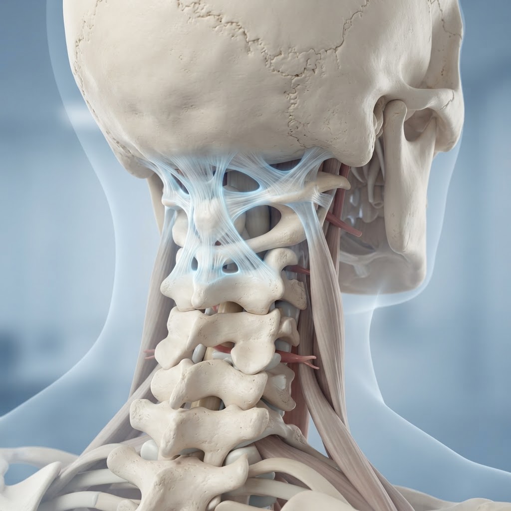
- Craniocervical instability (CCI) occurs when the ligaments supporting the skull and upper spine become too lax to maintain structural integrity.
- While chronic neck pain is a hallmark sign, CCI symptoms often manifest neurologically, affecting vision, balance, and cognitive function.
- There is a well-documented clinical connection between EDS and CCI due to systemic collagen defects.
- Ligament laxity can stem from connective tissue disorders, physical trauma, or inflammatory processes that degrade tissue stability.
- Diagnosing CCI is notoriously complex, often requiring dynamic imaging rather than standard static X-rays or MRIs.
For those living with persistent, unexplained neurological struggles and persistent upper cervical discomfort, the journey toward a diagnosis can feel like a labyrinth. Craniocervical instability, often referred to as CCI, is a condition that exists at the intersection of structural orthopedics and complex neurology. It represents a failure of the body’s primary support system for the head, leading to a cascade of systemic issues that can diminish quality of life. As awareness grows, more individuals are seeking to understand how their structural alignment impacts their total health, searching for answers beyond conventional pain management. In this guide, the healthauthoritylife Editorial Team explores the intricacies of CCI—from the mechanical failures of the cervical spine to the pathways toward natural relief—to empower you with the knowledge needed to advocate for your own well-being.
Understanding Craniocervical Instability: What It Is
Craniocervical instability is a mechanical condition involving the junction where the skull meets the cervical spine, specifically at the C0-C1 (atlanto-occipital) and C1-C2 (atlanto-axial) joints. In a healthy, well-functioning neck, a complex network of ligaments, including the alar and transverse ligaments, acts as a system of cables and anchors. These structures ensure that the heavy mass of the skull remains balanced securely on the delicate spinal column, protecting the brainstem and spinal cord from excessive movement during everyday activities like walking, turning the head, or even swallowing.
When someone experiences CCI, these ligaments become overly loose—a state clinically known as ligament laxity. This loss of tension allows the skull to shift abnormally on the cervical vertebrae. Think of it as a loose hinge on a heavy door; because the hinge is not tight, the door sags and creates friction, misalignment, and stress on the surrounding hardware. In the human body, this “sagging” can place undue pressure on the medulla oblongata, the brainstem, and the delicate cranial nerves that exit the skull base. Because this area is the command center for involuntary bodily functions, the consequences of this instability are rarely localized just to the neck.
Understanding the anatomy is vital to grasping why the condition feels so systemic. The upper cervical spine is unique in its mobility; it is designed to allow for rotation and nodding, but it lacks the bony locking mechanisms found in the lower lumbar spine. Consequently, it relies almost entirely on soft tissue for stability. When these soft tissues lose their inherent tension, the nervous system enters a state of constant high alert, attempting to stabilize the head using muscles that were never intended for such a heavy load. This leads to muscle spasms, myofascial trigger points, and a cycle of secondary pain that can make a localized problem feel like a full-body health crisis.
Many experts emphasize that CCI is not merely a spinal issue but a structural foundation issue. When the skull is not properly supported, the entire vertebral column may attempt to compensate, leading to pelvic tilts, scoliosis, or secondary issues in the thoracic spine. Identifying CCI requires moving beyond standard diagnostic models that focus on bone density or disc herniation and instead looking at how the skull and spine behave during physical movement. Whether the cause is an inherent weakness in collagen or a traumatic event that overstretched the ligaments, the fundamental reality remains the same: the mechanical anchor of the nervous system is no longer functioning as intended.
Common Symptoms of CCI: More Than Just Neck Pain
The clinical presentation of craniocervical instability is notoriously broad, which is why patients often visit a dozen different specialists before finding a name for their experience. While chronic neck pain relief is the primary goal for many, the neurological symptoms of CCI are often what impact day-to-day functionality the most. Because the brainstem is situated directly at the craniocervical junction, any mechanical shift in the skull can compress or irritate these nerve tracts, leading to symptoms that mimic other conditions like dysautonomia, vestibular disorders, or even primary headaches.
One of the most frequent CCI symptoms reported is postural orthostatic tachycardia syndrome (POTS) or other forms of autonomic dysfunction. Patients may notice that their heart rate spikes upon standing, or they may experience cold, clammy extremities and blood pressure instability. This occurs because the autonomic nervous system, which regulates heart rate and digestion, passes through the very area that is being compressed by the shifting skull base. When the “cables” are loose, the messaging system between the brain and the body experiences signal interference.
Vision and balance are also frequently compromised. Many people with CCI report “visual snow,” double vision, or a feeling of being detached from their body, a phenomenon often described as brain fog. This happens because the brainstem acts as a relay station for visual information and proprioception—the body’s ability to sense its position in space. If the brainstem is irritated, the signals from the inner ear and the eyes are processed incorrectly, leading to a persistent sense of dizziness or an inability to focus on moving objects. These symptoms are often worse after periods of physical exertion or prolonged screen time, as the neck muscles fatigue and lose their protective support.
Other common manifestations include:
- Headache patterns: Specifically, occipital neuralgias or tension-type headaches that radiate from the base of the skull toward the eyes.
- Dysphagia: Difficulty swallowing, or a feeling of a “lump in the throat” caused by proximity of the pharyngeal nerves to the unstable vertebrae.
- Tinnitus: Persistent ringing or buzzing in the ears, often linked to the flow of cerebrospinal fluid or nerve irritation near the auditory pathways.
- Sleep disturbance: Many patients find they cannot fall asleep unless their head is positioned in a very specific way, as the neck feels “too heavy” when lying down.
It is important to note that these symptoms are often episodic. They may flare up after a long day or during periods of systemic inflammation and then subside with rest. This fluctuation is one of the most confusing aspects for patients, as it can make them feel like their symptoms are “all in their head.” However, the mechanical reality of the lax ligaments remains, regardless of whether the symptoms are present at any given moment.
The Link Between Ehlers-Danlos Syndrome and CCI
The correlation between EDS and CCI is one of the most discussed topics in modern physical medicine. Ehlers-Danlos Syndrome (EDS) is a group of genetic disorders that affect the body’s connective tissue. Connective tissue is essentially the “glue” that holds the body together, composed primarily of collagen. In individuals with EDS, the collagen is structurally flawed, making the ligaments, tendons, and skin much more stretchy and fragile than average. Because the ligaments in the upper neck are the primary stabilizers of the head, it is easy to see why those with connective tissue disorders are highly susceptible to developing craniocervical instability.
When an individual has EDS and CCI, their body lacks the structural “toughness” to maintain the alignment of the C1 and C2 vertebrae. For the general population, a minor whiplash injury might lead to temporary stiffness; for an individual with EDS, that same injury can cause permanent ligament laxity because the tissue lacks the elasticity to “snap back” to its original length. This condition is often referred to as hypermobility, but when it affects the spine, it becomes a distinct clinical challenge that requires a more nuanced approach than standard physical therapy.
The management of this specific intersection requires caution. Traditional aggressive manual adjustments (often called “cracking” the neck) can be potentially hazardous for someone with EDS and associated instability, as the ligaments are already compromised. Instead, experts generally suggest focusing on isometric stabilization and gentle, targeted strengthening of the deep neck flexors. Understanding that the instability is not a muscular weakness—but a tissue-integrity failure—is the first step toward creating a safer, more sustainable management plan.
| Approach | Focus | Best For |
|---|---|---|
| Isometric Stabilization | Building muscle tension without movement | Acute phase, early strengthening |
| Cervical Collars | External structural support | Severe symptom relief during flares |
| Proprioceptive Training | Retraining nerve-to-muscle pathways | Chronic, long-term alignment support |
| Manual Therapy (Gentle) | Myofascial release and fluid flow | Reducing compensatory muscle spasms |
Living with the combination of these conditions requires a paradigm shift. One must learn to listen to the body’s signals of instability—such as a sudden feeling of heaviness in the head or a brief loss of balance—and respond with rest rather than pushing through. By focusing on maintaining a neutral spine and utilizing supportive aids during high-activity periods, many individuals find that they can successfully manage the symptoms without needing more invasive interventions. The key is consistent, low-impact habits that prioritize the protection of the cervical junction above all else.
Major Causes of Ligament Laxity in the Upper Neck
Ligament laxity, the root cause of craniocervical instability, does not occur in a vacuum. While genetic predisposition like EDS plays a significant role, many cases are acquired through a combination of environmental factors, trauma, and chronic systemic issues. Understanding these neck instability causes is essential for developing a personalized strategy for natural relief. When we look at why ligaments lose their ability to support the head, we must look at both mechanical and biochemical contributors.
Trauma remains a leading cause of acquired CCI. This is not restricted to high-impact injuries like car accidents or contact sports. While those events are obvious triggers for “whiplash-associated disorders,” repetitive micro-trauma can be just as damaging over time. For example, individuals who spend excessive hours in a forward-head posture—often called “text neck”—place constant, abnormal tension on the posterior ligaments of the cervical spine. Over years, this consistent strain can cause the ligaments to stretch and lose their resting tone, effectively creating a state of mechanical instability without a single, dramatic accident.
Another major contributor is chronic systemic inflammation. Ligaments are not static; they are living tissues that require a stable internal environment to maintain their integrity. Conditions such as rheumatoid arthritis, ankylosing spondylitis, or even chronic undiagnosed systemic infections can degrade the quality of collagen within the ligaments. When the body is in a state of systemic inflammation, the enzymes responsible for tissue turnover can become overactive, leading to the breakdown of collagen fibers faster than the body can repair them. This process, often referred to as “ligamentous laxity due to inflammatory insult,” can leave the upper cervical spine vulnerable even in the absence of a direct injury.
Furthermore, lifestyle factors such as nutritional deficiencies can impact how the body repairs its connective tissue. Vitamin C, for instance, is a critical cofactor for collagen synthesis. Similarly, magnesium and collagen-building amino acids are required to maintain the structural health of all soft tissues. A diet high in processed foods and deficient in these micronutrients may impair the body’s ability to recover from minor, daily strains. When you combine this lack of building blocks with the modern sedentary lifestyle, the cervical spine loses its natural resiliency. It is a slow, silent progression, but it is one that can be mitigated through proactive lifestyle adjustments and targeted nutritional support, which remain fundamental components of any natural CCI treatment plan.
Diagnostic Challenges and When to See a Specialist
One of the primary frustrations for those investigating craniocervical instability is the difficulty of achieving a clinical diagnosis. Standard imaging protocols, such as static X-rays, CT scans, and MRIs, are performed while the patient is lying completely still. While these tests are excellent for ruling out tumors, fractures, or disc herniations, they are often insufficient for identifying CCI. Because CCI is a dynamic condition—meaning it occurs when the head moves into certain positions—the instability is frequently invisible on a static scan.
This diagnostic gap is why many patients feel gaslit by the medical system. A technician may perform a standard MRI and conclude that the cervical spine looks “normal,” while the patient is still suffering from severe neurological symptoms. To accurately assess the health of the craniocervical junction, specialists often need to employ dynamic imaging, such as upright or flexion-extension X-rays and MRIs. These allow the clinician to see exactly how the vertebrae shift when the head is tilted or rotated, which can reveal the telltale signs of instability that occur only during movement.
When should you consider seeking a specialist? It is generally advised to pursue a consultation with a neurologist, neurosurgeon, or a physiatrist who specializes in hypermobility or cervical spine disorders if you experience the following:
- Symptoms that worsen significantly when you are upright but improve when you lie down.
- A history of unexplained autonomic symptoms, such as sudden heart rate changes or fainting.
- Persistent “clunking” or “grinding” sensations in the neck, especially during rotational movements.
- Neurological symptoms that do not respond to physical therapy or chiropractic adjustments and instead seem to flare up after such interventions.
- A known diagnosis of a connective tissue disorder like EDS or Marfan syndrome paired with chronic, refractory neck pain.
Seeking out a provider who understands the nuance of CCI is crucial. You want to work with someone who asks about your specific triggers and considers your functional capacity rather than just looking at the shape of your bones on a film. If a specialist seems unfamiliar with the concept of dynamic imaging for cervical stability, it may be time to seek a second opinion. Because the craniocervical junction is so close to the brainstem, it is always best to prioritize practitioners who emphasize non-invasive, conservative management first, using surgical intervention only as a measure of last resort when stability cannot be achieved through other means. Empowerment begins with finding a medical partner who views your symptoms as a legitimate, measurable physiological challenge rather than a mystery to be ignored.
Natural Strategies to Support Neck Stability
When dealing with craniocervical instability, the primary goal is to minimize mechanical stress on the ligaments that connect the skull to the upper cervical spine. Natural management is rarely about “curing” the structural laxity, but rather about creating an environment where your remaining structural integrity is preserved and your neurological system is protected from irritation. A multi-modal approach is often the most successful strategy.
The first natural strategy involves proprioceptive training. Individuals with CCI often experience a disconnect between their brain and the positioning of their head in space. This “proprioceptive deficit” can lead to erratic muscle movements or bracing patterns that further strain the cervical ligaments. Working with specialized physical therapists on gentle cervical proprioception—such as learning to hold the head in a neutral “stacked” position—can reduce the need for the muscles to over-contract in a futile attempt to compensate for ligamentous laxity.
Thermal therapy, when applied correctly, can be a potent tool. While systemic inflammation is a hallmark of EDS and related collagen disorders, targeted thermal application can help manage the secondary muscular guarding that occurs when the neck becomes unstable. Many find that cold therapy (ice packs) is superior for acute flare-ups where inflammation is driving the pain, while moist heat is better for chronic, low-grade muscle tension. However, it is vital to avoid aggressive cervical traction or heavy heat packs that can cause the neck muscles to become too relaxed, potentially leading to further instability.
Additionally, the use of targeted cervical orthotics or soft collars—under the supervision of a healthcare provider—can be a natural intervention to provide temporary external support. Rather than relying on them 24/7, which can lead to muscle atrophy, using them during high-exertion activities or long periods of sitting can allow the damaged ligaments to recover by preventing end-range shearing movements.
The Role of Posture and Ergonomics in Managing CCI
For someone with CCI, the “text neck” phenomenon is not just a nuisance; it is a mechanical threat. Every inch that the head migrates forward from its neutral alignment increases the load on the cervical spine by several pounds. When your ligaments are already lax, this increased weight creates a “hinge” effect at the craniocervical junction, leading to the hallmark CCI symptoms like dizziness, vision disturbances, and headaches.
Optimizing your environment is essential. Your monitor should be positioned so that the top third of the screen is at eye level. This prevents the tendency to tilt the chin downward or extend the neck forward. If you use a laptop, a separate keyboard and a laptop stand are non-negotiable. Furthermore, your chair should provide robust lumbar support. When the lower back loses its natural curve (slumping), the head naturally follows, pushing the cervical spine into a vulnerable position.
Sleeping posture is another critical factor often overlooked. Those with neck instability often find that sleeping with excessive pillows forces the neck into a flexed position all night long. A cervical pillow designed to support the neck’s natural lordosis, or even a rolled-up towel placed specifically under the neck, can keep the cervical spine neutral while sleeping on your back. Side sleeping requires a pillow thick enough to keep the neck aligned with the spine, preventing a lateral tilt that could irritate the C1-C2 joint.
Safe Exercises for Strengthening the Neck Muscles
Strengthening for CCI is a process of nuance, not intensity. Traditional “neck curls” or heavy resistance training can be counterproductive, potentially causing a traumatic subluxation if the ligaments are severely compromised. Instead, the focus must be on deep cervical flexors and scapular stabilization. The deep neck flexors—the muscles located in the front of the throat—are responsible for stabilizing the head. When these muscles are weak, the larger, more superficial neck muscles (the trapezius and sternocleidomastoid) take over, leading to chronic tightness and pain.
The “chin tuck” is the gold standard exercise, but it must be performed correctly. While lying flat on your back, gently draw your chin inward as if making a double chin, while keeping the back of your head in contact with the surface. The goal is not to lift the head, but to shorten the muscles in the front of the neck. Hold for a few seconds and release. This isometric contraction strengthens the stabilizers without putting the cervical spine through an unsafe range of motion.
Scapular retraction is equally important. Your shoulder blades act as the foundation for your neck. If your shoulders are rounded forward, your neck is structurally disadvantaged. Exercises that focus on pulling the shoulder blades together and downward—like rows with light resistance bands—can unload the cervical spine by improving overall thoracic posture.
| Exercise Type | Primary Benefit | Best For |
|---|---|---|
| Isometric Chin Tucks | Stabilizes deep neck flexors | Beginners/Acute symptoms |
| Scapular Retraction | Unloads tension from the upper traps | Improving thoracic alignment |
| Eye-Movement Training | Improves vestibular-cervical control | Reducing dizziness/vertigo |
| Seated Posture Correction | Prevents end-range cervical shear | Long-term daily stability |
Nutritional Support for Ligament and Collagen Health
Ligament laxity is often exacerbated by systemic nutritional deficiencies that impair collagen synthesis. While diet cannot “tighten” a loose ligament, providing the body with the necessary building blocks is a fundamental step in supporting the connective tissue you have left. Collagen is the most abundant protein in the body, and it relies heavily on specific amino acids and vitamins for its structural integrity.
Vitamin C is perhaps the most critical nutrient for collagen cross-linking. Without sufficient Vitamin C, the body cannot effectively bind collagen fibers together, which may contribute to the fragility seen in those with hypermobility spectrum disorders. Increasing intake of citrus fruits, bell peppers, and strawberries is a simple, natural way to support tissue health.
Additionally, bone broth has become a staple for those with connective tissue issues because it contains naturally occurring collagen, proline, and glycine. These amino acids are the primary components of ligaments and tendons. Some experts also suggest that maintaining adequate hydration is vital; ligaments are largely composed of water and proteins, and dehydration can lead to increased stiffness and decreased elasticity, making the tissue more prone to micro-tears.
Magnesium supplementation is also frequently discussed, as it plays a role in muscle relaxation and the prevention of the muscle spasms that often plague CCI patients. A magnesium-rich diet or transdermal magnesium (via Epsom salt baths) can help calm the nervous system and reduce the hyper-tonicity of the neck musculature, creating a more stable foundation for the cervical spine.
Lifestyle Adjustments to Prevent Further Neck Strain
Managing CCI requires a permanent shift in how you interact with your environment. High-impact activities are generally contraindicated for those with documented neck instability. Activities like contact sports, heavy lifting, or even activities that involve repetitive jarring, such as running on hard surfaces, can create significant shear forces on the cervical vertebrae.
Learning to move your body as a single unit is a crucial skill. For example, if you need to turn to look at something behind you, don’t just rotate your neck. Turn your entire torso. This “en-bloc” turning minimizes the amount of rotation required at the atlantoaxial joint, protecting the vulnerable ligaments from twisting forces.
Furthermore, managing the digital load is essential. Using voice-to-text software for long emails or documents can significantly reduce the amount of time you spend looking down at a screen. If you must use a phone, bring the device up to eye level rather than lowering your head to the device. These small, incremental changes to your daily habits can prevent the accumulation of micro-trauma that leads to chronic neck pain relief being so elusive.
Frequently Asked Questions
Can physical therapy cure craniocervical instability?
Physical therapy is generally not considered a “cure” for structural ligamentous laxity, especially in cases related to conditions like EDS. However, it is an essential management tool. By strengthening the deep stabilizers of the neck, physical therapy can often reduce the severity of symptoms and prevent further degradation of the cervical alignment, allowing many individuals to improve their quality of life significantly.
What are the first signs that I might have CCI?
Early CCI symptoms often include a heavy feeling in the head, frequent “cracking” or popping sensations in the neck, persistent headaches that start at the base of the skull, and sudden episodes of dizziness or lightheadedness when changing positions. Many people also report a feeling that they need to physically support their head with their hand when sitting for long periods.
Is surgery the only option for someone with CCI?
Surgery, such as a cervical fusion, is typically considered a last resort for severe cases where neurological damage is imminent or quality of life is profoundly impacted despite comprehensive conservative management. Most experts recommend exhausting all natural, non-invasive, and regenerative medicine options before considering surgical intervention, as cervical fusion carries risks and long-term mechanical implications.
How does Ehlers-Danlos Syndrome (EDS) affect my neck?
EDS is a group of genetic disorders that affect the connective tissue. Because the ligaments in the body are largely composed of collagen, those with EDS have ligaments that are more prone to stretching or tearing. In the cervical spine, this systemic ligament laxity means the ligaments holding the skull to the spine are often too loose to keep the vertebrae in proper alignment, which is why CCI is common in the EDS community.
Are there specific diagnostic tests for CCI?
Diagnosing CCI can be difficult because traditional static imaging, like a standard MRI or X-ray while lying down, often fails to show the instability. Doctors typically look for dynamic imaging, such as an Upright MRI or Digital Motion X-ray (DMX), which captures the cervical spine while the head is in motion or under gravity. These tests are far better at identifying the degree of “slide” or instability between the vertebrae.
Can I lead a normal life with craniocervical instability?
Yes, many people with CCI lead fulfilling lives by making intentional lifestyle adjustments. While it may require giving up high-impact activities and prioritizing ergonomic health, learning how to stabilize the neck through exercise and supportive habits allows many individuals to manage their symptoms successfully and maintain a high level of daily function.
Conclusion
Craniocervical instability is a complex and often misunderstood condition, but it is one that can be managed with the right combination of patience, lifestyle discipline, and informed guidance. By understanding the mechanical causes of your neck instability—whether related to collagen disorders like EDS or simple wear and tear—you can move from a state of reactive pain management to a proactive strategy of stabilization and protection. Remember that your cervical spine is a delicate structure, and the small, consistent efforts you make today—through posture, targeted exercise, and nutritional support—are the best insurance against further decline. Take charge of your movement, listen to your body’s signals, and work closely with healthcare providers who understand the nuances of the craniocervical junction.
If you suspect you are struggling with neck instability, start by tracking your symptoms and discussing them with a specialist who understands cervical biomechanics. You do not have to settle for chronic discomfort; informed action is your greatest tool for relief.
By healthauthoritylife Editorial Team
This article is for informational purposes only and is not a substitute for professional medical advice. Always consult your doctor before making changes to your diet, exercise routine, or treatment plan.
Leave a Reply