
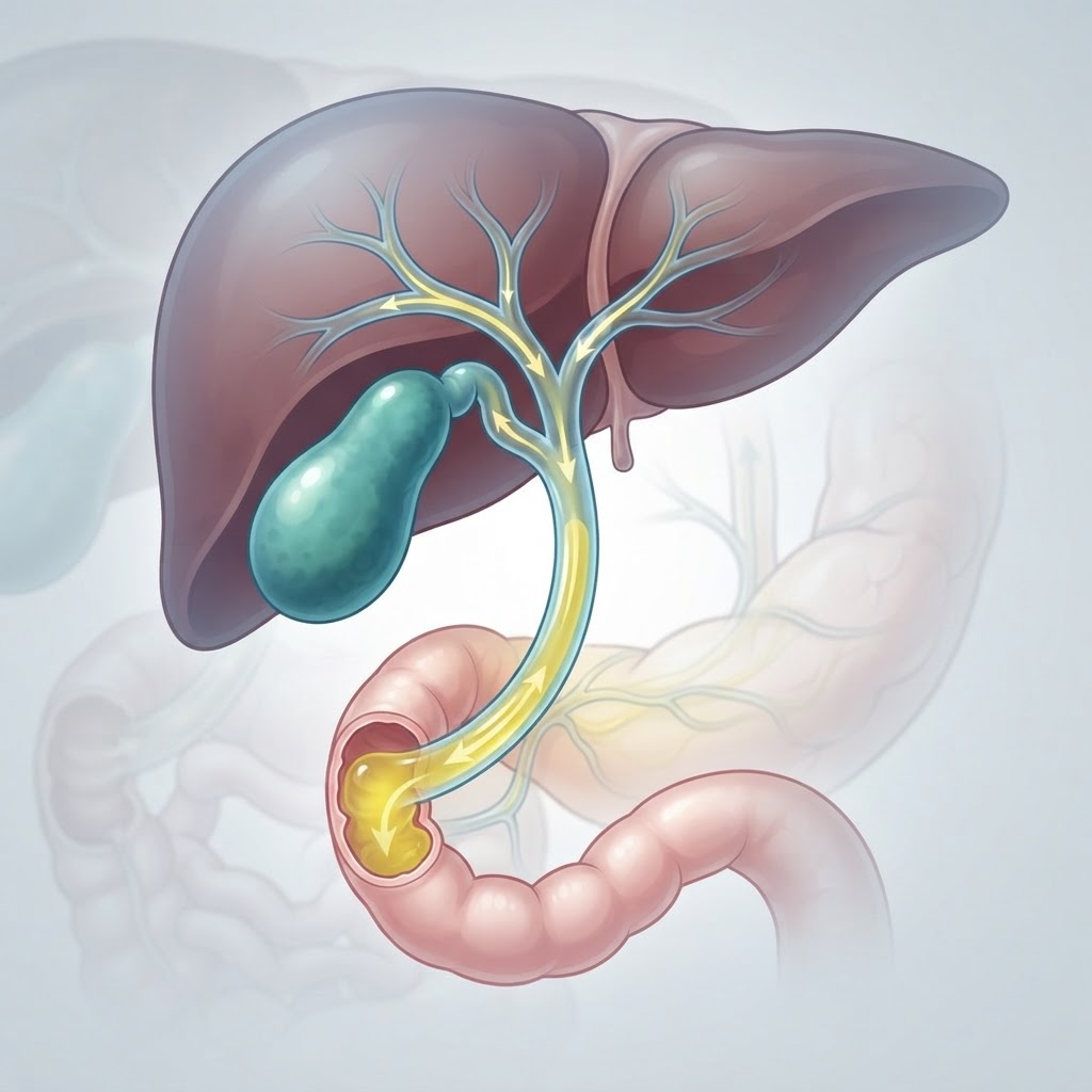
- Gallbladder dyskinesia is a functional disorder where the gallbladder fails to empty bile effectively, despite the absence of gallstones.
- Key biliary dyskinesia symptoms often mimic traditional gallbladder disease, including upper right quadrant pain and nausea after meals.
- The gallbladder ejection fraction is the primary diagnostic metric used to determine how well the organ contracts during a HIDA scan.
- Common biliary pain causes include chronic inflammation, sphincter of Oddi dysfunction, and metabolic imbalances that disrupt bile flow.
- Diagnosis is complex and requires ruling out other digestive pathologies, often leading to a multidisciplinary approach to symptom management.
When digestive distress strikes, it is easy to assume the culprit is simply a heavy meal or a temporary bout of indigestion. However, for many individuals experiencing chronic, stabbing pain in the upper abdomen, the root cause is far more persistent. Gallbladder dyskinesia represents a silent, functional challenge that frequently eludes traditional diagnostic imaging like standard ultrasounds. Because this condition involves a gallbladder that is structurally “normal”—lacking the tell-tale presence of stones—it is often misunderstood, misdiagnosed, or dismissed. Understanding the nuances of this biliary disorder is the first step toward reclaiming digestive comfort and addressing the underlying mechanics of your internal health.
What Is Gallbladder Dyskinesia?
To understand what is gallbladder dyskinesia, one must first view the gallbladder as more than just a storage pouch. It is a dynamic, muscular organ that plays a critical role in the digestive system. Located beneath the liver, its primary job is to store and concentrate bile—a fluid produced by the liver that is essential for emulsifying fats and aiding in the absorption of fat-soluble vitamins. Under healthy conditions, when we consume a meal containing fat, the brain signals the gallbladder to contract, pushing the bile through the cystic duct and into the small intestine to facilitate digestion.
Gallbladder dyskinesia occurs when this precise muscular coordination fails. In the medical community, “dyskinesia” refers to abnormal or impaired movement. In this context, the gallbladder becomes sluggish or fails to contract with sufficient force to empty its contents effectively. Unlike cholelithiasis, where hard stones physically block the flow of bile, this condition is purely functional. The plumbing is technically clear, but the “pump” is not working as it should.
Because the gallbladder is failing to empty properly, the bile can become stagnant or excessively concentrated. This stagnation can create a feedback loop of irritation within the organ walls. Over time, this chronic lack of efficiency can lead to the very biliary pain causes that plague patients daily. Many experts classify this as a motility disorder, similar in nature to functional dyspepsia or irritable bowel syndrome, but localized specifically to the biliary tree.
The complexity of this condition lies in its invisibility. If a patient visits an urgent care clinic with abdominal pain, an initial ultrasound will likely show a stone-free gallbladder, leading clinicians to clear the patient of typical gallbladder disease. This leaves the patient in a state of diagnostic limbo. Understanding that this is a disorder of function rather than structure is vital for patients seeking answers. It confirms that the symptoms are not “all in their head,” but rather the result of a physiological system struggling to perform its basic duty. Recognizing the existence of this functional impairment is the pivot point where many patients finally move from recurring frustration to finding viable relief strategies, whether through lifestyle modification, medical intervention, or targeted nutritional support.
Common Symptoms of Biliary Dyskinesia
The hallmark of biliary dyskinesia symptoms is their predictability—or, more accurately, their correlation with the intake of dietary fats. Because the gallbladder is stimulated to empty specifically when fats enter the duodenum, patients often report that their discomfort reaches a peak shortly after consuming high-fat, fried, or rich foods. This pain is not random; it is a physiological response to a demand the gallbladder is unable to meet.
The most frequently reported sensation is a sharp, aching, or cramping pain located in the right upper quadrant (RUQ) of the abdomen, just below the rib cage. However, biliary pain is notorious for being referred. It is common for this sensation to radiate to the back, particularly near the right shoulder blade or between the shoulder blades. Patients often describe a feeling of fullness, bloating, or a “heavy” sensation in the upper abdomen that can linger for several hours.
Beyond localized pain, nausea is a frequent companion to biliary dyskinesia. This nausea may strike immediately after eating or persist as a low-level background sensation throughout the day. Some individuals experience significant digestive fragility, where even small amounts of fat or certain fibrous vegetables trigger a flare-up. Burping, gas, and a general feeling of intolerance to heavy meals are also staples of the clinical picture.
It is important to note that these symptoms can vary significantly in intensity. Some people describe their episodes as “attacks” that are debilitating and require bed rest, while others live with a constant, nagging discomfort that impacts their quality of life over months or years. Because these symptoms overlap heavily with other conditions—such as gastritis, acid reflux, or even food sensitivities—keeping a detailed food and symptom diary is one of the most effective tools for patients. Noting exactly what was eaten, the time delay before symptoms appeared, and the nature of the pain can provide a healthcare provider with the necessary clues to suspect a gallbladder motility issue rather than a standard gastric problem.
Furthermore, because the bile is not entering the intestines effectively, some individuals may experience subtle changes in stool color or consistency, reflecting the lack of adequate bile for proper lipid digestion. While this is less commonly the primary reason for a clinic visit, it remains a clinical marker of the body’s struggle to process fats. Ultimately, recognizing the pattern of these biliary dyskinesia symptoms is essential for moving toward an accurate diagnosis and avoiding years of unnecessary restrictive dieting or ineffective gastrointestinal treatments.
Understanding the Role of the Gallbladder Ejection Fraction
The diagnostic gold standard for identifying gallbladder dyskinesia is the HIDA scan, also known as a cholescintigraphy. This scan is unique because it assesses the function—not just the anatomy—of the organ. During the procedure, a radioactive tracer is injected, which travels to the liver and is subsequently excreted into the bile. A gamma camera tracks the flow of this tracer into the gallbladder and eventually into the small intestine.
Once the gallbladder is filled with the tracer, a hormone called cholecystokinin (CCK) is typically administered. CCK acts as a natural signal to the gallbladder to contract and push the tracer out. The crucial metric measured during this phase is the gallbladder ejection fraction (GBEF). The GBEF is essentially the percentage of bile (represented by the tracer) that the gallbladder successfully ejects upon stimulation.
While clinical standards can vary between institutions, a low GBEF is generally considered the primary marker for biliary dyskinesia. A common threshold for dysfunction is often cited as anything below 35% to 40%, though clinicians must interpret this in the context of the patient’s actual symptoms. A low ejection fraction indicates that the muscular walls of the gallbladder are too weak, or perhaps too stiff, to respond appropriately to the signal to evacuate.
However, it is crucial to understand that the GBEF is not a perfect diagnostic indicator. There are cases where a patient may have a “low” ejection fraction but remain entirely asymptomatic, or conversely, a patient with a “normal” ejection fraction may still experience severe biliary-like pain. This highlights that biliary motility is a complex interplay of hormones, nervous system signaling, and structural integrity. The GBEF should be viewed as one piece of a larger diagnostic puzzle rather than an absolute rule.
Patients should also be aware that the GBEK test is essentially a “stress test” for the gallbladder. By using a standardized agent to trigger contraction, doctors can artificially recreate the conditions that cause the patient’s pain. If the patient reports their characteristic abdominal pain during the GBEF measurement phase, it is a very strong clinical indicator that the gallbladder is indeed the source of the distress. This functional testing is the definitive way to bridge the gap between “I feel pain” and “We know why.” It moves the conversation away from speculation and toward a targeted therapeutic plan, whether that involves conservative management or surgical consultation.
| Diagnostic Approach | Focus | Best For |
|---|---|---|
| Standard Ultrasound | Visual Anatomy | Detecting physical stones or wall thickening. |
| HIDA Scan with GBEF | Gallbladder Function | Confirming dyskinesia and motility issues. |
| Endoscopic Ultrasound | Detailed Imaging | Finding “sludge” or micro-stones that regular imaging misses. |
| Symptom Tracking | Patient History | Identifying specific dietary triggers for biliary pain. |
Primary Causes and Triggers for Gallbladder Dysfunction
Defining the specific biliary pain causes is an area of ongoing medical research, as “dyskinesia” is often a label for a wide spectrum of functional issues. Unlike gallstone disease, which has clear risk factors like obesity or rapid weight loss, the causes of gallbladder dyskinesia can be more elusive. Experts generally agree that a combination of factors—ranging from neurological imbalances to hormonal shifts—often plays a role in preventing the gallbladder from contracting correctly.
One major contributor is chronic inflammation of the gallbladder wall, known as cholecystitis. Even without visible stones, long-term irritation from “biliary sludge”—a thick, mucus-like suspension of cholesterol crystals—can weaken the muscular wall of the gallbladder. Over time, this chronic low-grade inflammation alters the gallbladder’s ability to respond to hormonal cues. The nerve cells within the wall of the gallbladder, which are responsible for triggering contraction, may also become desensitized, essentially “tuning out” the signals from the digestive tract.
Another significant factor is Sphincter of Oddi dysfunction. The sphincter is a small muscular valve that controls the flow of bile from the common bile duct into the small intestine. If this valve does not open fully or relaxes at the wrong time, it creates a “backup” effect. This back-pressure can hinder the gallbladder’s ability to empty effectively, leading to that characteristic feeling of fullness and pain. Because the gallbladder and the sphincter work in a coordinated, rhythmic dance, any lack of synchronization between them can result in clinical symptoms of dyskinesia.
Metabolic and hormonal factors also cannot be overlooked. High levels of circulating estrogen, for example, have been linked to changes in bile composition, making it thicker and more difficult to move. Pregnancy, hormone replacement therapy, or fluctuations in gut health can all influence the delicate chemistry of bile. Furthermore, the gut-brain axis plays a substantial role. We know that stress and nervous system dysregulation can slow down motility throughout the entire digestive tract; it is highly probable that similar mechanisms apply to the biliary tree, where sympathetic nervous system dominance (the “fight or flight” response) can dampen the normal digestive movements that should be governed by the parasympathetic system.
Finally, dietary habits are the most common trigger. A diet chronically lacking in healthy fats can lead to “lazy” gallbladder syndrome. Because the gallbladder is stimulated to empty primarily by fat, a persistent low-fat diet means the organ is rarely called upon to exercise its muscles. While this might seem protective, it can actually lead to the accumulation of stagnant bile, increasing the risk of sludge formation and eventual dyskinesia. Finding the balance—providing enough stimulation for the gallbladder to work, but avoiding excess fat that overwhelms a sensitive system—is the key to managing the triggers of biliary pain.
How Doctors Diagnose Gallbladder Dyskinesia
Diagnosing gallbladder dyskinesia is a process of elimination. Because symptoms overlap with everything from peptic ulcers to pancreatic issues and IBS, physicians must work systematically to rule out other pathologies before landing on a diagnosis of functional biliary disease. The process typically begins with a thorough clinical history. A provider will ask about the timing of pain relative to meals, the nature of the pain, and whether the patient has experienced associated symptoms like jaundice, fever, or weight loss, which would point toward more serious or infectious concerns.
Physical examination is the next step, though in cases of dyskinesia, it is frequently unremarkable. The physician will check for “Murphy’s sign”—a technique where they press on the right upper quadrant and ask the patient to inhale deeply. If the patient stops their inhalation due to sudden pain, it suggests inflammation of the gallbladder. However, many people with dyskinesia do not have a positive Murphy’s sign, which again highlights the functional rather than acute inflammatory nature of the condition.
Laboratory testing, including blood work, is used to rule out infection, liver enzyme abnormalities, or pancreatic inflammation. Elevated liver enzymes or bilirubin would typically suggest that bile is physically obstructed by a stone or other mass, rather than simply moving slowly due to poor motility. In the case of pure dyskinesia, blood results are usually, frustratingly, normal. This normal lab profile is often a source of relief for the patient but also a source of confusion, as it confirms there is no “emergency” while simultaneously failing to explain the intense pain.
Imaging follows the lab work. A transabdominal ultrasound is the standard first-line diagnostic tool. It is excellent at detecting stones, polyps, or sludge. If the ultrasound is clear but symptoms persist, the physician will likely order a HIDA scan with cholecystokinin (CCK) stimulation. This remains the most valuable test for diagnosing the functional capacity of the gallbladder. By observing how much bile is ejected during the scan, doctors can calculate the gallbladder ejection fraction. A finding below the established institutional baseline, coupled with the reproduction of the patient’s familiar pain during the scan, is the strongest indicator of a diagnosis.
It is important to emphasize that this is a diagnosis of exclusion. Doctors will often perform an endoscopy (EGD) to ensure the patient does not have an ulcer or gastritis, as these can mimic gallbladder pain perfectly. In some cases, a doctor might suggest a trial of medication or dietary changes to see if symptoms improve. If the diagnosis of gallbladder dyskinesia is confirmed, the conversation then shifts toward management strategies. This is a critical juncture where the focus moves from “What is wrong?” to “How do we make life manageable?” Whether that path involves exploring natural biliary support or, in some cases, surgical consultation, the process of diagnosis provides the roadmap for moving forward.
Lifestyle Adjustments for Managing Biliary Pain
Managing the discomfort associated with gallbladder dyskinesia often requires a holistic approach that extends beyond the kitchen. Because the gallbladder is a dynamic organ that responds to both hormonal shifts and physical movement, lifestyle modifications can play a pivotal role in reducing the frequency and intensity of biliary pain. One of the most effective strategies involves stress management. The gallbladder is intricately linked to the autonomic nervous system. When the body enters a state of chronic stress, the “fight or flight” response can inhibit proper digestive motility, including the contraction and relaxation cycles of the gallbladder. Incorporating daily mindfulness practices, such as deep-breathing exercises, meditation, or restorative yoga, can help keep the nervous system in a parasympathetic state, which is essential for optimal digestion.
Physical activity is another cornerstone of biliary health. Moderate, consistent movement, such as walking or low-impact aerobics, helps stimulate overall gastrointestinal transit. When the body is sedentary, digestion can become sluggish, potentially leading to bile stagnation. However, it is vital to balance activity levels. Overexertion can sometimes trigger physical stress that worsens digestive symptoms. Aiming for gentle, rhythmic movement after meals can be particularly beneficial. Rather than engaging in high-intensity interval training immediately after eating, consider a light stroll. This gentle motion assists the gut in moving food through the system and encourages the gallbladder to empty bile in a regulated, rhythmic manner.
Furthermore, maintaining a healthy weight through balanced lifestyle habits is critical. Rapid weight loss, often triggered by “crash diets,” is a known stressor for the gallbladder and can lead to the formation of biliary sludge or stones, complicating existing dyskinesia. If weight management is a goal, it should be approached with a sustainable, slow-and-steady mindset. Finally, prioritizing sleep hygiene is often overlooked but remains vital. Poor sleep quality is associated with systemic inflammation, which can affect the smooth muscle function of the biliary tract. Creating a consistent bedtime routine ensures that the body has the necessary recovery time to maintain digestive homeostasis.
Dietary Changes to Reduce Gallbladder Stress
When gallbladder dyskinesia is present, the organ struggles to eject bile efficiently in response to high-fat stimuli. Therefore, the primary objective of a dietary shift is to reduce the “biliary load”—the total amount of work the gallbladder must perform per meal. Rather than eliminating all fats, which are essential for nutrient absorption, the strategy should focus on the quality and distribution of fat intake.
Fractional Feeding: Instead of consuming two or three large, heavy meals, transition to five or six smaller, nutrient-dense meals throughout the day. Smaller volumes of food are significantly easier for the biliary system to manage, as they require less intense bile secretion cycles. This approach prevents the gallbladder from being “overwhelmed” by a massive influx of fatty content at once.
Fat Quality Matters: Not all fats are created equal regarding gallbladder stress. Highly processed oils, trans fats, and saturated animal fats can be particularly difficult to digest. Focus instead on anti-inflammatory, easily digestible fats such as those found in avocados, extra virgin olive oil, and raw nuts (in moderation). These fats are less likely to trigger the intense gallbladder contractions that cause pain compared to deep-fried foods or fatty cuts of red meat.
Focus on Fiber: Soluble fiber, found in oats, beans, lentils, and certain fruits like apples, helps regulate digestive speed and binds to excess bile acids in the gut, which can ease the burden on the entire digestive system. A diet rich in whole, unprocessed plant foods provides the structural support needed for a smooth digestive process.
| Dietary Modification | Mechanism of Relief | Best For |
|---|---|---|
| Smaller, Frequent Meals | Prevents biliary over-contraction | Acute symptom flare-ups |
| Plant-Based Fats | Lowers inflammatory gallbladder load | Long-term bile maintenance |
| High Soluble Fiber | Assists in bile acid sequestration | Preventing biliary sludge |
| Warm Herbal Teas | Promotes smooth muscle relaxation | Immediate post-meal discomfort |
Natural Supplements That May Support Biliary Function
While diet is the primary foundation, specific natural supplements may offer additional support for those navigating biliary challenges. The goal of these supplements is generally twofold: to improve bile fluidity and to provide gentle support for the liver and gallbladder tissues. It is essential to consult with a healthcare professional before adding these to your regimen, as individual responses vary.
Ox Bile: For some individuals, the issue is not just the speed of the gallbladder, but the production or concentration of bile itself. Supplemental ox bile can provide temporary relief by assisting with the breakdown of fats during a meal, effectively “taking the load off” the gallbladder until it can be managed through other means. This is often used during a transition period while dietary habits are being refined.
Digestive Enzymes: Broad-spectrum digestive enzymes, specifically those containing lipase, can assist the pancreas and gallbladder in breaking down lipids. By aiding the chemical breakdown of fats before they reach the duodenum, enzymes can reduce the “emergency signal” that triggers the gallbladder to contract forcefully, potentially reducing pain.
Bitter Herbs: Traditional wellness practices have long utilized “bitters” (such as gentian root, dandelion, or milk thistle) to support digestive health. These substances stimulate the production of digestive juices, including bile. Milk thistle, in particular, is frequently cited for its role in supporting liver function, which is the precursor to healthy bile production. By maintaining a healthy liver, you ensure that the bile entering the gallbladder is of optimal consistency.
Magnesium: Magnesium is a well-known muscle relaxant. Because the biliary tract relies on smooth muscle cells to regulate flow, ensuring adequate magnesium intake may help prevent excessive spasms in the bile ducts. Many individuals find that a magnesium glycinate supplement helps support a relaxed state, which can be beneficial for managing chronic, dull biliary pain.
When Surgery Is Considered for Chronic Dyskinesia
In cases where lifestyle modifications, dietary changes, and supportive therapies fail to provide adequate relief, or if symptoms progress to the point of significantly hindering daily life, medical professionals may discuss surgical intervention. The standard procedure for chronic gallbladder dysfunction is a cholecystectomy, or the surgical removal of the gallbladder.
Surgery is typically considered when there is documented evidence of a low ejection fraction during a HIDA scan, coupled with persistent, classic biliary symptoms. It is rarely the first line of defense; most experts prefer to attempt conservative management for several months to see if the gallbladder can return to a functional state. However, if a patient is experiencing repeated episodes of biliary colic, has developed complications such as inflammation or infection of the bile ducts, or if the pain is severe enough to cause weight loss due to fear of eating, surgery becomes a viable pathway.
It is important to understand the implications of life without a gallbladder. While the body is quite resilient and most people adapt well, the absence of the “storage tank” means that bile drips continuously into the small intestine rather than being released in concentrated bursts. This can necessitate long-term dietary adaptations, as the body may struggle to process large amounts of fat in a single sitting. The decision for surgery is a balance between the risk of continuing chronic pain and the adjustment period required post-procedure.
Long-Term Outlook for Digestive Health and Wellness
The long-term prognosis for individuals diagnosed with gallbladder dyskinesia is generally positive, provided that proactive measures are taken. Even for those who undergo surgery, the focus remains on supporting the liver’s production of bile and maintaining a healthy gut microbiome. Digestion is a highly adaptable process. By shifting toward an anti-inflammatory, whole-foods-based diet and managing internal stress, most people find that they can return to a high quality of life.
Long-term wellness involves viewing the gallbladder not in isolation, but as part of an integrated digestive system. Ongoing communication with healthcare providers ensures that any new symptoms are addressed immediately, preventing minor issues from becoming chronic conditions. Whether through dietary vigilance or surgical recovery, the journey toward digestive balance is a process of learning the body’s specific requirements and honoring those needs with consistency and care.
Frequently Asked Questions
Can gallbladder dyskinesia heal on its own?
Some individuals may find that their symptoms subside with significant lifestyle and dietary interventions that reduce the workload on the gallbladder. While the organ’s structure may not change, its functional performance can often be stabilized, leading to long periods without pain or discomfort.
Is a HIDA scan the only way to diagnose this condition?
While the HIDA scan with CCK stimulation is the gold standard for assessing gallbladder ejection fraction, it is not the only diagnostic tool. Doctors typically use ultrasounds to rule out gallstones first. The diagnosis is a clinical one, relying on a combination of specific symptoms, imaging results, and the exclusion of other digestive disorders.
What does a biliary attack feel like?
Biliary pain typically presents as a dull, steady ache or a sharp, cramping sensation in the upper right quadrant of the abdomen, just below the rib cage. The pain may radiate to the right shoulder or the area between the shoulder blades and often intensifies following a fatty meal.
Should I avoid all fat if I have dyskinesia?
No, avoiding all fat is generally not recommended. Your body requires healthy fats to absorb essential fat-soluble vitamins like A, D, E, and K. Instead of total elimination, the focus should be on small, frequent amounts of high-quality fats that are easier for the digestive system to process.
Are there specific exercises that hurt the gallbladder?
High-impact activities or those that involve intense abdominal twisting may exacerbate pain during a flare-up. If you are experiencing active discomfort, it is best to stick to gentle, low-impact movements like walking or restorative yoga until the inflammation subsides.
How long after a meal do symptoms usually occur?
Symptoms typically manifest within 30 minutes to two hours after eating. This delay corresponds to the time it takes for food to reach the small intestine, which triggers the hormonal signal for the gallbladder to contract and release bile.
Conclusion
Gallbladder dyskinesia is a complex and often misunderstood condition that requires a personalized approach to management. By understanding the functional nature of the gallbladder—its role as a bile reservoir and the importance of its rhythmic contraction—you can take actionable steps to reclaim your digestive health. From adjusting your dietary habits to incorporating mindfulness and targeted supplementation, there are many tools available to help mitigate biliary pain. Remember, patience is essential in this journey; the digestive system often responds slowly to change. By focusing on consistency and working closely with your healthcare providers, you can manage your symptoms effectively and foster long-term wellness. If you are experiencing persistent abdominal pain, do not wait—seek professional guidance today to create a strategy tailored to your unique health needs.
By healthauthoritylife Editorial Team
This article is for informational purposes only and is not a substitute for professional medical advice. Always consult your doctor before making changes to your diet, exercise routine, or treatment plan.
Leave a Reply