
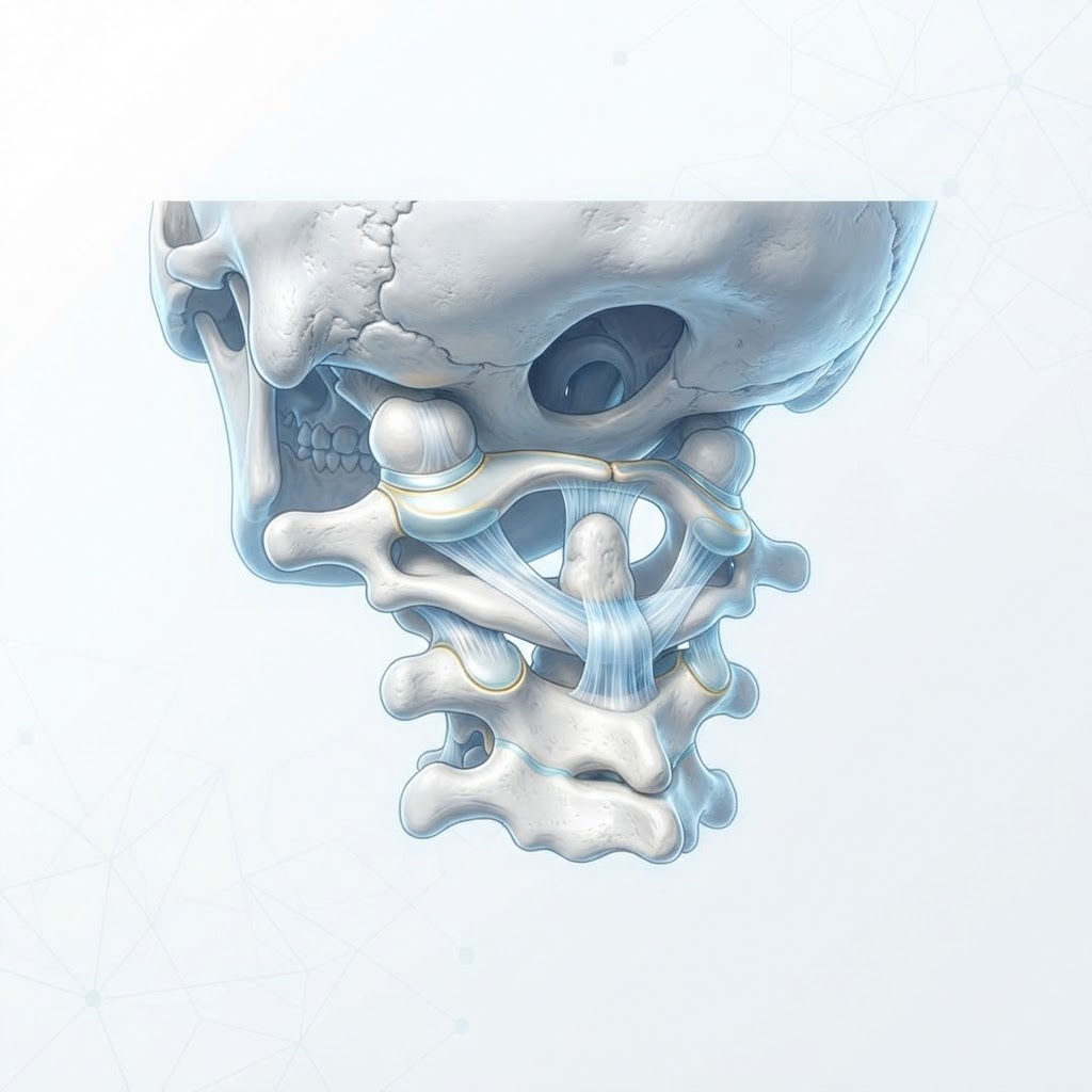
- Craniocervical instability (CCI) occurs when the ligaments connecting the skull to the spine become lax, leading to excessive motion and potential neurological compression.
- CCI symptoms are often diverse and systemic, frequently masquerading as other conditions like fibromyalgia, dysautonomia, or chronic fatigue syndrome.
- Ligamentous laxity, often linked to connective tissue disorders, is a primary driver of instability in the upper cervical spine.
- Nervous system disruption is a hallmark of the condition, as the brainstem and spinal cord are highly sensitive to even minor structural misalignments.
- While surgical intervention is sometimes necessary for severe cases, many individuals explore natural management for neck instability to improve stabilization and quality of life.
For those navigating the complex landscape of chronic pain, unexplained neurological glitches, and persistent headaches, the term craniocervical instability (CCI) is increasingly appearing in clinical conversations. While once considered a niche structural issue, a growing body of anecdotal evidence and clinical focus suggests that many individuals suffer from upper cervical structural weaknesses without ever receiving a formal diagnosis. At its core, CCI represents a fundamental failure of the body’s “anchor point”—the junction where the skull meets the spinal column. When this area loses its structural integrity, the consequences ripple throughout the entire central nervous system, creating a challenging diagnostic puzzle for both patients and healthcare providers. By understanding the mechanical, neurological, and connective tissue components of this condition, we can better identify the path toward stability and relief.
What is Craniocervical Instability?
Craniocervical instability is a clinical condition characterized by excessive movement or “laxity” between the skull (the cranium) and the top two vertebrae of the neck, known as the atlas (C1) and the axis (C2). Under normal circumstances, a complex network of ligaments—specifically the alar, transverse, and apical ligaments—functions as a sophisticated tethering system. These ligaments are responsible for holding the head in a stable position while allowing for the wide range of motion the neck requires. In a state of health, this system ensures that the skull remains perfectly aligned with the spinal cord, protecting the brainstem and the vital nerves passing through the foramen magnum, which is the large opening at the base of the skull.
When these ligaments become overstretched, torn, or developmentally loose, the “anchor” fails. The result is a mechanical instability that allows the skull to shift abnormally on the cervical spine. This is not merely a localized neck issue; it is a structural failure that creates a cascade of physiological stresses. Because the craniocervical junction is the highest point of the spine, any misalignment here acts like a tilt at the base of a tower—even a small shift at the bottom translates into significant, amplified motion at the top. This instability can cause the vertebrae to move into positions that put mechanical pressure on the spinal cord, nerve roots, and the dura mater, the protective sheath surrounding the brain and spinal cord.
Experts generally agree that CCI is a spectrum disorder. Some individuals experience minor, sub-clinical laxity that only becomes symptomatic during physical exertion or certain head positions, while others face severe instability that threatens the integrity of their neural pathways. Unlike a standard “stiff neck,” which is often a muscular reaction to tension, CCI is rooted in the degradation of passive stabilizers—the ligaments themselves. Because these tissues have a limited blood supply compared to muscles, they often do not heal efficiently once injured. Over time, the muscles surrounding the neck, such as the trapezius and sternocleidomastoid, may go into chronic spasm as they attempt to “splint” the unstable joint. This constant state of muscular overactivity often leads to the mistaken assumption that the problem is purely muscular, when in reality, the muscles are merely compensating for the underlying skeletal instability.
Common Symptoms of Upper Cervical Issues
Because the craniocervical junction acts as a “bottleneck” for neurological traffic, CCI symptoms are famously broad and often systemic. Many people report that their symptoms fluctuate based on gravity—they may feel significantly worse after sitting or standing for long periods and experience some relief when lying flat. This gravity-dependent nature is a major clue that the issue is mechanical rather than strictly inflammatory or neurological. Chronic headache causes are often investigated through the lens of migraine or tension patterns, yet for those with CCI, these headaches are frequently the result of the skull putting mechanical pressure on the nerves exiting the base of the brain.
The hallmark of CCI is often a profound, heavy feeling in the head, colloquially referred to as “bobblehead” syndrome, where the neck feels incapable of supporting the weight of the skull. This is often accompanied by cervicogenic dizziness or a sense of disequilibrium, as the proprioceptors—the sensors in the neck that tell the brain where the body is in space—become unreliable due to the joint laxity. When these sensors fire inaccurate data, the brain struggles to coordinate balance, leading to persistent lightheadedness.
Other symptoms include auditory and visual disturbances. Patients may report tinnitus (ringing in the ears), visual blurring, or even light sensitivity. Because the vertebral arteries pass through the cervical spine, instability can also cause intermittent blood flow fluctuations, contributing to “brain fog,” cognitive processing difficulties, and fatigue. Some individuals describe an intense, burning sensation at the base of the skull or radiating pain into the shoulders and arms. Swallowing difficulties or a sensation of a lump in the throat (globus sensation) can also occur, as the soft tissues of the throat are in close proximity to the C1 and C2 vertebrae. Importantly, CCI symptoms often intensify with neck flexion (looking down), which stretches the already strained ligaments and potentially narrows the spinal canal, further compressing sensitive neural structures.
| Approach | Mechanism | Best For |
|---|---|---|
| Postural Stabilization | Strengthening deep neck flexors to support ligaments. | Mild instability and prevention. |
| Cervical Bracing | External rigid/soft support to restrict motion. | Acute flare-ups and short-term relief. |
| Upper Cervical Chiropractic | Gentle, precise alignment of the atlas (C1). | Functional alignment improvement. |
| Regenerative Therapies | Injections to encourage ligamentous thickening. | Chronic laxity non-responders. |
Primary Causes and Risk Factors
Understanding CCI causes requires looking at both traumatic events and developmental vulnerabilities. For many, the trigger is a significant physical trauma, such as a motor vehicle accident (specifically whiplash), a fall, or a sports injury. During a high-velocity impact, the ligaments are stretched beyond their physiological limit, resulting in micro-tears that may never fully regain their original tension. This is why many patients can trace their symptoms back to a specific event that occurred months or even years prior to the full manifestation of their condition.
However, trauma is not the only path to upper cervical spine health degradation. Repetitive strain also plays a critical role. In our modern era, the “text neck” phenomenon—constant forward-head posture while looking at mobile devices—places an enormous amount of mechanical leverage on the craniocervical junction. While a healthy neck can tolerate occasional flexion, holding the head in a forward, downward position for several hours a day can lead to a phenomenon known as “creep,” where the ligaments gradually stretch out over time, similar to a rubber band that has been left under tension for too long.
Genetics and developmental anatomy are also major risk factors. Some individuals are born with anatomical variations that predispose them to instability, such as a flatter atlas or a shorter dens (the “peg” of the C2 vertebra that helps stabilize the joint). These structural variations mean that the person has less “bony” support and relies more heavily on ligamentous health. When this inherent anatomical predisposition is combined with environmental factors, the threshold for instability is significantly lowered. Furthermore, conditions that affect bone density or tissue quality can weaken the osseous and ligamentous environment of the upper neck, making it easier for minor injuries to cascade into chronic issues. Recognizing these risk factors early is essential for implementing preventative measures before symptoms become debilitating.
The Connection Between CCI and Collagen Disorders
One of the most significant emerging areas of research in the field of upper cervical instability is the clear association between CCI and connective tissue disorders. Collagen is the most abundant protein in the human body, acting as the “glue” that holds our tissues together. It provides the structural scaffolding for our ligaments, tendons, skin, and blood vessels. For individuals with hypermobility syndromes, the body’s collagen architecture is often less rigid or “stretchy” than that of the general population.
When an individual has a collagen disorder, their ligaments are inherently more susceptible to laxity. In the context of the craniocervical junction, this means the natural “cables” intended to hold the head in place have a higher tendency to elongate under normal daily loads. This creates a situation where even common, everyday movements—like turning the head to look behind you or even sleeping in an awkward position—can cause the joints to shift too far. This systemic hypermobility often means that the instability is not limited to the neck; patients may also report issues with their shoulders, hips, knees, or ankles, as the same collagen structural weakness affects joints throughout the body.
Because the ligaments in the craniocervical area are vital for neurological protection, individuals with connective tissue disorders often struggle with more severe or early-onset CCI symptoms. Managing these cases is particularly complex because simple exercise protocols that might work for a typical patient could be counterproductive for someone with hypermobility, as they may inadvertently push their joints further into a state of subluxation. Clinicians are increasingly focusing on a multidisciplinary approach for these patients, emphasizing the need for stabilizers that don’t rely solely on joint tension. Understanding that CCI can be a manifestation of a systemic connective tissue issue is life-changing for many patients, as it shifts the perspective from “my neck is weak” to “my body has a fundamental, genetic difference in how it holds itself together,” which allows for a more personalized and effective approach to natural management for neck instability.
How CCI Affects Nervous System Function
To understand why CCI causes such a wide variety of symptoms, we must look at the neuro-anatomy of the craniocervical junction. This region is the bridge between the brain and the body, housing the medulla oblongata, which is part of the brainstem. The brainstem is the control center for many autonomic functions, including heart rate, blood pressure, breathing, and digestion. When there is excessive motion at the C1/C2 level, it creates a “chaotic” environment for the spinal cord and the surrounding nerve roots.
The first way CCI disrupts the nervous system is through mechanical compression. As the skull shifts, it can decrease the space available for the spinal cord, or irritate the dura mater—the sensitive, pain-sensitive lining that covers the brain and spine. Even slight irritation of the dura can trigger a massive sensory response, often resulting in widespread, non-localized pain patterns. This explains why some people with CCI experience pain that feels like it is moving around the body or why they have extreme sensitivity to touch or light.
Secondly, the cervical spine is densely populated with proprioceptors. These are specialized sensory receptors that provide the brain with constant feedback about the head’s position. In a healthy spine, this system is quiet and efficient. In an unstable spine, the brain is bombarded with contradictory or noisy signals, leading to what is sometimes called “proprioceptive confusion.” This constant, erroneous signal traffic forces the brain to devote excessive cognitive energy to balance and stabilization, leaving fewer resources for higher-level cognitive tasks—the classic “brain fog.”
Finally, there is the connection to the autonomic nervous system. The upper cervical region is in close proximity to the superior cervical ganglion, a major hub for the sympathetic nervous system. Chronic irritation in this area can trigger a state of “sympathetic dominance,” often referred to as a “fight or flight” response. This helps explain why many people with CCI report symptoms such as tachycardia (rapid heart rate), gastrointestinal distress, temperature regulation issues, and poor sleep quality. The nervous system is essentially locked in a defensive posture, unable to return to the calm, restorative “rest and digest” state because it is constantly receiving alarm signals from the structurally compromised neck. This neurological “noise” can be just as debilitating as the physical pain itself, making the restoration of stability a top priority for calming the entire autonomic system.
Diagnostic Challenges and When to See a Specialist
Diagnosing craniocervical instability (CCI) is notoriously complex. Because the craniocervical junction—the point where the skull meets the spinal column—is a region of high kinetic complexity, standard diagnostic imaging often fails to capture the nuance of instability. Many individuals with CCI struggle for years, labeled with various misdiagnoses ranging from fibromyalgia and chronic fatigue syndrome to idiopathic migraines, simply because traditional resting MRIs do not show the pathology in action.
The primary diagnostic hurdle is that many cases of CCI are dynamic rather than static. A standard MRI is performed while the patient is lying completely still, often in a supine position. However, CCI typically manifests when the head is in motion or under the stress of gravity. Consequently, providers who specialize in upper cervical health often utilize upright or positional MRIs. These allow for imaging while the head is in various positions—flexion, extension, and rotation—to see if the brainstem or spinal cord is being compressed by the shifting of the atlas or axis vertebrae.
Furthermore, digital motion X-ray (DMX) has become a valuable tool in assessing ligamentous laxity. By taking a continuous stream of X-ray images while the patient performs specific neck movements, clinicians can observe the vertebrae moving in real-time. This can reveal “gapping” or abnormal translation that would remain invisible in a snapshot.
When to See a Specialist:
You should seek a specialist—ideally a neurosurgeon or an orthopedic spine surgeon with a sub-specialty in craniocervical junction disorders—if you experience specific “red flag” symptoms. These include, but are not limited to, the feeling that your head is “too heavy” for your neck to support, frequent sensations of the head “slipping” or clicking, sudden neurological deficits such as loss of coordination, difficulty swallowing, or episodes of dizziness triggered specifically by changing the position of your head. If your neck pain does not respond to traditional physical therapy or if your symptoms worsen despite conservative care, it is time to move beyond primary care and seek out experts who understand the unique structural nature of the upper cervical spine.
Natural Strategies for Managing Neck Stability
While severe cases of instability may require surgical intervention, many individuals manage their symptoms through targeted, conservative strategies that prioritize stabilizing the musculature surrounding the junction. The goal is to offload the stressed ligaments by strengthening the deep neck flexors and proprioceptive muscles that hold the vertebrae in place.
Isometrics are often considered the gold standard for initiating stability. Unlike traditional neck strengthening exercises that involve movement, isometrics involve applying resistance against the head without allowing the neck to move. This builds strength while minimizing the risk of overstretching already lax ligaments. Small, repetitive movements using a neutral spine position are also encouraged to improve proprioception—your brain’s awareness of where your head is in space.
In addition to strengthening, managing inflammation is critical. Inflammation can soften connective tissues, further exacerbating laxity. Implementing anti-inflammatory lifestyle habits, such as identifying food sensitivities or managing systemic stress, can reduce the “foggy” sensations and muscle spasms that often accompany CCI. Many also find that cold therapy applied to the base of the skull helps manage acute flare-ups, while heat therapy applied to the upper traps can help relax the compensatory muscle guarding that occurs when the neck is trying to “hold on” to an unstable head.
The Role of Posture and Ergonomics in Relief
In an era defined by screen time, the “tech neck” phenomenon is a significant contributor to neck instability. For someone with underlying CCI, forward head posture acts as a lever, exponentially increasing the force applied to the craniocervical junction. For every inch the head moves forward of the shoulders, the weight of the head on the cervical spine increases significantly, placing an immense burden on the ligaments at the top of the neck.
Optimizing ergonomics is not merely about comfort; it is a structural intervention. Screens should be at eye level to prevent the constant flexion required to look down at phones or laptops. Chairs should provide adequate lumbar support to maintain the natural curve of the lower spine, which in turn helps support the cervical spine. If the lumbar spine slumps, the thoracic spine compensates by rounding, which forces the cervical spine into a forward, precarious position.
Ergonomic Support Table: Choosing the Right Tools
| Tool/Strategy | Function | Best for |
|---|---|---|
| Adjustable Monitor Arm | Elevates screen to eye level | Preventing forward head posture |
| Lumbar Support Pillow | Maintains spinal curvature | Reducing thoracic kyphosis |
| Cervical Traction Pillow | Provides gentle, passive stretch | Decompressing the junction while sleeping |
| Standing Desk | Encourages movement variety | Breaking up long periods of static posture |
Beyond the workspace, consider how you sleep. Using a pillow that supports the natural curve of the neck without pushing the head into excessive flexion is vital. Some individuals find that a “cervical roll” placed inside a pillowcase offers better support than a standard bed pillow, as it fills the gap between the neck and the mattress without altering the alignment of the jaw or skull.
Nutritional Support for Connective Tissue Health
Because CCI involves the compromise of ligaments and collagenous structures, nutritional support is aimed at optimizing the body’s ability to repair connective tissue. Collagen, the primary structural protein in the body, is the foundation of ligaments. While supplemental collagen is popular, it must be paired with the correct cofactors to be effectively synthesized into strong, resilient tissue.
Vitamin C is perhaps the most critical nutrient in this process, as it is a necessary cofactor for collagen cross-linking. Without sufficient Vitamin C, the body cannot assemble collagen fibers into strong, stable structures. Additionally, nutrients like copper, manganese, and zinc play essential roles in the enzymes that stabilize these collagen fibers. Many individuals also explore the use of supplements that may support joint health, such as glucosamine, chondroitin, or omega-3 fatty acids, which help maintain the fluidity of the joints and reduce systemic inflammation.
Hydration also plays an underrated role in spinal health. The intervertebral discs and the connective tissues surrounding the neck rely on water content for shock absorption. Dehydration can lead to a decrease in the volume of these tissues, potentially narrowing the space around the nerves and blood vessels at the craniocervical junction. Prioritizing mineral-rich hydration—often with added electrolytes—can ensure that tissues remain supple and capable of performing their protective functions.
When Lifestyle Changes Are Not Enough
It is important to acknowledge that there is a limit to what conservative management can achieve. If the ligaments are significantly damaged, or if the instability leads to progressive neurological symptoms—such as drop attacks, worsening dysautonomia, or severe compression of the brainstem—lifestyle changes, physical therapy, and nutritional support may be insufficient to maintain safety and quality of life.
When conservative measures fail, medical providers may discuss interventional options. These can range from regenerative medicine injections, such as Prolotherapy or Platelet-Rich Plasma (PRP) therapy, which aim to trigger a healing response in the lax ligaments, to surgical stabilization. Surgical fusion of the craniocervical junction is typically considered a last resort. It is a major procedure that fixes the vertebrae in place, eliminating the instability but also permanently limiting the range of motion in the neck.
The decision to move toward surgical intervention should involve a multidisciplinary team. It is essential to have an honest conversation with your medical team about the expected outcomes versus the risks, as well as the long-term implications of having a fused upper cervical spine. Never assume that “natural” methods will be enough for advanced structural failure; recognize that while lifestyle is the foundation of health, the structural reality of the spine sometimes requires surgical support to prevent permanent damage.
Frequently Asked Questions
Is Craniocervical Instability reversible through exercise?
While exercise is a vital part of managing CCI, it is generally not considered “curable” in the sense that lax, overstretched ligaments rarely return to their original, taut state. However, many people achieve significant functional recovery by building a “muscular corset” around the neck that holds the vertebrae in place, effectively bypassing the need for surgical intervention.
Can stress make CCI symptoms worse?
Yes, stress is a major trigger for CCI symptoms. When the body is under stress, the sympathetic nervous system triggers muscle guarding and tension, particularly in the shoulders and neck. This tension pulls on the vertebrae, potentially worsening the instability of the craniocervical junction and leading to an increase in headaches and neurological discomfort.
Why do people with CCI often have autonomic nervous system issues?
The craniocervical junction is a high-density area for nerves that feed into the brainstem and the vagus nerve. When the vertebrae in the upper neck are unstable, they can cause micro-compression or irritation of these pathways. This often results in dysautonomia, which may manifest as heart rate fluctuations, temperature regulation issues, and digestive problems.
Is it safe to see a chiropractor if I have suspected CCI?
You must exercise extreme caution. Traditional high-velocity, low-amplitude (HVLA) adjustments—the “popping” or “cracking” type of manipulation—are generally contraindicated for individuals with proven or suspected craniocervical instability. The forceful nature of these adjustments could cause significant harm. Always seek out an upper cervical specialist who uses gentle, non-force techniques if you choose to pursue chiropractic care.
How do I know if my headache is from CCI or something else?
Headaches associated with CCI are often “cervicogenic,” meaning they originate from the neck. They typically present at the base of the skull, often referred to as “coat hanger” pain that travels up the back of the head and behind the eyes. A key differentiator is that CCI-related headaches often worsen with physical activity or changes in head position and may improve slightly when lying down.
What is the role of the Vagus Nerve in CCI?
The vagus nerve passes through the jugular foramen near the craniocervical junction. Because this area is prone to shifting in CCI, the vagus nerve can become physically compressed or irritated. Since the vagus nerve controls the parasympathetic nervous system (the “rest and digest” system), its irritation can lead to systemic issues including anxiety, nausea, and poor heart rate variability.
Conclusion
Navigating the world of craniocervical instability is a journey that requires patience, advocacy, and a deep commitment to understanding the mechanics of your own body. While the symptoms of CCI can feel overwhelming and life-altering, a comprehensive approach that combines smart ergonomics, targeted physical therapy, and nutritional support can provide a roadmap to stability and improved quality of life. By focusing on strengthening the deep supportive musculature and minimizing the mechanical stress placed on the upper cervical spine, many people find they can significantly reduce their pain and regain their daily function.
If you suspect you are dealing with instability, start by tracking your symptoms alongside your head positions and physical activities. Seek out specialists who prioritize functional, motion-based diagnostics and never be afraid to ask for a second opinion. You are the expert on your own experience; ensure that your care team treats you as an active partner in your healing process. Take the first step today by auditing your workspace and speaking with a qualified professional about your concerns.
By healthauthoritylife Editorial Team
This article is for informational purposes only and is not a substitute for professional medical advice. Always consult your doctor before making changes to your diet, exercise routine, or treatment plan.
Leave a Reply