
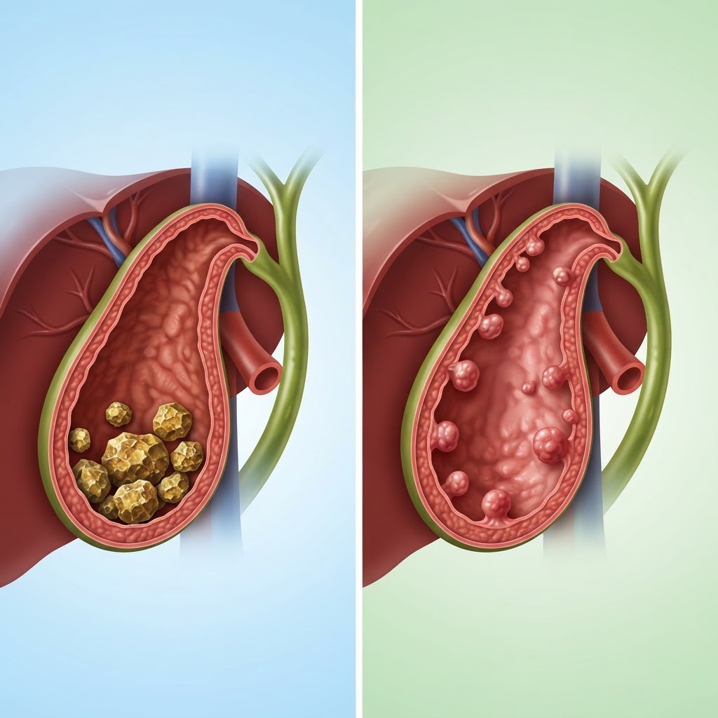
- Gallstones are hardened deposits of digestive fluid, whereas gallbladder polyps are abnormal tissue growths on the gallbladder lining.
- Diagnostic imaging, particularly gallbladder ultrasound results, is the primary method for distinguishing between these two conditions.
- While gallstones are frequently associated with biliary pain and inflammation, polyps are often asymptomatic and discovered incidentally.
- The risk of malignancy is the primary clinical concern with polyps, necessitating monitoring for those that exceed specific size thresholds.
- Understanding the clinical distinctions between gallstones vs gallbladder polyps is essential for determining appropriate follow-up and treatment strategies.
Receiving news of an abnormality in your gallbladder during a routine check-up or after experiencing abdominal discomfort can be an unsettling experience. For many, the diagnostic process involves looking at gallbladder ultrasound results that mention either “stones” or “polyps.” While these terms are frequently discussed in the context of gallbladder health, they represent fundamentally different physiological phenomena. Navigating the nuances of gallstones vs gallbladder polyps is crucial for patients and providers alike, as the management pathways—ranging from watchful waiting to surgical intervention—differ significantly. This article explores the clinical distinctions, diagnostic markers, and risk factors associated with each, providing the clarity needed to make informed decisions about your digestive health.
1. Understanding Gallstones: Causes and Common Symptoms
Gallstones, medically known as cholelithiasis, are hardened deposits of bile that form within the gallbladder. The gallbladder is a small, pear-shaped organ located beneath the liver, serving as a reservoir for bile—a digestive fluid produced by the liver that helps break down fats. When the chemical balance of this bile is disrupted, or if the gallbladder does not empty properly, the components of bile can crystallize into stones. These stones can range in size from as small as a grain of sand to as large as a golf ball, and they vary in composition, typically being categorized as cholesterol stones or pigment stones.
Cholesterol stones are by far the most common variety. They generally develop when the bile contains an excess of cholesterol, which the liquid bile can no longer dissolve. Over time, these microscopic crystals aggregate, forming solid masses. Pigment stones, on the other hand, are typically darker and composed of bilirubin—a byproduct of red blood cell breakdown. These are more often associated with specific underlying conditions, such as liver disease or blood disorders.
The primary concern regarding gallstones is not necessarily the presence of the stones themselves, but rather their potential to cause obstruction. Biliary pain—often referred to as a gallbladder attack—occurs when a stone moves and becomes lodged in one of the ducts leading out of the gallbladder. This prevents bile from flowing, causing the organ to swell and leading to intense, cramping pain in the upper right quadrant of the abdomen. This pain can radiate to the back or under the right shoulder blade.
Symptoms often manifest after eating, particularly after consuming a meal high in fat, as this triggers the gallbladder to contract and squeeze bile into the digestive tract. If a stone blocks the duct, the pain can last for several hours. Aside from pain, other signs of symptomatic gallstones may include nausea, vomiting, and, in more severe cases, jaundice—a yellowing of the skin and eyes—if the bile duct remains blocked, preventing bile from reaching the intestines. Not everyone with gallstones experiences symptoms; many individuals go their entire lives without realizing they have them. However, when symptoms do occur, they frequently signal a need for medical consultation to prevent complications such as inflammation (cholecystitis) or infection.
2. What Are Gallbladder Polyps and Why Do They Form?
Unlike gallstones, which are essentially “sludge” or crystals that have solidified, gallbladder polyps are abnormal tissue growths that project from the inner lining (mucosa) of the gallbladder wall. While gallstones are mobile and typically shift with the body’s position, polyps are fixed. They are common findings during routine abdominal imaging, and their discovery often leads to a period of careful assessment to determine their nature and potential for growth.
The majority of gallbladder polyps are benign, meaning they are non-cancerous. They are often classified into several types based on their histological structure. Cholesterol polyps are the most common; these are not true neoplasms (tumors) but rather localized accumulations of cholesterol-laden cells within the lining of the gallbladder wall. Because they are closely related to the same dietary and metabolic factors that cause gallstones, they are sometimes found in patients who also have cholelithiasis. Another common type is the adenomyoma, which is a benign overgrowth of the gallbladder muscle and mucosal lining. While less common, inflammatory polyps can also occur, usually as a response to chronic irritation or infection within the gallbladder.
Why exactly these polyps form is not always clear, but experts generally agree that a combination of factors, including age, metabolic health, and perhaps genetic predispositions, plays a role. Unlike gallstones, which are essentially a physical manifestation of stagnant or supersaturated bile, polyps are a structural change to the gallbladder wall itself. They do not typically cause the classic biliary pain associated with gallstones because they do not “block” the ducts in the same way, unless they are exceptionally large or situated near the cystic duct, which is rare.
The clinical challenge with polyps is that they are rarely symptomatic on their own. Most patients are entirely unaware of their presence until they undergo an ultrasound for an unrelated reason, such as bloating or vague abdominal discomfort. Because symptoms are rarely localized to the polyp itself, differentiating a gallbladder polyp vs stone symptoms becomes a primary focus for clinicians. The primary directive when managing polyps is to ensure they are not precancerous or malignant. While most polyps remain small and stable, clinicians keep a watchful eye on those that exhibit rapid growth or reach a certain size, as larger polyps are more statistically associated with a higher risk of malignancy.
3. Key Differences Between Stones and Polyps on Ultrasound
Diagnostic imaging is the cornerstone of distinguishing between these two gallbladder abnormalities. When a patient undergoes an ultrasound, the radiologist looks for specific visual “signatures” to differentiate between stones and polyps. These gallbladder ultrasound results are interpreted based on how the ultrasound waves interact with the objects inside the gallbladder.
The most definitive sign of a gallstone is “acoustic shadowing.” Because gallstones are solid, dense structures, the ultrasound waves hit the stone and are either reflected or absorbed, leaving a dark, empty space—a shadow—behind the stone on the screen. Furthermore, gallstones are gravity-dependent. If the patient changes their position during the exam—such as moving from lying on their back to lying on their side—the gallstones will typically “roll” or shift to the dependent part of the gallbladder. This movement is a hallmark characteristic that confirms the presence of mobile stones.
| Feature | Gallstones | Gallbladder Polyps | Best For |
|---|---|---|---|
| Mobility | Shifts with body position | Fixed to gallbladder wall | Determining status |
| Ultrasound Shadow | Distinct acoustic shadow | No shadow (usually) | Visual ID |
| Primary Risk | Obstruction and Biliary Pain | Malignancy potential | Clinical focus |
In contrast, polyps appear as small, stationary projections that grow from the gallbladder wall into the interior space (lumen). Because they are usually composed of soft tissue or cholesterol deposits rather than dense crystalline minerals, they typically do not produce the acoustic shadowing seen with gallstones. Crucially, they do not move when the patient changes position. They are tethered to the wall, and this fixed position is the clearest indicator for a radiologist to differentiate them from stones.
However, diagnostic ambiguity can still occur. For example, a small stone might become trapped in a fold of the gallbladder wall, mimicking a fixed polyp. Conversely, a large, calcified polyp might occasionally produce a faint shadow, complicating the assessment. In these instances, clinicians may rely on other imaging modalities or follow-up ultrasound scans to observe whether the object remains stable over time. Understanding these ultrasound nuances allows patients to better discuss their results with their primary care physician. If a report shows “hyperechoic foci without shadowing,” it is the technical way of describing a potential polyp; if the report mentions “shadowing” and “gravity-dependent movement,” it is the hallmark of gallstones.
4. Symptom Comparison: How to Tell the Difference
When comparing gallstones vs gallbladder polyps, the most striking difference is how they affect the daily life of the patient. Gallstones are notorious for their symptomatic nature, specifically the acute onset of biliary pain. This pain is often predictable, occurring after high-fat meals, and can be severe enough to require emergency care. The underlying mechanism is simple: the stone physically blocks the flow of bile. This obstruction increases pressure within the gallbladder, leading to the intense, spasmodic pain that characterizes a “gallbladder attack.”
Conversely, gallbladder polyps are generally silent. Because they are essentially bumps on the wall of the gallbladder, they rarely interfere with the storage or release of bile. Most people with polyps have no idea they have them until they undergo imaging for an unrelated issue. When a patient with polyps reports pain, it is often coincidental. Many patients found to have polyps also have concurrent gallstones, which may be the actual source of their discomfort. Distinguishing between gallbladder polyp vs stone symptoms requires a careful history—does the pain feel like a classic biliary colic, or is it more of a persistent, dull ache?
However, there are rare exceptions where polyps can become symptomatic. If a polyp is large enough and located very close to the neck of the gallbladder, it could potentially obstruct the cystic duct, leading to symptoms similar to gallstones. Additionally, if a polyp is associated with cholecystitis—inflammation of the gallbladder wall—it may be accompanied by fever, chills, and localized tenderness. These scenarios are far less common than the typical obstruction caused by a free-floating gallstone.
Because the symptoms of polyps are so subtle or entirely absent, the medical approach is markedly different from the approach for symptomatic stones. For gallstones, the presence of regular pain is often an indication for surgery (cholecystectomy) to prevent future attacks or complications like pancreatitis. For polyps, the lack of symptoms means the focus shifts almost entirely to monitoring for growth. If you are experiencing digestive issues, it is essential not to assume the cause is a polyp without professional confirmation. Digestive symptoms such as bloating, gas, and abdominal pain are common to many conditions, including gallbladder stones, gastritis, and irritable bowel syndrome. A professional diagnosis ensures you are treating the correct underlying cause rather than relying on guesswork.
5. Assessing the Cancer Risk: When Polyps Become Concerning
The conversation surrounding gallbladder polyps often centers on the “fear of the unknown,” specifically the concern regarding gallbladder cancer. While the overwhelming majority of gallbladder polyps are benign, they represent a small category of tissue growth that, if left unmonitored, could theoretically harbor malignant cells or progress toward cancer. This potential for malignancy is the primary reason why polyps are treated with more diagnostic caution than stones.
The size of the polyp is the most significant indicator for clinical concern. Experts generally agree that polyps smaller than 5 millimeters have a very low risk of becoming cancerous and often require no further intervention beyond periodic monitoring. As polyps grow beyond 10 millimeters, the statistical risk of malignancy increases. These larger polyps are considered more concerning because they have a higher likelihood of being true neoplasms (adenomas) rather than simple cholesterol deposits. For these cases, doctors may recommend more frequent imaging or even a surgical consultation to discuss whether the risks of the polyp outweigh the risks of gallbladder removal.
Beyond size, the rate of growth is another critical factor in assessing risk. If a series of scans over a period of months shows that a polyp is steadily increasing in size, it is a red flag that warrants closer evaluation. A stagnant, small polyp is typically viewed with less concern than one that demonstrates rapid expansion. Furthermore, the number of polyps matters. A single, large polyp is often viewed with more scrutiny than multiple, tiny polyps, as the latter are frequently consistent with benign cholesterol deposits.
While the cancer risk associated with gallbladder polyps is low in the general population, it is not zero. This is why the medical community emphasizes the importance of following up on any imaging report that mentions “gallbladder polyps.” Ignoring these findings is not recommended, as the early detection of growth is the best way to prevent potential complications. For patients with identified polyps, the standard of care usually involves a schedule of “surveillance imaging.” This ensures that if the lesion displays concerning characteristics, it can be addressed before it presents a significant health risk. In this context, diagnostic imaging acts as a safety net, providing the necessary data to determine when a condition that was once “watch and wait” needs to move to “proactive management.”
Diagnostic Procedures for Differentiating Growths
When you receive a gallbladder ultrasound report indicating an abnormality, it is common to experience anxiety. However, diagnostic imaging has become remarkably precise in distinguishing between gallstones and gallbladder polyps. The primary goal of any diagnostic workup is to determine the composition of the growth—whether it is a mobile, mineralized stone or an attached, tissue-based polyp—and to assess the risk of malignancy.
The first line of defense is almost always the transabdominal ultrasound. Radiologists look for specific markers to differentiate these two conditions. Gallstones typically exhibit “acoustic shadowing,” which means the sound waves cannot pass through the dense stone, creating a dark void behind it. Furthermore, stones are usually mobile; they follow the pull of gravity when the patient changes position. In contrast, a gallbladder polyp is a soft-tissue projection attached to the inner wall. Polyps do not cast an acoustic shadow, and they remain stationary regardless of how the patient moves.
When an ultrasound result is inconclusive, or if the polyp size approaches a clinical threshold of concern, physicians often escalate to more advanced imaging. Endoscopic Ultrasound (EUS) is considered the gold standard for high-resolution gallbladder imaging. By placing an ultrasound probe at the end of an endoscope inside the digestive tract, clinicians can get a much closer look at the gallbladder wall, allowing them to see the layered structure of the tissue. This helps determine if a growth is a true polyp or a pseudopolyp, such as cholesterol deposits that have attached to the wall.
Computed Tomography (CT) scans or Magnetic Resonance Imaging (MRI), specifically MR Cholangiopancreatography (MRCP), may also be utilized. While MRCP is primarily used to look at the bile ducts for blockage, it can provide additional data regarding the nature of a gallbladder mass. These procedures help rule out complications like inflammation, wall thickening, or evidence of cancer, which are vital for formulating a management plan.
| Diagnostic Method | Primary Capability | Best For |
|---|---|---|
| Transabdominal Ultrasound | Detecting mobility and acoustic shadows | Initial screening of suspected stones |
| Endoscopic Ultrasound (EUS) | High-resolution wall analysis | Differentiating small polyps from cancer |
| MRCP | Mapping the biliary tree anatomy | Identifying duct obstructions |
| HIDA Scan | Measuring gallbladder ejection fraction | Assessing functional gallbladder disease |
Treatment Options for Gallstones: Surgical vs Non-Surgical
Treatment for gallstones depends largely on whether the patient is experiencing biliary pain, commonly referred to as “biliary colic.” Many people harbor “silent” gallstones that never cause symptoms and require no intervention. However, once stones trigger repeated bouts of pain or inflammation (cholecystitis), medical professionals typically recommend a proactive approach.
The definitive treatment for symptomatic gallstones is a cholecystectomy, which is the surgical removal of the gallbladder. This procedure is performed using laparoscopic techniques in the vast majority of cases, involving small incisions and a rapid recovery time. Because the gallbladder is not an essential organ—the liver continues to produce bile even in its absence—most patients resume a normal lifestyle quickly after recovery.
Non-surgical options exist, though they are generally reserved for patients who are not candidates for surgery due to severe underlying health conditions. Oral dissolution therapy involves taking prescription medications containing bile acids to slowly dissolve cholesterol-based stones. This process can take months or even years and has a significant recurrence rate, as the gallbladder remains in place and continues to produce the conditions favorable to stone formation. Another method, extracorporeal shock wave lithotripsy, uses sound waves to shatter stones into smaller fragments that can then pass through the bile ducts. This method is rarely used today because the risk of a fragment becoming lodged in the bile duct and causing pancreatitis is a significant clinical concern.
Management Strategies for Monitoring Gallbladder Polyps
Managing gallbladder polyps requires a different philosophy than managing stones. Because most small polyps are benign, the strategy is typically one of “watchful waiting.” Physicians monitor polyps based on their size and growth trajectory. Small polyps—typically those under 6 millimeters—are often benign, and the recommendation may be to repeat an ultrasound after a specific interval to ensure they remain stable.
If a polyp is larger than 10 millimeters, the recommendation often shifts toward surgical intervention. As size increases, so does the statistical risk of malignancy. Clinicians monitor for “growth,” defined as a change in size over a six-to-twelve-month period. If a polyp is found to be rapidly expanding or if the patient is over the age of 50 (a demographic correlate for increased risk), doctors may suggest preventative removal.
The management plan also considers the presence of concurrent risk factors. Patients with Primary Sclerosing Cholangitis (PSC) or those who have had abnormal ultrasound results showing a broad-based polyp (sessile) rather than a narrow-necked one (pedunculated) are often monitored more aggressively. It is critical to maintain consistency with follow-up appointments, as serial imaging is the only way to track whether a polyp remains a benign observation or requires surgical removal to mitigate cancer risk.
Lifestyle Habits to Support Long-Term Gallbladder Health
While some risk factors for gallbladder issues, such as genetics, age, and biological sex, are beyond our control, lifestyle adjustments can significantly influence biliary health. The goal is to maintain a healthy bile composition and ensure regular gallbladder contractions, preventing the stagnation that leads to stone formation.
Dietary fiber is an essential component of gallbladder health. A diet high in soluble and insoluble fiber helps regulate cholesterol levels and promotes healthy digestion. By maintaining a stable weight and avoiding rapid, drastic weight loss—which is known to increase the risk of gallstone formation by altering bile concentration—you can protect your system. Gradual, sustainable weight loss is far safer for the biliary tract than crash dieting.
Healthy fats, such as those found in olive oil, avocados, nuts, and fatty fish, are preferable to saturated and trans fats. Consuming moderate amounts of healthy fats actually helps the gallbladder contract and empty regularly, which prevents bile from lingering too long and forming sludge or stones. Staying well-hydrated is equally important, as water is a key component of bile. Regular physical activity also helps lower cholesterol levels, reducing the likelihood of cholesterol-rich bile that facilitates stone growth.
Frequently Asked Questions
Are all gallbladder polyps dangerous?
No, the vast majority of gallbladder polyps are benign. Most are either cholesterol polyps, which are essentially small accumulations of cholesterol, or adenomyomatosis, a non-cancerous overgrowth of the gallbladder wall. However, larger polyps have a higher potential for malignancy, which is why doctors monitor them closely.
Can I dissolve a gallbladder polyp like I can a gallstone?
Generally, no. While certain bile acid medications are used in specific cases to dissolve cholesterol gallstones, these do not work for gallbladder polyps. Because polyps are tissue-based growths attached to the gallbladder wall, they do not respond to dissolution therapy.
What does it feel like to have a “gallbladder attack”?
Biliary pain typically manifests as a sudden, sharp, or cramping pain in the upper right quadrant of the abdomen, just below the rib cage. The pain often radiates to the right shoulder or back and can last anywhere from thirty minutes to several hours. It is frequently triggered by eating a fatty meal.
How often should I get an ultrasound if I have a small polyp?
The follow-up schedule is determined by your physician, but common practice involves repeating the ultrasound in six to twelve months to check for stability. If the polyp remains unchanged over multiple visits, your doctor may suggest lengthening the time between screenings or stopping surveillance altogether.
Does a gallbladder polyp eventually turn into a stone?
No, these are two distinct pathologies. A polyp is a growth attached to the tissue wall, while a stone is a hardened mass of bile ingredients that forms within the cavity of the gallbladder. One does not typically “turn into” the other, although they can coexist in the same gallbladder.
Is surgery always required for gallbladder polyps?
Surgery is not required for all polyps. It is typically reserved for polyps that exceed 10 millimeters, show rapid growth over time, or cause persistent symptoms such as pain or inflammation. If a polyp is small and stable, monitoring is often the preferred strategy.
Conclusion
Understanding the distinction between gallstones and gallbladder polyps is the first step toward effective management. While both can be detected during routine or diagnostic ultrasound imaging, their underlying mechanisms and clinical implications differ significantly. Gallstones are often tied to diet, metabolic health, and biliary flow, while gallbladder polyps require careful observation to rule out the potential for malignancy based on size and growth rate.
If you have been diagnosed with a gallbladder growth, do not panic. Most conditions are manageable with the right guidance. Whether your doctor suggests a conservative “watch and wait” approach or recommends surgical intervention, adhering to your follow-up schedule and adopting heart-healthy, high-fiber lifestyle habits will put you in the best position for long-term health. Take charge of your digestive wellness today by discussing these findings with your healthcare provider and ensuring you understand the specific nature of your diagnosis.
By healthauthoritylife Editorial Team
This article is for informational purposes only and is not a substitute for professional medical advice. Always consult your doctor before making changes to your diet, exercise routine, or treatment plan.
Leave a Reply